Medulla oblongata
The medulla oblongata is the caudal brainstem segment regulating vital autonomic functions and serving as a relay for cranial nerves.
The medulla oblongata forms the caudal portion of the brain stem, which collectively includes the midbrain, pons, and medulla. In embryological terms, the medulla is known as the myelencephalon. The term “myelencephalon” derives from the significant white matter found in this region, especially on its exterior, which is continuous with the white matter of the spinal cord. The tegmentum of the midbrain and pons continues into the medulla because this gray matter is responsible for processing cranial nerve information. A diffuse region of gray matter throughout the brain stem, known as the reticular formation, is related to sleep and wakefulness, such as general brain activity and attention. [1]
Figure 1Gray700
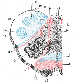
Long description
The formatio reticularis of the medulla oblongata, shown by a transverse section passing through the middle of the olive. (Testut.) 1. Anterior median fissure . 2. Fourth ventricle . 3. Formatio reticularis , with 3’, its internal part ( reticularis alba ), and 3’’, its external part ( reticularis g

Figure 1: The formatio reticularis of the medulla oblongata, shown by a transverse section passing through the middle of the olive ((testut.) 1).
Figure 2Gray681
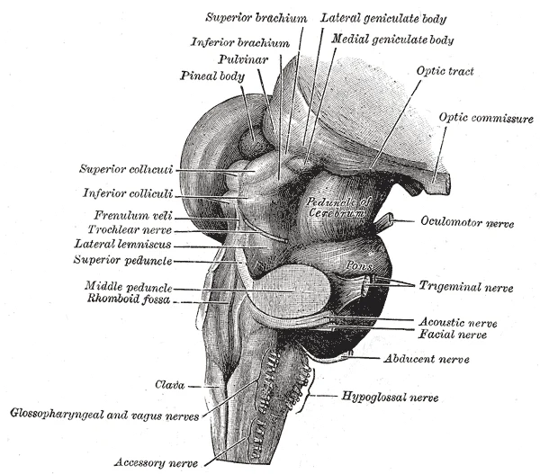
Long description
Hind- and mid-brains; postero-lateral view.

Figure 2: Hind- and mid-brains; postero-lateral view.
The nucleus tractus solitarius is a key sensory nucleus located in the medulla. It receives visceral sensory information from the cranial nerves, particularly the glossopharyngeal and vagus nerves. This nucleus is essential for monitoring blood pressure and oxygen levels, as well as taste sensation. It acts as the primary relay station for autonomic reflexes involving the cardiovascular and respiratory systems.
The nucleus ambiguus contains the cell bodies of motor neurons that supply the muscles of the pharynx, larynx, and soft palate. These muscles are innervated by the glossopharyngeal and vagus nerves. The nucleus is critical for swallowing, phonation, and coughing. Lesions in this area can lead to dysphagia and hoarseness, as the motor output to the striated muscles of the throat is disrupted.
It contains the cell bodies of parasympathetic preganglionic neurons. These neurons project to postganglionic neurons in the thorax and abdomen, regulating the heart rate, gastric motility, and secretion. It is the main source of parasympathetic outflow to the viscera.
The pons and the medulla regulate several crucial functions, including the cardiovascular and respiratory systems and rates. Fibers from this nucleus enter the cerebellum and are compared with the descending commands from the cerebrum. If the primary motor cortex sends a command to the spinal cord, a copy is sent to the cerebellum. The cerebellum compares these inputs to correct motor commands for coordination. [1]
| Nucleus | Primary Function | Nerve Association |
|---|---|---|
| Nucleus tractus solitarius | Visceral sensory relay | CN IX, CN X |
| Nucleus ambiguus | Motor to pharynx/larynx | CN IX, CN X |
| Dorsal motor nucleus of vagus | Parasympathetic preganglionic | CN X |
| Inferior olive | Motor coordination relay | Cerebellar input |
DefinitionMyelencephalon
The embryonic term for the medulla, named for its prominent external white matter continuous with the spinal cord.
Clinical pearlAutonomic Control
The medulla regulates cardiovascular and respiratory rates, making it vital for life-sustaining autonomic functions.
It is continuous with the spinal cord inferiorly and the pons superiorly. Understanding its gross anatomy requires examining both its external surface features and its internal nuclear organization, particularly those cranial nerves emerging from its lateral aspects. [1]
Figure 3Gray687
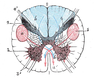
Long description
Section of the medulla oblongata through the lower part of the decussation of the pyramids. (Testut.) 1. Anterior median fissure. 2. Posterior median sulcus. 3. Anterior column (in red), with 3’, anterior root. 4. Posterior column (in blue), with 4’, posterior roots. 5. Lateral cerebrospinal fascicu

Figure 3: Section of the medulla oblongata through the lower part of the decussation of the pyramids ((testut.) 1).
) 1). Flanking this fissure are the pyramids, which contain the descending corticospinal tracts. At the lower end of the pyramids, most fibres decussate to form the pyramidal decussation. Lateral to the pyramids lie the olives, which are rounded elevations containing the inferior olivary nucleus. [1]
Figure 4Gray699
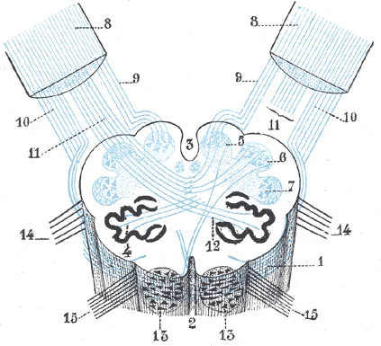
Long description
Diagram showing the course of the arcuate fibers. (Testut.) 1. Medulla oblongata anterior surface. 2. Anterior median fissure. 3. Fourth ventricle. 4. Inferior olivary nucleus, with the accessory olivary nuclei. 5. Gracile nucleus. 6. Cuneate nucleus. 7. Trigeminal. 8. Inferior peduncles, seen from

Figure 4: Diagram showing the course of the arcuate fibers ((testut.) 1).
) 1). The pyramids are prominent longitudinal ridges located on the anterior surface, containing the descending corticospinal tracts. Lateral to the pyramids lie the olives, which are oval bulges containing the inferior olivary nucleus. The inferior olivary nucleus is involved in comparing descending commands with sensory feedback. [1]
The medulla contains the nuclei for cranial nerves IX (glossopharyngeal), X (vagus), XI (accessory), and XII (hypoglossal). These nuclei are located in the floor of the fourth ventricle or in the parenchyma of the medulla. The hypoglossal nucleus lies near the midline in the floor of the fourth ventricle. The dorsal motor nucleus of the vagus is situated lateral to the hypoglossal nucleus. [1]
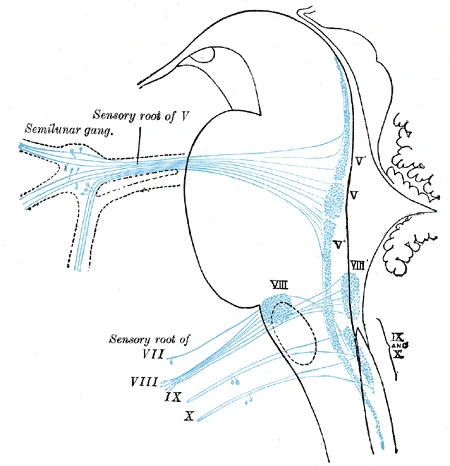
Figure 5Gray698
Long description
Primary terminal nuclei of the afferent (sensory) cranial nerves schematically represented; lateral view. The olfactory and optic centers are not represented.

Figure 5: Primary terminal nuclei of the afferent (sensory) cranial nerves schematically represented; lateral view.
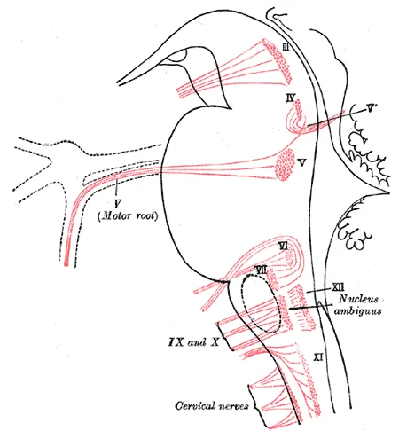
Figure 6Gray697
Long description
Nuclei of origin of cranial motor nerves schematically represented; lateral view.

Figure 6: Nuclei of origin of cranial motor nerves schematically represented; lateral view.
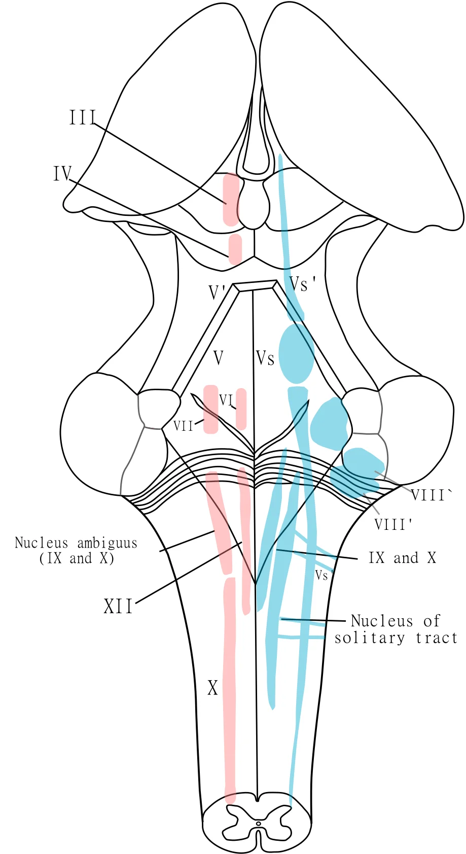
Figure 7Gray696
Long description
The cranial nerve nuclei schematically represented; dorsal view. Motor nuclei in red; sensory in blue. (The olfactory and optic centers are not represented.)

Figure 7: The cranial nerve nuclei schematically represented; dorsal view.
Lateral medullary syndrome, also known as Wallenberg syndrome, results from occlusion of the posterior inferior cerebellar artery or the vertebral artery. This lesion affects the lateral part of the medulla. Clinical features include ipsilateral loss of pain and temperature sensation on the face due to involvement of the spinal trigeminal nucleus. There is also contralateral loss of pain and temperature sensation on the body due to involvement of the spinothalamic tract. Other signs include ipsilateral Horner syndrome, ataxia, dysphagia, and hoarseness. [1]
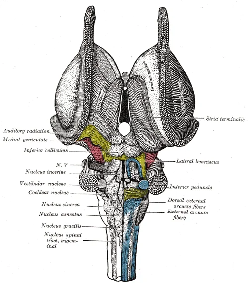
Figure 8Gray691
Long description
Dissection of brain-stem. Dorsal view. The nuclear masses of the medulla are taken from model by Weed, Carnegie Publication, No. 19.

Figure 8: Dissection of brain-stem (dorsal view).
Figure 9Gray686
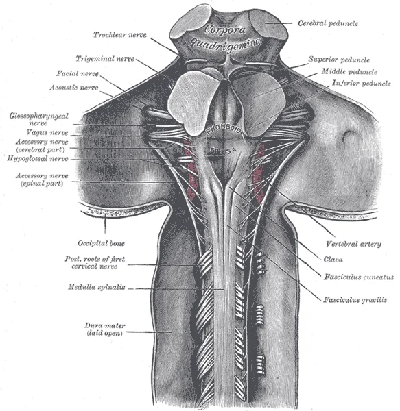
Long description
Upper part of medulla spinalis and hind- and mid-brains; posterior aspect, exposed in situ.

Figure 9: Upper part of medulla spinalis and hind- and mid-brains; posterior aspect, exposed in situ.
The medulla is related to the foramen magnum inferiorly and the pons superiorly. The cerebellum lies posterior to the medulla. The subarachnoid space surrounds the medulla, containing cerebrospinal fluid.
DefinitionMedulla Oblongata
The most caudal part of the brainstem, connecting the spinal cord to the pons and containing vital autonomic centres.
| Nerve | Nucleus | Function |
|---|---|---|
| IX | Nucleus ambiguus | Motor to pharynx and larynx |
| X | Dorsal motor nucleus | Parasympathetic to thoracic and abdominal viscera |
| XI | Nucleus ambiguus | Motor to sternocleidomastoid and trapezius |
| XII | Hypoglossal nucleus | Motor to tongue muscles |
The medulla oblongata forms the most inferior part of the brain stem, connecting directly to the spinal cord below and the pons above. It serves as a critical conduit for major ascending and descending pathways between the spinal cord and the higher brain centres, specifically the cerebrum. Additionally, it houses control centres that regulate vital autonomic functions, including the cardiovascular and respiratory systems. The cranial nerves connect through the brain stem, providing the brain wi [1]
Figure 10Gray709
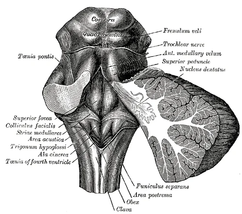
Long description
Rhomboid fossa. Labelled illustration from Wikimedia Commons, used under its open licence.

Figure 10: Rhomboid fossa.
Figure 11Gray695
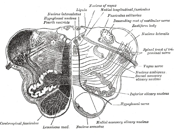
Long description
Transverse section of medulla oblongata below the middle of the olive.

Figure 11: Transverse section of medulla oblongata below the middle of the olive.
DefinitionInternal Structure
The medulla is divided into open and closed parts, containing distinct nuclei, laminae, and tracts that facilitate neural transmission and reflex control.
In cross-section, the internal architecture is defined by the arrangement of grey matter nuclei and white matter tracts. The white matter is separated into columns, analogous to the spinal cord. Ascending tracts carry sensory information up to the brain, while descending tracts carry motor commands from the brain. Between the two posterior horns of grey matter are the posterior columns. The anterior columns are bounded by the axons of motor neurons [1]
Figure 12Gray688
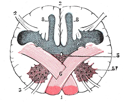
Long description
Section of the medulla oblongata at the level of the decussation of the pyramids. (Testut.) 1. Anterior median fissure. 2. Posterior median sulcus. 3. Motor roots. 4. Sensory roots. 5. Base of the anterior column, from which the head (5’) has been detached by the lateral cerebrospinal fasciculus. 6.

Figure 12: Section of the medulla oblongata at the level of the decussation of the pyramids ((testut.) 1).
The pyramids are prominent longitudinal ridges located on the anterior surface, containing the descending corticospinal tracts. Lateral to the pyramids lie the olives, which are oval bulges containing the inferior olivary nucleus. The inferior olivary nucleus is involved in comparing descending commands with sensory feedback.
The grey matter is organized into specific nuclei that correspond to the cranial nerves. The hypoglossal nucleus (XII) is located near the midline, giving rise to fibres that exit anteriorly. The dorsal motor nucleus of the vagus (X) lies lateral to the hypoglossal nucleus, providing parasympathetic output. These nuclei are arranged in a columnar fashion, reflecting their functional roles in autonomic and somatic control.
The white matter tracts include the medial lemniscus, which carries fine touch and proprioception, and the spinothalamic tract, which carries pain and temperature sensation. These tracts ascend through the medulla, decussating at various levels to reach the thalamus. The corticospinal tracts descend through the pyramids, with most fibres crossing at the medullary-spinal junction. This organization ensures that motor commands and sensory inputs are correctly routed to and from the brain.
| Nucleus | Cranial Nerve | Primary Function |
|---|---|---|
| Hypoglossal | XII | Motor innervation to tongue muscles |
| Nucleus Ambiguus | IX, X, XI | Motor innervation to pharynx and larynx |
| Dorsal Motor Nucleus | X | Parasympathetic output to thoracic and abdominal viscera |
| Nucleus Solitarius | IX, X | Visceral sensory input from taste and baroreceptors |
Understanding the internal structure of the medulla is essential for localizing lesions. Damage to the pyramids results in contralateral hemiparesis. Lesions affecting the medial lemniscus cause loss of proprioception and vibration sense. Involvement of the cranial nerve nuclei leads to specific deficits such as dysphagia or tongue deviation. The precise anatomical knowledge of these structures allows for accurate clinical diagnosis and management.
The medulla oblongata serves as the critical junction between the brainstem and the spinal cord, facilitating the passage of both ascending sensory and descending motor pathways. Understanding its connections requires tracing the trajectory of these tracts as they enter, exit, or relay within this structure. [1]
Afferent pathways bring sensory information from the periphery to the higher centres. The primary sensory tracts involved are the dorsal columns and the spinothalamic tract.
The dorsal columns carry fine touch, vibration, and proprioception. These fibres enter the spinal cord and ascend ipsilaterally in the fasciculus gracilis and fasciculus cuneatus. They terminate in the nucleus gracilis and nucleus cuneatus, located in the caudal medulla. From these nuclei, second-order neurons decussate as internal arcuate fibres and ascend as the medial lemniscus.
Figure 13Gray693
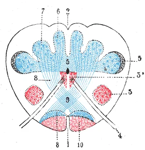
Long description
Transverse section passing through the sensory decussation. (Schematic.) 1. Anterior median fissure. 2. Posterior median sulcus. 3, 3. Head and base of anterior column (in red). 4. Hypoglossal nerve. 5. Bases of posterior columns. 6. Gracile nucleus. 7. Cuneate nucleus. 8, 8. Lemniscus. 9. Sensory d

Figure 13: Transverse section passing through the sensory decussation (Schematic.) 1.
The spinothalamic tract carries pain and temperature sensation. First-order neurons synapse in the dorsal horn of the spinal cord. Second-order neurons decussate in the anterior white commissure and ascend contralaterally through the medulla.
Efferent pathways transmit motor commands from the cortex to the spinal cord and brainstem nuclei. The principal descending tract is the corticospinal tract.
The corticospinal tract originates in the motor cortex. Fibres descend through the internal capsule, cerebral peduncles, and pons. In the caudal medulla, approximately 85-90% of fibres decussate at the pyramidal decussation. These crossed fibres form the lateral corticospinal tract, which descends in the lateral funiculus of the spinal cord. The uncrossed fibres continue as the anterior corticospinal tract, decussating at the spinal level of termination.
Other efferent pathways include the corticobulbar tract, which terminates in cranial nerve nuclei within the brainstem, including those in the medulla. These nuclei control muscles of the face, head, and neck. [1]
Figure 14Gray679
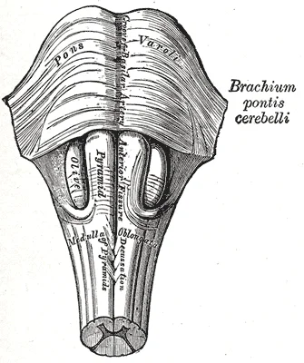
Long description
Medulla oblongata and pons. Anterior surface.

Figure 14: Medulla oblongata and pons (anterior surface).
Posterior to the pyramids are the olives, housing the inferior olivary nucleus. Lateral to the olives are the inferior cerebellar peduncles.
DefinitionPyramidal Decussation
The point in the caudal medulla where the majority of corticospinal fibres cross from one side to the other.
| Tract | Type | Course | Termination |
|---|---|---|---|
| Dorsal Columns | Afferent | Ascends ipsilaterally | Nucleus gracilis/cuneatus |
| Spinothalamic | Afferent | Ascends contralaterally | Thalamus |
| Corticospinal | Efferent | Decussates in medulla | Spinal cord |
| Corticobulbar | Efferent | Terminates in brainstem | Cranial nerve nuclei |
The medulla oblongata receives its vascular supply primarily from the vertebral arteries and their branches, with minor contributions from the anterior spinal artery and the posterior spinal arteries. Understanding this supply requires tracing the course of these vessels as they enter the cranial cavity and distribute blood to the distinct tracts and nuclei within the medulla.
Figure 15Gray694
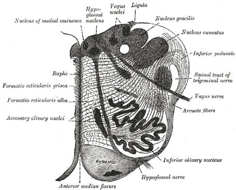
Long description
Detail of the Medulla Oblongata in the human brain

Figure 15: Detail of the Medulla Oblongata in the human brain.
The anterior spinal artery, formed by the union of branches from both vertebral arteries, runs along the anterior median fissure. The posterior inferior cerebellar arteries (PICA) branch off the vertebral arteries and supply the lateral medulla. The anterior spinal artery lies anterior to the pyramids. [1]
The anterior spinal artery supplies the medial medulla. It gives off paramedian branches that penetrate the anterior median fissure to supply the medial lemniscus, the corticospinal tract, and the hypoglossal nerve nucleus. Occlusion of these branches leads to medial medullary syndrome. This syndrome is characterised by contralateral hemiparesis due to corticospinal tract involvement, contralateral loss of proprioception and vibration sense due to medial lemniscus damage, and ipsilateral tongue weakness due to hypoglossal nerve nucleus injury.
They run along the posterolateral sulcus and give off branches to the gracile and cuneate nuclei. These nuclei are located in the dorsal medulla and are responsible for transmitting proprioceptive and fine touch information from the lower and upper limbs, respectively.
The lateral medulla is supplied by the posterior inferior cerebellar artery (PICA) and sometimes by direct branches from the vertebral artery. The PICA arises from the vertebral artery and courses posteriorly to supply the inferior cerebellum. Before reaching the cerebellum, it gives off lateral medullary branches. These branches supply the lateral part of the medulla, including the spinothalamic tract, the nucleus ambiguus, the vestibular nuclei, and the descending sympathetic fibres. Occlusion of the PICA or its lateral medullary branches results in lateral medullary syndrome, also known as Wallenberg syndrome. This syndrome presents with ipsilateral ataxia, Horner syndrome, loss of pain and temperature sensation on the ipsilateral face, and contralateral loss of pain and temperature sensation on the body.
The development of this vascular network begins early in embryogenesis. These arteries obtain blood from carotid-vertebrobasilar anastomoses, including the trigeminal, otic, hypoglossal, and proatlantal arteries.
Clinical pearlMedial vs Lateral
Medial medullary syndrome affects the corticospinal tract and medial lemniscus. Lateral medullary syndrome affects the spinothalamic tract and vestibular nuclei.
| Syndrome | Vessel | Structures Affected | Key Deficits |
|---|---|---|---|
| Medial | Anterior Spinal Artery | Corticospinal tract, Medial lemniscus, Hypoglossal nerve | Contralateral hemiparesis, Contralateral proprioception loss, Ipsilateral tongue weakness |
| Lateral | PICA or Vertebral Artery | Spinothalamic tract, Nucleus ambiguus, Vestibular nuclei, Descending sympathetic | Contralateral pain/temp loss, Dysphagia, Horner syndrome, Ataxia |
The medulla oblongata serves as the critical junction between the brainstem and the spinal cord, housing the nuclei for several cranial nerves and controlling vital autonomic functions. It is the lowest part of the brainstem, situated directly above the spinal cord and below the pons. The structure is responsible for regulating life-sustaining processes, including respiration, cardiovascular function, and reflex actions such as swallowing, vomiting, and coughing. Understanding its function requi [1]
The pyramids are prominent longitudinal ridges located on the anterior surface, containing the descending corticospinal tracts. Lateral to the pyramids lie the olives, which are oval bulges containing the inferior olivary nucleus. The inferior olivary nucleus is involved in comparing descending commands with sensory feedback.
DefinitionVital Centers
The medulla contains vital centers for respiration and cardiovascular regulation, which are essential for survival.
The respiratory center in the medulla regulates the rate and depth of breathing. It consists of the dorsal respiratory group and the ventral respiratory group. The dorsal respiratory group is primarily responsible for inspiration, while the ventral respiratory group is involved in both inspiration and expiration. These centers respond to changes in blood pH and carbon dioxide levels, adjusting breathing accordingly. [1]
The cardiovascular center regulates heart rate and blood pressure. [1]
| Nucleus | Cranial Nerve | Function |
|---|---|---|
| Hypoglossal | XII | Motor to tongue muscles |
| Dorsal motor nucleus of vagus | X | Parasympathetic to thoracic and abdominal viscera |
| Nucleus ambiguus | IX, X, XI | Motor to pharynx, larynx, and upper esophagus |
| Solitary nucleus | VII, IX, X | Sensory for taste and visceral afferents |
| Gracile nucleus | - | Sensory for proprioception and fine touch from lower body |
| Cuneate nucleus | - | Sensory for proprioception and fine touch from upper body |
Lesions of the medulla can result in severe deficits, including respiratory failure, cardiac arrhythmias, and loss of cranial nerve function. Medullary syndromes, such as Wallenberg syndrome, result from vascular occlusion and present with specific patterns of sensory and motor deficits. Understanding the anatomy and function of the medulla is crucial for diagnosing and managing these conditions. [1]
The medulla oblongata serves as the critical junction between the brainstem and the spinal cord, housing nuclei that regulate vital autonomic functions. Lesions in this region produce distinct clinical syndromes depending on whether the damage is medial or lateral. Understanding these patterns requires a precise anatomical localisation of the affected tracts and nuclei.
) 1). The lesion is typically caused by occlusion of the posterior inferior cerebellar artery or the vertebral artery. The inferior olivary nucleus is centrally located and serves as a landmark for the medial-lateral distinction.
DefinitionLateral Medullary Syndrome
Also known as Wallenberg syndrome, this condition results from infarction of the lateral medulla, typically due to vertebral artery occlusion.
The clinical features of lateral medullary syndrome are diverse and reflect the multiple structures involved. Patients often present with vertigo, nausea, and vomiting due to vestibular nucleus involvement. Ipsilateral Horner syndrome occurs from disruption of the descending sympathetic fibres. Dysphagia and hoarseness result from damage to the nucleus ambiguus, which supplies the pharyngeal and laryngeal muscles. Loss of pain and temperature sensation on the ipsilateral face is due to spinal trigeminal nucleus damage. Contralateral loss of pain and temperature sensation in the body occurs from spinothalamic tract involvement. Ataxia and dysmetria arise from inferior cerebellar peduncle damage. Hiccups are a common and distinctive feature.
| Structure Affected | Clinical Deficit |
|---|---|
| Vestibular nuclei | Vertigo, nystagmus, nausea |
| Descending sympathetic fibres | Ipsilateral Horner syndrome |
| Nucleus ambiguus | Dysphagia, hoarseness, loss of gag reflex |
| Spinal trigeminal nucleus | Ipsilateral loss of pain/temperature on face |
| Spinothalamic tract | Contralateral loss of pain/temperature on body |
| Inferior cerebellar peduncle | Ipsilateral ataxia, dysmetria |
Medial medullary syndrome, in contrast, involves the medial structures. It is characterised by contralateral hemiparesis due to pyramidal tract damage. Contralateral loss of proprioception and vibration sense occurs from medial lemniscus involvement. Ipsilateral tongue weakness and deviation result from hypoglossal nucleus damage. This syndrome is typically caused by occlusion of the anterior spinal artery.
The differential diagnosis of brainstem lesions is crucial. Lateral medullary syndrome must be distinguished from other causes of vertigo and dysphagia. Imaging, such as MRI, is essential for confirmation. The clinical presentation provides strong localising value. Early recognition improves management and outcomes.
The medulla oblongata is the most caudal part of the brainstem, continuous inferiorly with the spinal cord and superiorly with the pons. It serves as a critical relay station and contains vital autonomic centres.
The medulla houses the nuclei for cranial nerves IX, X, XI, and XII.
- Hypoglossal nerve (CN XII): Motor supply to the tongue muscles. The nucleus lies in the floor of the fourth ventricle. Lesions cause ipsilateral tongue deviation towards the side of the lesion.
- Vagus nerve (CN X): Parasympathetic and motor supply to the pharynx, larynx, and viscera. The nucleus ambiguus provides motor fibres.
- Glossopharyngeal nerve (CN IX): Sensory from the posterior tongue and pharynx; parasympathetic to the parotid gland.
- Accessory nerve (CN XI): Motor to sternocleidomastoid and trapezius. The spinal root ascends through the foramen magnum.
The medulla contains the cardiac, respiratory, and vasomotor centres.
- Cardiac centre: Located in the reticular formation. It modulates heart rate via the vagus nerve (parasympathetic) and sympathetic fibres.
- Respiratory centre: Includes the dorsal and ventral respiratory groups. It generates the basic rhythm of breathing.
- Vasomotor centre: Regulates blood pressure by controlling the diameter of arterioles via sympathetic outflow.
The anterior surface of the medulla features the pyramids, containing corticospinal tract fibres.
- Decussation of pyramids: Approximately 85-90% of fibres cross to the opposite side at the lower medulla. This explains why a left-sided brain lesion causes right-sided body weakness.
Sensory pathways ascend through the medulla.
- Medial lemniscus: Carries fine touch, vibration, and proprioception from the body. Fibers from the nucleus gracilis (lower body) and nucleus cuneatus (upper body) decussate as internal arcuate fibres.
- Spinothalamic tract: Carries pain and temperature. These fibres have already crossed in the spinal cord and ascend in the lateral medulla.
- Medial Medullary Syndrome (Dejerine): Caused by occlusion of the anterior spinal artery. Presents with contralateral hemiparesis (corticospinal tract), contralateral loss of proprioception (medial lemniscus), and ipsilateral tongue deviation (CN XII).
- Lateral Medullary Syndrome (Wallenberg): Caused by occlusion of the posterior inferior cerebellar artery (PICA) or vertebral artery. Presents with ipsilateral Horner’s syndrome, ataxia, loss of pain/temperature on the ipsilateral face and contralateral body, dysphagia, and hoarseness.
- Anterior: Foramen magnum, tectorial membrane.
- Posterior: Fourth ventricle, inferior medullary velum.
- Lateral: Inferior cerebellar peduncle.
Exam trapDecussation Timing
Remember that the corticospinal tract decussates in the medulla, but the spinothalamic tract decussates in the spinal cord. This is a common exam trap.
DefinitionInternal Arcuate Fibres
These are the axons of the nucleus gracilis and nucleus cuneatus that cross the midline to form the medial lemniscus.
| Syndrome | Vessel | Key Deficits |
|---|---|---|
| Medial | Anterior Spinal Artery | Contralateral hemiparesis, contralateral proprioception loss, ipsilateral tongue deviation |
| Lateral | PICA or Vertebral Artery | Ipsilateral Horner’s, ataxia, facial pain/temp loss, contralateral body pain/temp loss, dysphagia |
Students frequently confuse the functional anatomy of the basal ganglia with the structural anatomy of the brainstem. A primary error is assuming that the substantia nigra pars compacta directly initiates movement. In reality, the substantia nigra pars compacta acts as a switch between pathways rather than a direct motor initiator. It projects to the striatum and releases dopamine, which modulates the direct and indirect pathways. Dopamine receptors are either excitatory (D1-type) or inhibitory (D2-type). The direct pathway is activated by dopamine, while the indirect pathway is inhibited by dopamine. When the substantia nigra pars compacta is firing, it signals an active state where movement is likely. Conversely, when it is silent, the body is in a passive state and movement is inhibited. For example, while a student sits listening to a lecture, the substantia nigra pars compacta is silent, making the student less likely to get up. [1]
Another common pitfall involves the structural identification of the pons. Students often mistake the entire pons for a uniform mass of white matter. The word pons comes from the Latin word for bridge, referring to the thick bundle of white matter on the anterior surface attached to the cerebellum. This bridge-like white matter is only the anterior surface; the gray matter beneath is a continuation of the tegmentum from the midbrain. Gray matter in the tegmentum contains neurons receiving descending input from the forebrain sent to the cerebellum. Confusing the anterior white matter with the posterior gray matter leads to errors in understanding sensory and motor relay functions. [1]
Students also misinterpret the scale and function of the cerebellum. It is described as the “little brain” because it is covered in gyri and sulci like the cerebrum, resembling a miniature version. It accounts for approximately 10 percent of the mass of the brain. Its primary role is comparing information from the cerebrum with sensory feedback from the periphery through the spinal cord. Failing to recognise this comparative function leads to an incomplete understanding of coordination disorders. [1]
A significant anatomical misconception concerns motor neuron size. Some of the largest neurons of the spinal cord are the multipolar motor neurons in the anterior horn. The fibers causing skeletal muscle contraction are the axons of these neurons. For instance, the motor neuron for the big toe is in the sacral cord, and its axon may be a meter long. The cell body maintaining this fiber must be large, possibly several hundred micrometers in diameter, making it one of the largest cells in the body. Underestimating this scale obscures the metabolic demands of long-range motor control. [1]
Vascular variants are often overlooked or misidentified. A persistent stapedial artery has a reported prevalence of 0.5%. It supplies the middle meningeal artery territory, leading to the absence of the foramen spinosum. Absence of the foramen spinosum is seen in up to 3% of skull-base CT studies. This absence is an indirect sign of a persistent stapedial artery but can also relate to an ophthalmic origin of the middle meningeal artery. True ICA agenesis, aplasia, or hypoplasia is reported in 0. [2]
Duplication of the PCOMM artery is a rare event with a prevalence of 1.8% in surgical dissection series. [2]
At the bedside, understanding the substantia nigra pars compacta is crucial for managing movement disorders. The switch between pathways is the substantia nigra pars compacta, which projects to the striatum and releases the neurotransmitter dopamine. Dopamine receptors are either excitatory (D1-type receptors) or inhibitory (D2-type receptors). The direct pathway is activated by dopamine, and the indirect pathway is inhibited by dopamine. When the substantia nigra pars compacta is firing, it signals to the basal nuclei that the body is in an active state, and movement will be more likely. When the substantia nigra pars compacta is silent, the body is in a passive state, and movement is inhibited. To illustrate this situation, while a student is sitting listening to a lecture, the substantia nigra pars compacta would be silent and the student less likely to get up and walk around. This physiological model explains the bradykinesia and rigidity seen in Parkinson’s disease, where dopaminergic neurons degenerate. [1]
In the operating theatre, knowledge of the pons is vital for neurosurgical approaches. The word pons comes from the Latin word for bridge. It is visible on the anterior surface of the brain stem as the thick bundle of white matter attached to the cerebellum. The pons is the main connection between the cerebellum and the brain stem. The bridge-like white matter is only the anterior surface of the pons; the gray matter beneath that is a continuation of the tegmentum from the midbrain. Gray matter in the tegmentum region of the pons contains neurons receiving descending input from the forebrain that is sent to the cerebellum. Surgeons must distinguish the anterior white matter tracts from the posterior tegmental gray matter to avoid disrupting cranial nerve nuclei and ascending sensory pathways. [1]
On imaging, recognising vascular variants prevents misdiagnosis. The reported prevalence of a persistent stapedial artery is 0.5%. The persistent stapedial artery supplies the middle meningeal artery territory, leading to the absence of the foramen spinosum. Absence of the foramen spinosum is seen in up to 3% of skull-base CT studies. It is an indirect sign of a persistent stapedial artery, but it can also be related to an ophthalmic origin of the middle meningeal artery. A true ICA agenesis, aplasia, or hypoplasia is reported in 0.01% of the population. Radiologists must identify these variants to avoid unnecessary interventions or misinterpretation of collateral circulation. [2]
Fetal origin of the PCA is the most frequent variant involving this artery. It has been reported in between 13%-15% of the population. It is typically described in only one PCA, but it can also occur in both PCA in up to 0.5% of the population. More commonly (in up to 10% of the cases), the P1 segment is hypoplastic, which renders a fetal-like circulation to the occipital lobes, although it is not technically a fetal PCA given the physical link of the PCOMM with the BA. Duplications of the PCA ( [2]
In examinations, students must recall specific anatomical details. Some of the largest neurons of the spinal cord are the multipolar motor neurons in the anterior horn. The fibers that cause contraction of skeletal muscles are the axons of these neurons. The motor neuron that causes contraction of the big toe, for example, is located in the sacral spinal cord. The axon that has to reach all the way to the belly of that muscle may be a meter in length. The neuronal cell body that maintains that long fiber must be quite large, possibly several hundred micrometers in diameter, making it one of the largest cells in the body. [1]
The cerebellum, as the name suggests, is the “little brain”. It is covered in gyri and sulci like the cerebrum, and looks like a miniature version of that part of the brain. The cerebellum is largely responsible for comparing information from the cerebrum with sensory feedback from the periphery through the spinal cord. It accounts for approximately 10 percent of the mass of the brain. [1]
Some plausible explanations for this presumed observation are the fact that smooth muscle cells and collagen are decreased at the proximal and distal end of the duplicated segment. [2]
The substantia nigra pars compacta projects to the striatum and releases the neurotransmitter dopamine. Dopamine receptors are either excitatory (D1-type receptors) or inhibitory (D2-type receptors). The direct pathway is activated by dopamine, and the indirect pathway is inhibited by dopamine. When the substantia nigra pars compacta is firing, it signals to the basal nuclei that the body is in an active state, and movement will be more likely. When the substantia nigra pars compacta is silent, the body is in a passive state, and movement is inhibited. The pons is the main connection between the cerebellum and the brain stem. Gray matter in the tegmentum region of the pons contains neurons receiving descending input from the forebrain that is sent to the cerebellum. The cerebellum accounts for approximately 10 percent of the mass of the brain.
Answer each one in your head first, then open the answer.
What neurotransmitter does the substantia nigra pars compacta release?
Show answerHide answer
DopamineWhich pathway is inhibited by dopamine?
Show answerHide answer
The indirect pathwayWhat is the reported prevalence of a persistent stapedial artery?
Show answerHide answer
0.5%What is the most frequent variant involving the PCA?
Show answerHide answer
Fetal origin of the PCAWhat percentage of the brain's mass does the cerebellum account for?
Show answerHide answer
10 percent
- Betts JG, Young KA, Wise JA, et al. The Central Nervous System. OpenStax, Anatomy and Physiology 2e (CC BY-NC-SA 4.0). Accessed .
- Menshawi K, Mohr JP, Gutierrez J. A Functional Perspective on the Embryology and Anatomy of the Cerebral Blood Supply. Journal of stroke 2015;17(2):144-158. doi:10.5853/jos.2015.17.2.144. Accessed .
Figure credits
- Figure 1: Henry Vandyke Carter, via Wikimedia Commons, Public domain, Public domain.
- Figure 2: Henry Vandyke Carter, via Wikimedia Commons, Public domain, Public domain.
- Figure 3: Henry Vandyke Carter, via Wikimedia Commons, Public domain, Public domain.
- Figure 4: Henry Vandyke Carter, via Wikimedia Commons, Public domain, Public domain.
- Figure 5: Henry Vandyke Carter, via Wikimedia Commons, Public domain, Public domain.
- Figure 6: Henry Vandyke Carter, via Wikimedia Commons, Public domain, Public domain.
- Figure 7: Gray696.png: User:mcstrother derivative work: Mcstrother, via Wikimedia Commons, Public domain, Public domain.
- Figure 8: Henry Vandyke Carter, via Wikimedia Commons, Public domain, Public domain.
- Figure 9: Henry Vandyke Carter, via Wikimedia Commons, Public domain, Public domain.
- Figure 10: Henry Vandyke Carter, via Wikimedia Commons, Public domain, Public domain.
- Figure 11: Henry Vandyke Carter, via Wikimedia Commons, Public domain, Public domain.
- Figure 12: Henry Vandyke Carter, via Wikimedia Commons, Public domain, Public domain.
- Figure 13: Henry Vandyke Carter, via Wikimedia Commons, Public domain, Public domain.
- Figure 14: Henry Vandyke Carter, via Wikimedia Commons, Public domain, Public domain.
- Figure 15: Henry Vandyke Carter, via Wikimedia Commons, Public domain, Public domain.
Educational content for medical students and doctors. It is not a substitute for clinical judgement or local guidelines.