Pons
The pons is a brainstem structure connecting the cerebellum to the rest of the brain, housing vital motor and autonomic pathways.
The term pons derives from the Latin word for bridge, reflecting its anatomical role as a connector. It is visible on the anterior surface of the brain stem as a thick bundle of white matter attached to the cerebellum. This bridge-like white matter constitutes only the anterior surface of the pons; the gray matter beneath this is a continuation of the tegmentum from the midbrain. The pons serves as the main connection between the cerebellum and the brain stem. [1]
DefinitionPontine Anatomy
The pons consists of an anterior basal portion and a posterior tegmentum, together forming the bridge between the midbrain and medulla.
The basal pons, or basal part, comprises the large anterior mass of transverse and longitudinal fibres. It acts as the primary relay station for information travelling between the cerebral cortex and the cerebellum. Gray matter in the tegmentum region of the pons contains neurons receiving descending input from the forebrain that is sent to the cerebellum. These pontine nuclei receive cortical input and send projections via the middle cerebellar peduncles to the contralateral cerebellar cortex. This pathway allows for the coordination of voluntary movements by comparing intended motor commands with actual sensory feedback.
Throughout the midbrain, pons, and medulla, the tegmentum contains the nuclei that receive and send information through the cranial nerves. [1]
NoteTegmental Function
The tegmentum is the posterior part of the brainstem, containing cranial nerve nuclei and ascending/descending tracts, distinct from the anterior basal pons.
A diffuse region of gray matter throughout the brain stem, known as the reticular formation, is related to sleep and wakefulness, such as general brain activity and attention. The brain stem controls the head and neck region of the body through the cranial nerves. There are control centers in the brain stem that regulate the cardiovascular and respiratory systems. The pontine respiratory group, located within the reticular formation, helps regulate the transition between inspiration and expiration, ensuring smooth breathing patterns. Damage to these areas can lead to irregular breathing patterns such as apneusis or ataxic breathing, highlighting the pons' critical role in autonomic control. [1]
Figure 1Gray679
Long description
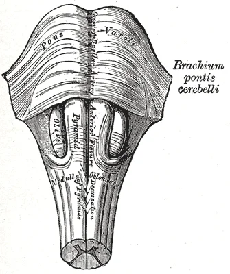
Medulla oblongata and pons. Anterior surface.

Figure 1: Medulla oblongata and pons (anterior surface).
The pons appears as a large, bulbous anterior prominence that bulges forward. This convexity is formed by the transverse pontine fibres, which sweep laterally to form the middle cerebellar peduncles. These peduncles serve as the primary connection between the pons and the cerebellum. The basilar groove runs longitudinally down the midline of the anterior surface, housing the basilar artery. The abducens nerve emerges from the pontomedullary junction, lateral to the midline. The facial and vestibulocochlear nerves emerge from the cerebellopontine angle, lateral to the abducens nerve. The trigeminal nerve is the largest cranial nerve emerging from the lateral aspect of the pons. Posteriorly, the pons forms the upper part of the floor of the fourth ventricle. The superior cerebellar peduncles connect the pons to the midbrain posteriorly. The inferior cerebellar peduncle connects the pons to the medulla. The pons acts as a relay station, connecting the cerebrum to the cerebellum and the spinal cord.
DefinitionPons
A major part of the brainstem that serves as a relay between the cerebrum and cerebellum, and contains nuclei for several cranial nerves.
The pons is integral to motor coordination. Descending fibers from the cerebrum have branches that connect to neurons in the pons. Those neurons project into the cerebellum, providing a copy of motor commands sent to the spinal cord. The cerebellum is connected to the brain stem, primarily at the pons, where it receives a copy of the descending input from the cerebrum to the spinal cord. It can compare this with sensory feedback input through the medulla and send output through the midbrain that [1][2]
The trigeminal system of the head and neck is the equivalent of the ascending spinal cord systems of the dorsal column and the spinothalamic pathways. Somatosensation of the face is conveyed along the nerve to enter the brain stem at the level of the pons. Synapses of those axons, however, are distributed across nuclei found throughout the brain stem. The chief nucleus, located in the pons, receives information about light touch as well as proprioceptive information about the mandible, which are both relayed to the thalamus and, ultimately, to the postcentral gyrus of the parietal lobe. [2]
| Nerve | Function | Relation to Pons |
|---|---|---|
| Trigeminal (V) | Sensation of face, motor to muscles of mastication | Emerges from lateral aspect |
| Abducens (VI) | Motor to lateral rectus muscle | Emerges at pontomedullary junction |
| Facial (VII) | Motor to facial muscles, taste | Emerges at cerebellopontine angle |
| Vestibulocochlear (VIII) | Hearing and balance | Emerges at cerebellopontine angle |
The pons contains the chief nucleus of the trigeminal nerve. This nucleus receives information about light touch as well as proprioceptive information about the mandible. These signals are relayed to the thalamus and, ultimately, to the postcentral gyrus of the parietal lobe. It is the sensory component of the jaw-jerk reflex, a stretch reflex of the masseter muscle. [2]
Clinical pearlClinical Pearl
Lesions in the pons can affect multiple cranial nerves and long tracts, leading to complex neurological deficits.
The pons is a critical hub for integrating motor and sensory information. It receives input from the cerebrum and sends output to the cerebellum. This integration allows for smooth and coordinated movement. The pons also houses nuclei for cranial nerves that control facial sensation and movement. Understanding the gross anatomy of the pons is essential for localizing lesions in the brainstem.
Figure 2Gray701
Long description
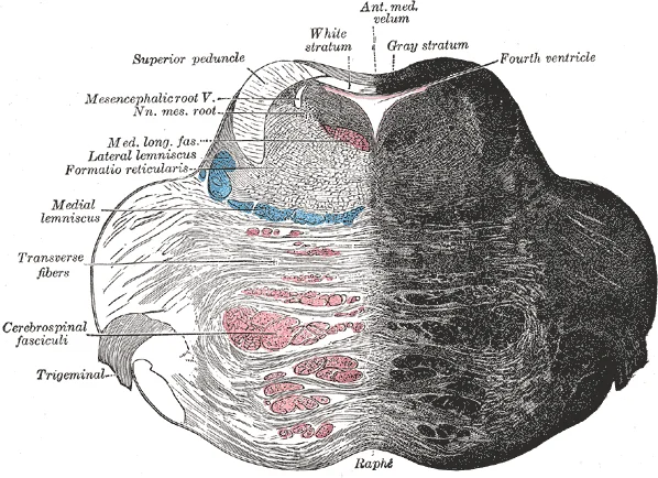
Coronal section of the pons, at its upper part.

Figure 2: Coronal section of the pons, at its upper part.
The basal pons contains the pontine nuclei and the transverse pontine fibres that form the middle cerebellar peduncles.
Cranial nerve nuclei V to VIII are embedded within the tegmentum. The motor nucleus controls the muscles of mastication, while the sensory nucleus processes touch and pain from the face. The abducens nucleus (VI) controls the lateral rectus muscle, enabling eye abduction. The facial nucleus (VII) innervates the muscles of facial expression. The vestibulocochlear nuclei (VIII) process auditory and balance information. These nuclei are closely packed, making them susceptible to simultaneous damage in vascular events. For instance, a lesion in the lateral pons may affect the facial nucleus and the vestibulocochlear nuclei, resulting in facial paralysis and hearing loss or vertigo.
Pontine syndromes often result from occlusion of the basilar artery or its branches. A classic example is locked-in syndrome, which occurs due to bilateral damage to the corticospinal and corticobulbar tracts in the basal pons. Patients retain consciousness and vertical eye movements but are unable to move any other voluntary muscles. This condition highlights the segregation of motor pathways in the pons. The tegmentum contains the reticular activating system, which is crucial for maintaining wakefulness. Damage here can lead to coma, whereas damage to the basal pons spares consciousness but abolishes motor output.
The pontocerebellar pathway is a key component of the cerebellar circuitry. It begins with corticopontine fibres from the cerebral cortex, which synapse on pontine nuclei. The axons of these nuclei cross the midline and enter the middle cerebellar peduncle, terminating in the cerebellar cortex. This pathway allows the cerebellum to receive information about intended movements, enabling precise coordination. Disruption of this pathway can result in dysmetria or intention tremor. The anatomical arrangement of these fibres in the basal pons ensures that motor plans are transmitted efficiently to the cerebellum for refinement.
DefinitionLocked-in Syndrome
A condition resulting from bilateral ventral pontine lesions, causing paralysis of all voluntary muscles except those controlling eye movement, while consciousness remains intact.
| Nerve | Nucleus Location | Primary Function |
|---|---|---|
| V (Trigeminal) | Lateral tegmentum | Motor to mastication; Sensory to face |
| VI (Abducens) | Dorsal tegmentum | Motor to lateral rectus |
| VII (Facial) | Ventral tegmentum | Motor to facial expression |
| VIII (Vestibulocochlear) | Dorsolateral tegmentum | Sensory for hearing and balance |
The pons serves as a critical relay station and conduit within the brainstem, linking the cerebrum with the cerebellum and the spinal cord. Its structural integrity is vital for the transmission of motor and sensory signals. The term 'pons' derives from the Latin for 'bridge', reflecting its role in connecting these major neural structures.
Afferent pathways bring information into the pons from various sources. The most significant input comes from the cerebral cortex via the corticopontine fibres. These fibres originate in the frontal, parietal, temporal, and occipital lobes. They travel through the internal capsule and cerebral peduncles. Upon reaching the pons, they synapse with pontine nuclei. These nuclei are located in the ventral part of the pons. The axons of the pontine nuclei cross the midline and form the middle cerebellar peduncle. This peduncle carries the information to the contralateral cerebellar hemisphere. This pathway is essential for coordinating voluntary movements.
Another important afferent input arises from the vestibular system. The vestibular nuclei, located at the junction of the pons and medulla, receive input from the vestibular nerve. These nuclei are involved in balance and spatial orientation. They also send projections to the cerebellum and spinal cord. As noted in clinical contexts, deficits in this reflex may relate to dorsal brain stem damage affecting eye movement nuclei or their connections through the medial longitudinal fasciculus (MLF). [2]
Efferent pathways carry signals away from the pons. The primary efferent output is the corticospinal and corticobulbar tracts. These fibres descend through the basis pontis. They are arranged in a specific topographical order. Fibres destined for the cervical spinal cord are located most laterally. Fibres for the thoracic cord are more medial. Fibres for the lumbar and sacral cord are located most medially. This arrangement is crucial for localizing lesions.
The cranial nerve nuclei within the pons also provide efferent output. The abducens nucleus (CN VI) controls lateral eye movement. The facial nucleus (CN VII) innervates the muscles of facial expression. The trigeminal motor nucleus (CN V) controls muscles of mastication. These nuclei send axons that exit the pons to form their respective cranial nerves.
The pons is situated above the medulla and below the midbrain. The tegmentum lies dorsal to the basis pontis. The corticospinal tracts are embedded within the basis pontis. The cranial nerve nuclei are located in the tegmentum. Understanding this anatomy is key to interpreting neurological deficits.
The pons receives its vascular supply from a rich network of arteries derived primarily from the basilar artery and the vertebral arteries. This dual supply ensures robust perfusion, yet specific territories are vulnerable to occlusion of their parent vessels. Understanding the hierarchical branching of these arteries is essential for localising lesions based on clinical deficits.
The basilar artery runs in the basilar sulcus on the anterior surface of the pons. It gives off paramedian, short circumferential, and long circumferential branches. These vessels penetrate the brainstem at right angles to supply the deep and lateral structures respectively.
DefinitionParamedian arteries
Small penetrating branches that supply the medial structures of the pons, including the corticospinal tract and medial lemniscus.
Paramedian arteries arise directly from the basilar artery. They penetrate the anterior pons to supply the medial tegmentum and basis pontis. Lesions here cause medial pontine syndrome, characterised by contralateral hemiparesis and contralateral loss of proprioception. [1]
Short and long circumferential branches wrap around the lateral aspect of the pons. They supply the lateral tegmentum and basis pontis. The superior cerebellar artery and anterior inferior cerebellar artery contribute significantly to this lateral supply. [1]
| Territory | Artery | Structures Supplied | Clinical Deficit |
|---|---|---|---|
| Medial | Paramedian | Corticospinal tract, Medial lemniscus | Contralateral hemiparesis, Contralateral proprioception loss |
| Lateral | AICA / SCA | Lateral lemniscus, Spinothalamic tract, Facial nucleus | Ipsilateral facial palsy, Contralateral pain/temperature loss |
Occlusion leads to lateral pontine syndrome, featuring ipsilateral facial paralysis and deafness.
The vertebral arteries join to form the basilar artery. Small paramedian branches arise from the vertebral arteries before this union. These supply the most inferior part of the pons and the medullary pyramids.
Clinical pearlClinical Pearl
Medial pontine syndrome results from paramedian basilar artery occlusion, sparing lateral structures like the facial nucleus.
The vertebral arteries also give off circumferential branches that anastomose with basilar branches. This collateral network provides some protection against ischaemia, although complete occlusion of a major branch still causes significant infarction.
Short circumferential arteries branch laterally from the basilar artery. They curve around the lateral edge of the pons. Long circumferential arteries extend further laterally towards the cerebellum. The superior cerebellar artery arises higher up. It also courses laterally. The paramedian arteries supply the medial basis pontis. The circumferential arteries supply the lateral tegmentum and basis pontis. Understanding this layout helps predict deficits from specific vascular occlusions. The basilar artery is the central source. Its branches distribute blood to the entire pons. The vertebral arteries contribute to the inferior supply. This vascular map is critical for neuroimaging interpretation.
The pons serves as a critical relay station and coordination centre within the brainstem, linking the cerebrum with the cerebellum and spinal cord. It is anatomically situated anterior to the medulla oblongata and inferior to the midbrain. The structure is named for its prominent bulging anterior surface, which contains transverse fibres connecting the two cerebellar hemispheres. These fibres facilitate the communication necessary for the coordination of voluntary movements. The pons also houses nuclei for several cranial nerves, integrating sensory and motor pathways.
The large, convex anterior portion is the basis pontis, composed of transverse pontine fibres that sweep laterally to enter the middle cerebellar peduncle. Posterior to this lies the tegmentum, which contains the cranial nerve nuclei and ascending/descending tracts. The facial nerve (CN VII) and abducens nerve (CN VI) nuclei are located within the tegmentum, near the floor of the fourth ventricle. The superior cerebellar peduncles connect the pons to the midbrain superiorly.
The pons contains nuclei for cranial nerves V, VI, VII, and VIII. The trigeminal nerve (CN V) nuclei are involved in facial sensation and mastication. The abducens nerve (CN VI) controls lateral eye movement. The facial nerve (CN VII) governs facial expression and taste. The vestibulocochlear nerve (CN VIII) is responsible for hearing and balance. These nuclei are embedded within the pontine tegmentum. The transverse pontine fibres in the basis pontis carry information from the cerebral cortex to the cerebellum, enabling the cerebellum to monitor and coordinate motor commands. This pathway is essential for smooth, coordinated movement.
DefinitionPontine Relay
The pons acts as a bridge, transmitting signals between the cerebral cortex and the cerebellum via transverse pontine fibres, and connecting the medulla to higher brain centres.
The reticular formation within the pons plays a vital role in regulating arousal and consciousness. It contains nuclei that modulate pain perception and autonomic functions. This centre works in conjunction with the apneustic centre in the lower pons to control breathing patterns. Damage to these areas can result in abnormal breathing patterns, such as apneusis.
| Nerve | Primary Function | Location in Pons |
|---|---|---|
| Trigeminal (V) | Facial sensation, mastication | Mid-pons, lateral tegmentum |
| Abducens (VI) | Lateral eye movement | Dorsal tegmentum, near midline |
| Facial (VII) | Facial expression, taste | Dorsal tegmentum, lateral to VI |
| Vestibulocochlear (VIII) | Hearing, balance | Pontomedullary junction |
Lesions in the pons can lead to specific neurological deficits depending on the affected structures. A lesion in the basis pontis may cause contralateral hemiparesis due to disruption of the corticospinal tract. A lesion in the tegmentum may affect cranial nerve nuclei, leading to facial weakness or eye movement abnormalities. The "locked-in" syndrome is a rare but severe condition resulting from bilateral ventral pontine lesions, where the patient is conscious but unable to move or speak, except for vertical eye movements. This highlights the pons' critical role in motor transmission and cranial nerve function.
The pons serves as a critical relay station and coordination centre for cranial nerve functions and motor pathways. Damage to this region produces distinct clinical syndromes that localise the lesion with precision. Understanding these patterns requires correlating anatomical structures with their specific physiological roles.
Lateral to the basis pontis lie the middle cerebellar peduncles. The tegmentum, located dorsally, contains the cranial nerve nuclei and the reticular formation. The medial longitudinal fasciculus (MLF) runs vertically through the tegmentum, connecting the oculomotor, trochlear, and abducens nuclei to allow for conjugate gaze.
Coordinated movement of both eyes through different nuclei requires integrated processing through the brain stem. In the midbrain, the superior colliculus integrates visual stimuli with motor responses to initiate eye movements. The paramedian pontine reticular formation (PPRF) will initiate a rapid eye movement, or saccade, to bring the eyes to bear on a visual stimulus quickly. These areas are connected to the oculomotor, trochlear, and abducens nuclei by the medial longitudinal fasciculus (MLF) that runs through the majority of the brain stem. The MLF allows for conjugate gaze, or the movement of the eyes in the same direction, during horizontal movements that require the lateral and medial rectus muscles. Control of conjugate gaze strictly in the vertical direction is contained within the oculomotor complex.
Lesions affecting the basis pontis can disrupt the corticospinal and corticobulbar tracts. Bilateral destruction of the ventral pons, sparing the tegmentum, results in locked-in syndrome. Patients retain consciousness and vertical eye movements but are quadriplegic and anarthric. They cannot move any voluntary muscles except those controlling vertical gaze and blinking. This condition highlights the separation of motor pathways from consciousness centres.
| Structure | Function | Lesion Effect |
|---|---|---|
| Corticospinal tract | Voluntary motor control | Contralateral hemiparesis |
| Corticobulbar tract | Cranial nerve motor control | Contralateral facial weakness |
| Abducens nucleus | Lateral rectus movement | Ipsilateral gaze palsy |
| Facial nerve nucleus | Facial expression | Ipsilateral facial paralysis |
| MLF | Conjugate gaze coordination | Internuclear ophthalmoplegia |
Neurons of the inferior colliculus project to the thalamus, which then sends auditory information to the cerebrum for the conscious perception of sound. Activity in the superior colliculus is related to orienting the eyes to a sound or touch stimulus. That is the correlation of auditory and visual maps.
Pontine haemorrhage often presents with sudden coma, pinpoint pupils, and quadriplegia. The blood typically originates from ruptured paramedian branches of the basilar artery. This catastrophic event underscores the vulnerability of the pons to vascular insults.
Clinical pearlLocked-in Syndrome
Bilateral ventral pontine lesion causing quadriplegia and anarthria with preserved consciousness and vertical eye movement.
Exam trapGaze Palsy
PPRF lesion causes ipsilateral horizontal gaze palsy, not contralateral.
The pons is a distinct component of the brain stem, situated anterior to the cerebellum and superior to the medulla oblongata. It serves as a critical relay station and contains the nuclei for several cranial nerves. Understanding its anatomy requires distinguishing it from the midbrain and medulla, particularly regarding the structures that traverse or emerge from it.
The pons houses the nuclei for cranial nerves V, VI, VII, and VIII. The abducens nucleus (CN VI) is located in the lower pons, near the midline. The facial nerve (CN VII) and vestibulocochlear nerve (CN VIII) emerge at the pontomedullary junction. The trigeminal nerve (CN V) emerges from the lateral aspect of the mid-pons.
DefinitionPontomedullary Junction
The boundary between the pons and the medulla, where CN VII and CN VIII emerge.
Though the two senses are not directly related, anatomy is mirrored in the two systems. Problems with balance, such as vertigo, and deficits in hearing may both point to problems with the inner ear. Within the petrous region of the temporal bone is the bony labyrinth of the inner ear. The vestibule is the portion for equilibrium, composed of the utricle, saccule, and the three semicircular canals. The cochlea is responsible for transducing sound waves into a neural signal. The sensory nerves fro
The pons contains transverse pontine fibers that connect the cerebral cortex to the cerebellum via the middle cerebellar peduncles. Longitudinal tracts, including the corticospinal and corticobulbar tracts, pass through the basis pontis.
Exam trapCN VIII Pathway
CN VIII does not synapse in the pons; it synapses in the medulla after passing through the internal auditory meatus.
Lesions in the pons can result in specific cranial nerve deficits. A lesion affecting the facial nerve nucleus or its fascicle can cause ipsilateral facial paralysis.
There are two sets of muscles of the tongue. The extrinsic muscles of the tongue are connected to other structures, whereas the intrinsic muscles of the tongue are completely contained within the lingual tissues. While examining the oral cavity, movement of the tongue will indicate whether hypoglossal function is impaired. The test for hypoglossal function is the "stick out your tongue" part of the exam. The genioglossus muscle is responsible for protrusion of the tongue P86. If the hypoglossal nerv [2]
The pons is supplied by branches of the basilar artery, including the pontine arteries.
NoteAICA Syndrome
Infarction of AICA can cause lateral pontine syndrome, involving CN VII, CN VIII, and the spinal trigeminal nucleus.
DefinitionVestibulo-ocular Reflex
A reflex that stabilizes gaze during head movement, involving CN III, IV, and VI.
Diplopia is not restricted to failure of the lateral rectus, because any of the extraocular muscles may fail to move one eye in perfect conjugation with the other. [2]
Exam trapDiplopia Cause
Diplopia can result from any extraocular muscle weakness, not just CN VI palsy.
As visual stimuli move closer to the face, the two medial recti muscles cause the eyes to move in the one nonconjugate movement that is part of gaze control. When the two eyes move to look at something closer to the face, they both adduct, which is referred to as convergence. To keep the stimulus in focus, the eye also needs to change the shape of the lens, which is controlled through the parasy
Clinical pearlConvergence and Accommodation
Convergence and accommodation are mediated by the oculomotor nerve (CN III) parasympathetic fibers.
Lateral flexion of the neck toward the shoulder tests both at the same time. Any difference on one side versus the other would suggest damage on the weaker side. These strength tests are common for the skeletal muscles controlled by spinal nerves and are a significant component of the motor exam. Deficits associated with the accessory nerve may h [2]
NoteAccessory Nerve Test
Shrugging shoulders against resistance tests the spinal accessory nerve (CN XI).
Students frequently confuse the anatomical location and functional role of the pons with adjacent brainstem structures, particularly the midbrain and medulla oblongata. A primary error is assuming that all sensory information bypasses the brainstem nuclei before reaching the cortex. In reality, the thalamus acts as the requisite synapse for all sensory pathways except olfaction, relaying information between the cerebral cortex and the periphery. While the pons contains critical relay nuclei, it [1]
Failure of one eye to abduct while the other adducts in a horizontal movement is referred to as internuclear ophthalmoplegia. It is crucial to remember that this condition results in diplopia, or double vision, [2]
Another frequent error is misunderstanding the vascular supply and its clinical correlates. The pons is supplied by paramedian and circumferential branches of the basilar artery. Occlusion of paramedian branches can lead to medial pontine syndromes, affecting the corticospinal tract and the abducens nerve nucleus. Students must not confuse this with lateral pontine syndromes, which involve the spinal trigeminal nucleus and vestibular nuclei. Additionally, the spinal cord regions correspond to th
Knowledge of the pons is essential at the bedside for localising neurological deficits. When a patient presents with facial weakness, the clinician must determine if the lesion is upper motor neuron (supranuclear) or lower motor neuron (nuclear or infranuclear). The facial nerve nucleus lies in the pons, and its lesion causes ipsilateral facial paralysis. Similarly, eye movement testing is a routine part of the neurological examination. Having the patient track a pen tip through the visual field allows the examiner to assess conjugate eye movements. Failure of one eye to abduct while the other adducts suggests internuclear ophthalmoplegia, pointing to a lesion in the MLF within the pons. This finding is critical in diagnosing multiple sclerosis or brainstem stroke.
In the operating theatre, particularly in neurosurgery, understanding the compact anatomy of the pons is vital. The pons contains the corticospinal tracts, which are responsible for voluntary motor control. Damage to these tracts during surgery can result in severe motor deficits. The pons also houses the nuclei for cranial nerves V, VI, VII, and VIII. Surgeons must navigate carefully around these structures to avoid causing facial paralysis, hearing loss, or corneal reflex abnormalities. The proximity of the pons to the fourth ventricle means that tumours in this region can cause hydrocephalus by obstructing cerebrospinal fluid flow.
On imaging, the pons is clearly visible on MRI scans. Radiologists look for signal abnormalities that indicate stroke, demyelination, or tumours. The characteristic "face of the giant panda" sign on T2-weighted images is seen in Wilson's disease, affecting the midbrain, but pontine lesions can show similar signal changes in other conditions. The pons is also the site of the basilar artery, and its occlusion can lead to "locked-in" syndrome, where the patient is conscious but unable to move or speak, except for vertical eye movements. This syndrome results from infarction of the ventral pons, sparing the reticular formation and the oculomotor nuclei.
In examinations, students are often tested on the localisation of lesions based on clinical signs. For example, a patient with ipsilateral facial paralysis and contralateral hemiplegia has a lesion in the pons, affecting the facial nerve nucleus and the corticospinal tract. This is known as Millard-Gubler syndrome. Another common exam question involves the differentiation of hearing loss types. The Rinne test compares air conduction and bone conduction. In conductive hearing loss, bone conduction is better than air conduction. In sensorineural hearing loss, air conduction is better than bone conduction. The Weber test lateralises to the affected ear in conductive loss and to the normal ear in sensorineural loss. Understanding these tests helps in localising the lesion to the peripheral auditory system or the central pathways, including the pons. [2]
Clinical pearlClinical Pearl
Internuclear ophthalmoplegia is a hallmark of MLF lesions, often seen in multiple sclerosis, causing diplopia due to disconjugate eye movements.
| Nerve | Function | Clinical Deficit |
|---|---|---|
| V (Trigeminal) | Sensation to face, mastication | Facial numbness, weak bite |
| VI (Abducens) | Lateral rectus movement | Inability to abduct eye |
| VII (Facial) | Facial expression, taste | Ipsilateral facial paralysis |
| VIII (Vestibulocochlear) | Hearing and balance | Hearing loss, vertigo |
The thalamus is the requisite synapse for all sensory pathways except olfaction. The cerebellum accounts for approximately 10 percent of the mass of the brain. [1] The spinal cord does not grow significantly longer after the first or second year. [1] Multipolar motor neurons in the anterior horn can have axons up to a meter in length. The Snellen chart tests visual acuity based on a visual angle of 5 minutes of an arc at 20 feet. [2] The Rinne test distinguishes between conductive and sensorineural hearing loss. Internuclear ophthalmoplegia involves failure of one eye to abduct while the other adducts. The spinal accessory nerve controls the sternocleidomastoid and trapezius muscles.
Answer each one in your head first, then open the answer.
Which sensory pathway does not synapse in the thalamus?
Show answerHide answer
OlfactionWhat percentage of the brain's mass does the cerebellum account for?
Show answerHide answer
10 percentWhat is the name of the bundle of nerves resembling a horse's tail?
Show answerHide answer
Cauda equinaWhat condition causes diplopia due to disconjugate eye movements?
Show answerHide answer
Internuclear ophthalmoplegiaIn the Rinne test, what indicates conductive hearing loss?
Show answerHide answer
Sound conducted better through the temporal bone than through the ossicles
- Betts JG, Young KA, Wise JA, et al. The Central Nervous System. OpenStax, Anatomy and Physiology 2e (CC BY-NC-SA 4.0). Accessed .
- Betts JG, Young KA, Wise JA, et al. The Cranial Nerve Exam. OpenStax, Anatomy and Physiology 2e (CC BY-NC-SA 4.0). Accessed .
Figure credits
Educational content for medical students and doctors. It is not a substitute for clinical judgement or local guidelines.