Midbrain
Midbrain is the upper part of brainstem between thalamus and pons.
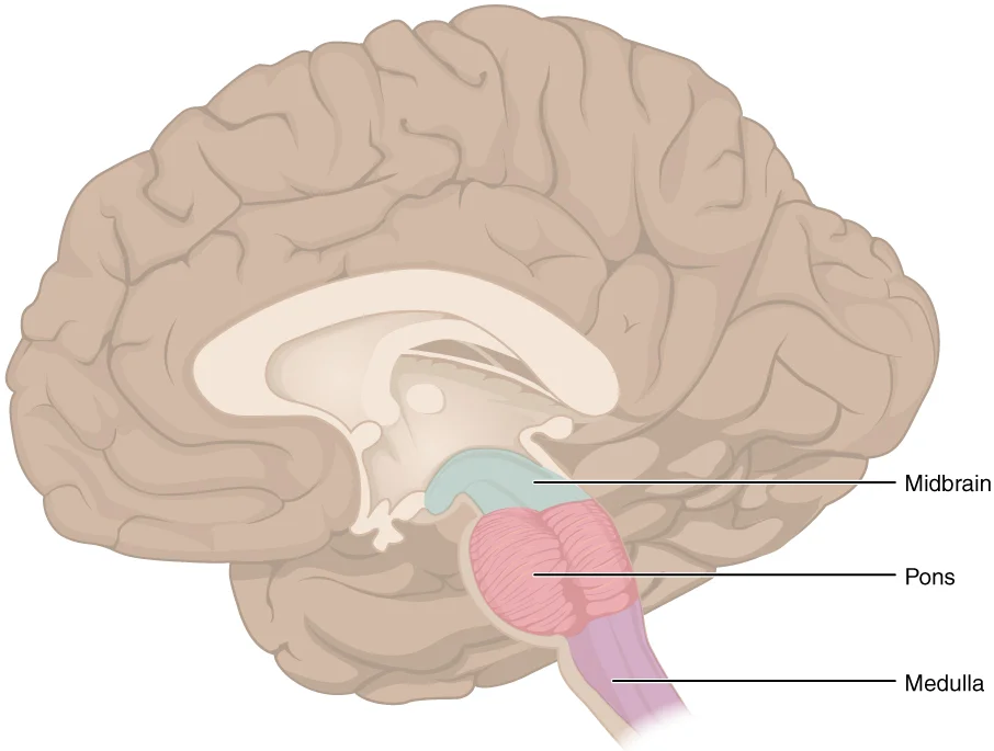
The midbrain is one of the original regions of the embryonic brain and is a small region located between the thalamus and pons. It is collectively referred to as part of the brain stem, along with the pons and medulla. [1]
Figure 11311 Brain Stem
Long description
The brain stem comprises three regions: the midbrain, the pons, and the medulla.

Figure 1: The brain stem comprises three regions: the midbrain, the pons, and the medulla.
The midbrain is separated into two main regions: the tectum and tegmentum. The term "tectum" comes from the Latin word for "roof," while "tegmentum" comes from the Latin word for "floor". [1]
The tegmentum is continuous with the gray matter of the rest of the brain stem. Throughout the midbrain, pons, and medulla, the tegmentum contains nuclei that receive and send information through the cranial nerves, as well as regions that regulate important functions such as those of the cardiovascular and respiratory systems. [1]
The tectum is composed of four bumps known as the colliculi (singular = colliculus), which means "little hill" in Latin. The inferior colliculus is the inferior pair of these enlargements and is part of the auditory brain stem pathway. Neurons of the inferior colliculus project to the thalamus, which then sends auditory information to the cerebrum for the conscious perception of sound. The superior colliculus is the superior pair and combines sensory information about visual space, auditory spac [1]
The midbrain also contains important structures such as the crus cerebri, red nucleus, substantia nigra, and periaqueductal grey matter. The substantia nigra is particularly important as it contains the pars compacta, which releases dopamine and helps regulate movement through its connections with the basal nuclei. Lesions in the midbrain can lead to specific syndromes such as Weber syndrome (involving the crus cerebri and red nucleus) and Parinaud syndrome (involving the superior colliculus). [1] [2]
The cerebral peduncles form the large, convex anterior bulge. They consist of the basis pedunculi, which contains descending motor fibres, and the tegmentum, which lies posterior to the substantia nigra. The interpeduncular fossa is a deep depression located between the two cerebral peduncles. This fossa contains the posterior perforated substance, where several arteries enter the brain. The oculomotor nerve (CN III) emerges from the medial aspect of the cerebral peduncle, just lateral to the interpeduncular fossa.
Posteriorly, the midbrain features the tectum, or roof. The tectum is composed of four rounded eminences known as the corpora quadrigemina. The superior colliculi are the upper pair and serve as visual reflex centres. The inferior colliculi are the lower pair and act as auditory reflex centres. The trochlear nerve (CN IV) is the only cranial nerve to exit from the dorsal aspect of the brainstem; it emerges just below the inferior colliculus.
Within the tegmentum, we find several important nuclei. The trochlear nucleus (cranial nerve IV) is located more caudally and innervates the superior oblique muscle. [1]
The midbrain is part of the mesencephalon. It does not have the extensive white matter exterior seen in the medulla, which is known as the myelencephalon in the embryonic brain. The initial portion of the name, “myel,” refers to the significant white matter found in this region—especially on its exterior, which is continuous with the white matter of the spinal cord. However, the midbrain retains a core of gray matter nuclei, including the substantia nigra and the red nucleus, which are crucial for motor control. [1]
DefinitionMidbrain Anatomy
The midbrain is the superior segment of the brainstem, characterized by cerebral peduncles anteriorly and the tectum posteriorly, serving as a conduit for motor and sensory pathways.
Lesions of the superior colliculus can impair visual tracking, while damage to the inferior colliculus may affect auditory reflexes. The emergence of CN III and CN IV provides key localising signs in clinical examination. [1]
| Structure | Location | Function |
|---|---|---|
| Cerebral Peduncles | Anterior | Descending motor pathways |
| Interpeduncular Fossa | Between peduncles | Entry for posterior cerebral arteries |
| Superior Colliculi | Posterior (Tectum) | Visual reflex centres |
| Inferior Colliculi | Posterior (Tectum) | Auditory reflex centres |
| Trochlear Nerve | Dorsal, below inf. colliculus | Motor supply to superior oblique muscle |
The midbrain is a small but crucial region of the brainstem that serves as a relay station between the forebrain and hindbrain. In cross-section, the midbrain can be divided into several key components that are essential for understanding its function.
The tectum is the dorsal portion of the midbrain and consists of four rounded swellings known as the corpora quadrigemina. These include the superior and inferior colliculi, which are important for visual and auditory reflexes respectively. [1]
The tegmentum lies ventral to the tectum and contains several important structures. The cerebral peduncles (crus cerebri) are prominent fiber bundles that contain descending motor tracts from the cerebral cortex. These tracts include the corticospinal, corticobulbar, and corticopontine fibers, which are crucial for voluntary movement.
Within the tegmentum, we find several important nuclei. The oculomotor nucleus (cranial nerve III) is located in the rostral midbrain. [1]
The red nucleus is a prominent pinkish structure located in the rostral midbrain tegmentum. It receives input from the contralateral motor cortex and cerebellum and projects to the spinal cord via the rubrospinal tract, which is involved in flexor muscle tone and fine motor control. [1]
It consists of two parts: the pars compacta, which contains dopamine-producing neurons that project to the striatum as part of the basal nuclei system, and the pars reticulata, which serves as an output nucleus of the basal nuclei. The substantia nigra plays a crucial role in regulating movement, and its degeneration is associated with Parkinson's disease. [1]
The Weber syndrome (midbrain syndrome) results from damage to the midbrain and can affect multiple structures. It typically involves the oculomotor nerve fibers, corticospinal tracts, and sometimes the red nucleus or substantia nigra. [1] [2]
Parinaud syndrome is another midbrain disorder caused by damage to the rostral midbrain, particularly affecting the pretectal area and superior colliculi. [1]
The tectum is located dorsally and includes the superior and inferior colliculi. The tegmentum lies ventral to the tectum and contains the cerebral peduncles, oculomotor nucleus, trochlear nucleus, red nucleus, and substantia nigra. The cerebral peduncles are the most ventral structures and contain the major descending motor tracts. The oculomotor nucleus is located in the rostral midbrain tegmentum, while the trochlear nucleus is more caudal. The red nucleus is positioned medial to the cerebral peduncles, and the substantia nigra is located just ventral to the red nucleus.
DefinitionMidbrain
The midbrain, or mesencephalon, is the most superior portion of the brainstem, acting as a critical relay centre for visual, auditory, and motor signals between the forebrain and the hindbrain.
The midbrain receives diverse sensory and motor inputs that allow it to integrate reflex responses and modulate voluntary movement.
Visual afferents reach the midbrain primarily via the superior colliculus. These fibres originate from the retina and the lateral geniculate nucleus, allowing the midbrain to coordinate head and eye movements in response to visual stimuli. This pathway is essential for the orientation reflex, where the head turns toward a sudden visual change.
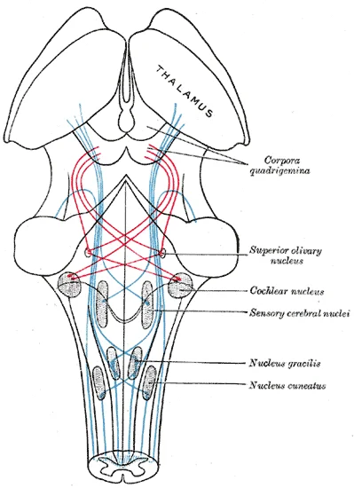
Auditory afferents are processed through the inferior colliculus. Signals ascend from the cochlear nuclei and the superior olivary complex via the lateral lemniscus. The inferior colliculus acts as the principal midbrain nucleus for auditory information, relaying these signals to the medial geniculate nucleus of the thalamus.
Figure 2Gray713
Long description
Scheme showing the course of the fibers of the lemniscus; medial lemniscus in blue, lateral in red.

Figure 2: Scheme showing the course of the fibers of the lemniscus; medial lemniscus in blue, lateral in red.
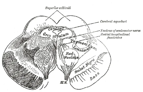
Figure 3Gray712
Long description
Transverse section of mid-brain at level of superior colliculi.

Figure 3: Transverse section of mid-brain at level of superior colliculi.
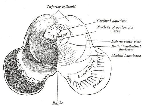
Figure 4Gray711
Long description
Transverse section of mid-brain at level of inferior colliculi.

Figure 4: Transverse section of mid-brain at level of inferior colliculi.
Descending motor afferents from the cerebral cortex travel through the midbrain via the corticospinal and corticobulbar tracts. These fibres descend through the internal capsule and enter the midbrain at the crus cerebri, where they are organised based on their destination in the brainstem or spinal cord.
Efferent connections from the midbrain are primarily concerned with the modulation of movement and the execution of cranial nerve functions.
These dopaminergic neurons project to the striatum of the basal ganglia. This pathway is fundamental for the initiation and smoothing of voluntary movements.
The tectum forms the posterior boundary, consisting of the superior colliculus (the upper rounded prominence) and the inferior colliculus (the lower rounded prominence). Anterior to the tectum is the tegmentum, the central core of the midbrain, which contains the red nucleus, appearing as a distinct circular mass. The aqueduct of Sylvius is the central canal separating the tectum from the tegmentum. Further anterior is the substantia nigra, a pigmented band of neurons that separates the tegmentum from the crus cerebri. The crus cerebri are the most anterior structures, forming the large bundles of the cerebral peduncles that carry descending cortical fibres.
Figure 5Gray714
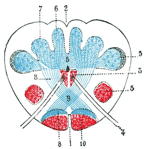
Long description
Transverse section passing through the sensory decussation. Schematic. (Testut.) 1. Anterior median fissure. 2. Posterior median sulcus. 3, 3’. Head and base of anterior column (in red). 4. Hypoglossal nerve. 5. Bases of posterior column. 6. Gracile nucleus. 7. Cuneate nucleus. 8, 8. Lemniscus. 9. S

Figure 5: Transverse section passing through the sensory decussation (schematic).
Figure 6Gray710
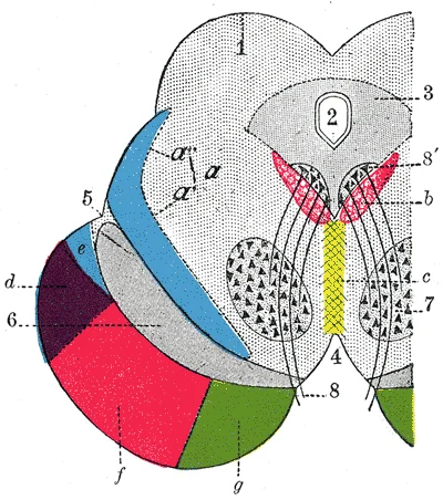
Long description
Coronal section through mid-brain. (Schematic.) (Testut.) 1. Corpora quadrigemina. 2. Cerebral aqueduct. 3. Central gray stratum. 4. Interpeduncular space. 5. Sulcus lateralis. 6. Substantia nigra. 7. Red nucleus of tegmentum. 8. Oculomotor nerve, with 8’, its nucleus of origin. a. Lemniscus (in blu

Figure 6: Coronal section through mid-brain (Schematic.) (Testut.) 1.
| Pathway | Origin | Termination | Primary Function |
|---|---|---|---|
| Nigrostriatal | Substantia nigra | Striatum | Dopaminergic motor modulation |
| Rubrospinal | Red nucleus | Spinal cord | Upper limb flexor tone |
| Corticospinal | Motor cortex | Spinal cord | Voluntary motor control |
| Tectospinal | Superior colliculus | Spinal cord | Visual-motor head orientation |
| Auditory relay | Inferior colliculus | Medial geniculate nucleus | Auditory processing |
Visual Orientation Reflex
- Visual StimulusRetinal activation
- Superior ColliculusIntegration of visual spatial data
- Tectospinal TractDescending motor signal
- Cervical Spinal CordActivation of neck muscles
- Head RotationOrientation toward stimulus
The development of the circulatory system begins with the formation of primitive branchial arch arteries, which undergo heavy modifications to supply the developing hindbrain. Specifically, the occipital lobe and brain stem growth stimulate the formation of the posterior circulation, initially involving the basilar artery (BA) and later the vertebral arteries (VA). At the 4-5 mm embryonic stage, the hindbrain is supplied by two parallel neural arteries, which obtain blood from carotid-vertebrobasilar anastomoses such as the trigeminal artery (TA). The BA itself forms during the 5-8 mm stage from the consolidation of these neural arteries. [2]
In the adult, the midbrain receives its primary blood supply from branches of the posterior cerebral artery (PCA) and the superior cerebellar artery (SCA). The PCA, a branch of the posterior division of the internal carotid artery (ICA), supplies the midbrain along with the posterior choroidal artery. The SCA, a branch of the BA, also contributes significantly to the midbrain vasculature. These arteries form a critical network that ensures perfusion to the midbrain structures.
NoteEmbryological Persistence
Adult individuals with persistence of fetal arterial configurations in the brains are typically asymptomatic, although they may have decreased collateral routes.
The posterior cerebral arteries arise from the terminal bifurcation of the basilar artery and wrap around the midbrain, supplying the inferior and medial aspects. The anterior choroidal artery, arising from the internal carotid artery, also contributes to the supply of the medial midbrain structures. The vertebral arteries join to form the basilar artery, providing the main inflow to this posterior circulation network. Understanding the spatial relationship between the basilar artery, the posterior cerebral arteries, and the superior cerebellar arteries is essential for localising lesions in the midbrain.
The degree of blood flow stagnation is influenced more by the ratio between the flows of the parent-to-daughter arteries than by the diameter ratio. Individuals with arterial configurations consisting of decreased collateral routes or discrepant diameters may be at a higher risk of developing atherosclerosis. [2]
| Artery | Origin | Supply Region |
|---|---|---|
| Posterior Cerebral Artery (PCA) | Basilar Artery | Inferior and medial midbrain |
| Superior Cerebellar Artery (SCA) | Basilar Artery | Superior midbrain |
| Anterior Choroidal Artery (AChA) | Internal Carotid Artery | Medial midbrain structures |
Variants in this supply system exist. The most typical MCA variants include an accessory MCA or a duplicated MCA, which are rare. The prevalence of an accessory MCA in DSA is about 0.3%. While these variants primarily affect the anterior circulation, they highlight the variability in cerebral arterial anatomy. More rarely, the trigeminal system arising from the ICA supplies the cerebellar arteries directly, bypassing the BA. This persistent TA variant is found in up to 0.2% of angiographic series. [2]
Although hypoplastic arteries can be found at birth, their prevalence increases with age. The persistence of carotid-vertebrobasilar anastomoses occasionally indicates a complete occlusion or agenesis of anterior circulation arteries. This highlights the importance of collateral pathways in maintaining blood supply to the midbrain and surrounding structures. [2]
Figure 7Midbrain
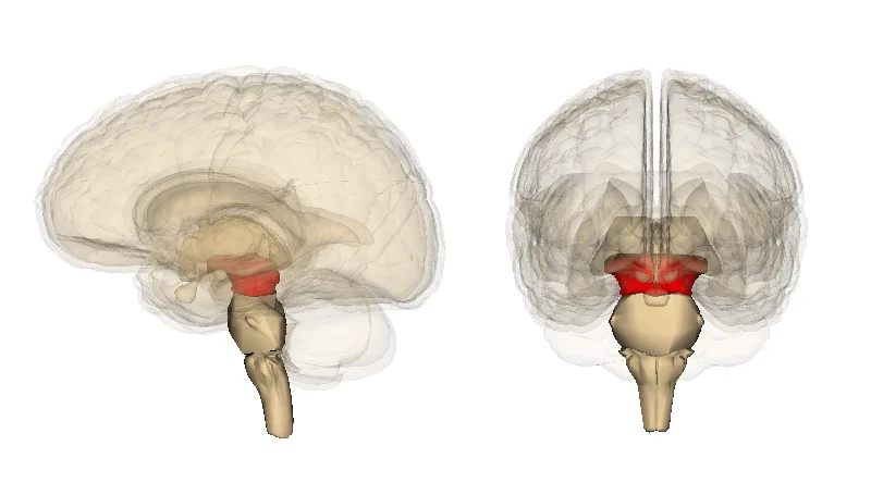
Long description
midbrain. Images are from Anatomography maintained by Life Science Databases(LSDB).

Figure 7: Midbrain.
The midbrain serves as a critical relay station and integration center for numerous neural pathways. It connects the forebrain with the hindbrain and contains several important nuclei and tracts that facilitate motor control, sensory processing, and reflexes.
The tectum, or "roof" of the midbrain, consists of the superior and inferior colliculi. The superior colliculi are involved in visual reflexes, particularly the control of eye movements and visual orientation. They receive input from the retina and project to the spinal cord to coordinate head and eye movements in response to visual stimuli. The inferior colliculi are part of the auditory pathway, receiving auditory information from the cochlear nuclei and relaying it to the medial geniculate bodies of the thalamus.
The tegmentum contains several important nuclei, including the red nucleus and substantia nigra. The red nucleus is involved in motor coordination, particularly integrating information from the cerebellum and motor cortex to facilitate limb movements. The substantia nigra contains dopaminergic neurons that project to the basal ganglia, playing a crucial role in regulating movement and motivation. Degeneration of these dopaminergic neurons is associated with Parkinson's disease. [1]
The cerebral peduncles, also known as the crus cerebri, contain the major descending motor tracts from the cerebral cortex to the spinal cord. These include the corticospinal tracts, which carry voluntary motor commands, and the corticobulbar tracts, which innervate cranial nerve nuclei. The midbrain also houses the nuclei for cranial nerves III (oculomotor) and IV (trochlear), which control most of the extraocular muscles responsible for eye movement. [1]
The superior colliculi are positioned dorsally, while the cerebral peduncles form the ventral aspect of the midbrain. The red nucleus and substantia nigra are located within the tegmentum, between the tectum and cerebral peduncles.
Midbrain lesions produce distinct clinical syndromes due to the compact arrangement of cranial nerve nuclei, long tracts, and the cerebral peduncles. Understanding these patterns requires correlating the anatomical location of the injury with the specific neurological deficits observed.
Weber's syndrome results from a lesion in the medial basis pedunculi. This area contains the corticospinal tract and the fibres of the oculomotor nerve (CN III) as they exit the midbrain. The classic presentation involves oculomotor palsy and hemiparesis. The oculomotor palsy manifests as ptosis, a dilated pupil, and the eye positioned 'down and out' due to unopposed action of the lateral rectus (CN VI) and superior oblique (CN IV). The hemiparesis affects the face, arm, and leg, reflecting damage to the descending motor fibres before they decussate in t
Benedikt’s syndrome arises from a lesion in the midbrain tegmentum, specifically affecting the red nucleus and the oculomotor nerve fibres. This syndrome presents with ipsilateral oculomotor palsy and contralateral tremor or choreoathetosis. The tremor results from disruption of the rubrospinal tract or connections between the red nucleus and the thalamus. Unlike Weber’s syndrome, motor strength is typically preserved, distinguishing it from lesions of the cerebral peduncles.
This rare syndrome causes ipsilateral lateral rectus palsy and contralateral hemiparesis. The abducens palsy leads to an inability to abduct the affected eye, resulting in horizontal diplopia. The contralateral weakness mirrors that seen in Weber’s syndrome but is accompanied by specific eye movement deficits rather than oculomotor involvement. [1]
| Syndrome | Lesion Site | Key Deficits |
|---|---|---|
| Weber’s | Medial basis pedunculi | Ipsilateral CN III palsy, contralateral hemiparesis |
| Benedikt’s | Tegmentum (red nucleus) | Ipsilateral CN III palsy, contralateral tremor |
| Raymond’s | Ventral tegmentum | Ipsilateral CN VI palsy, contralateral hemiparesis |
Clinical pearlClinical Pearl
Always assess pupillary response in midbrain lesions. A dilated, non-reactive pupil suggests compression of the parasympathetic fibres on the outside of CN III, often seen in uncal herniation.
The midbrain, or mesencephalon, is the most rostral part of the brainstem, situated between the diencephalon above and the pons below. It is characterised by the cerebral peduncles anteriorly and the tectum posteriorly. The cerebral aqueduct of Sylvius runs through its centre, connecting the third and fourth ventricles. [1]
The tectum consists of the superior and inferior colliculi, which form the corpora quadrigemina. These structures are crucial for visual and auditory reflexes respectively. The tegmentum lies ventral to the aqueduct and contains the red nucleus and the substantia nigra.
DefinitionMidbrain Division
The midbrain is anatomically divided into the tectum (posterior), tegmentum (middle), and basis pedunculi (anterior).
Two cranial nerve nuclei are located in the midbrain: the oculomotor nucleus (CN III) and the trochlear nucleus (CN IV). It gives rise to fibres that pass through the interpeduncular fossa. Its fibres are unique as they exit dorsally from the brainstem. [1] [2]
| Nerve | Nucleus Location | Function |
|---|---|---|
| Oculomotor (CN III) | Tegmentum, superior colliculus level | Eye movement, pupil constriction |
| Trochlear (CN IV) | Tegmentum, inferior colliculus level | Superior oblique muscle movement |
The midbrain receives its blood supply from branches of the posterior cerebral artery and the basilar artery. Key arteries include the posterior communicating artery, the superior cerebellar artery, and paramedian branches from the basilar artery. Occlusion of these vessels can lead to specific syndromes. [2]
Lesions of the midbrain can result in Weber’s syndrome, which involves ipsilateral oculomotor nerve palsy and contralateral hemiparesis due to involvement of the cerebral peduncle. Another classic presentation is Parinaud’s syndrome, which can lead to vertical gaze palsy and light-near dissociation. [2]
Exam trapTrochlear Nerve Exit
Remember that the trochlear nerve (CN IV) is the only cranial nerve to exit from the dorsal aspect of the brainstem.
Clinical pearlMidbrain Syndrome
Weber’s syndrome presents with ipsilateral CN III palsy and contralateral hemiplegia, localising the lesion to the cerebral peduncle.
Students frequently confuse the anatomical regions of the brain, particularly when distinguishing the midbrain from the cerebellum. A common error is attributing the function of comparing cerebrum information with sensory feedback to the midbrain. This function actually belongs to the cerebellum, which accounts for approximately 10 percent of the mass of the brain. The midbrain is part of the brainstem and is primarily involved in motor control, vision, hearing, and arousal, not the fine-tuning of movement via sensory comparison. [1]
Another frequent mistake involves directional terminology. Students often struggle with the interchangeability of dorsal/ventral and posterior/anterior, especially in the context of the spinal cord and brainstem. It is essential to learn to be comfortable with both sets of terms, as dorsal corresponds to posterior and ventral to anterior in these structures. Failing to recognise this synonymy can lead to errors in describing lesion locations or tract pathways.
Confusion also arises regarding the size and location of neurons. Students may incorrectly assume that all motor neurons are small. In reality, the largest neurons in the spinal cord are the multipolar motor neurons in the anterior horn. For instance, the motor neuron controlling the big toe is located in the sacral spinal cord, and its axon can be a meter in length. The cell body maintaining this long fiber must be quite large, possibly several hundred micrometers in diameter, making it one of the largest cells in the body. While this is a spinal cord fact, it highlights the importance of understanding neuronal scale, which differs significantly from the compact nuclei of the midbrain. [1]
Vascular variants are another source of error. Students might assume that the posterior communicating artery (PCOMM) is always present and robust. However, a hypoplastic PCOMM is overall the most common arterial variant reported in the brain. Furthermore, a fetal origin of the posterior cerebral artery (PCA) is the most frequent variant involving this artery, reported in between 13%-15% of the population. Students must not confuse a fetal PCA with a hypoplastic P1 segment; technically, a fetal PCA by definition is always accompanied by agenesis of the ipsilateral PCOMM, but in fact it is the P1 segment of the PCA that failed to form. [2]
Finally, students often overlook the clinical significance of arterial duplications and fenestrations. These anomalies are associated with the development of brain saccular aneurysms. Plausible explanations include decreased smooth muscle cells and collagen at the proximal and distal ends of the duplicated segment. In patients with non-aneurysmatic, unexplained subarachnoid hemorrhage, up to 95% of these patients have fenestrations close to the site of hematoma. Recognising these variants on imaging is crucial for risk stratification. [2]
Knowledge of the midbrain and its associated neurovascular structures is critical at the bedside for localising lesions. A lesion affecting the midbrain can present with specific cranial nerve deficits, such as oculomotor nerve palsy, which is often vascular in origin. Understanding the vascular supply helps clinicians predict the extent of damage. For example, the anterior cerebral artery (ACA) and middle cerebral artery (MCA) have distinct developmental origins. At the 11-12 mm embryological stage, the MCA is first identified as small buds originating proximal to the ACA on the anterior division of the primitive internal carotid artery. At this stage, although the MCA is still plexiform and not a true artery, it is the major blood source for the cerebral hemispheres. By the 16-18 mm stage, the MCA becomes more prominent, the plexi fuse into a single artery and further branches pierce the cerebral hemisphere. [2]
In the operating theatre and on imaging, recognising vascular anomalies is vital. The persistent stapedial artery is a rare anomaly with a reported prevalence of 0.5%. This vessel supplies the middle meningeal artery territory, leading to the absence of the foramen spinosum. Absence of the foramen spinosum is seen in up to 3% of skull-base CT studies, and it is an indirect sign of a persistent stapedial artery, but it can also be related to an ophthalmic origin of the middle meningeal artery. Surgeons must be aware of this to avoid iatrogenic injury during skull base procedures. [2]
On imaging, identifying the origin of the posterior cerebral artery is key. A fetal origin of the PCA is typically described in only one PCA, but it can also occur in both PCA in up to 0.5% of the population. More commonly, in up to 10% of the cases, the P1 segment is hypoplastic, which renders a fetal-like circulation to the occipital lobes, although it is not technically a fetal PCA given the physical link of the PCOMM with the basilar artery. Duplications of the PCA are extremely rare, occurring in 1.0 % of cases.
In examinations, students are often tested on the embryological development of cerebral arteries. The stem of the ACA gives rise to the olfactory artery at the embryological 18-mm stage. The ACA then continues growing medially towards the contralateral ACA, eventually leading to the formation of the ACOMM at the 21-24 mm embryological stage. Distally, the small arteries dive into the cerebral cortex in a radially-oriented pattern toward the wall of the developing lateral ventricles forming anastomoses among them.
Clinical pearlVascular Anomalies
Fenestrations and duplications of intracranial arteries are associated with aneurysm formation due to weakened arterial walls at the ends of the duplicated segment.
Vascular Variant Assessment
- Identify PCA OriginCheck for fetal pattern
- Question: Fetal PCA present?
Yes
- Confirm PCOMM agenesisP1 segment failed to form
No
- Check P1 segmentHypoplastic in 10% of cases
The cerebellum accounts for approximately 10 percent of the mass of the brain and compares cerebrum information with sensory feedback. Dorsal and ventral are interchangeable with posterior and anterior, particularly in reference to nerves and spinal cord structures. [1] The largest neurons in the spinal cord are multipolar motor neurons in the anterior horn, some with axons up to a meter in length. [1] A hypoplastic PCOMM is the most common arterial variant reported in the brain. A fetal PCA is reported in between 13%-15% of the population and is defined by agenesis of the ipsilateral PCOMM. [2] Absence of the foramen spinosum is seen in up to 3% of skull-base CT studies and may indicate a persistent stapedial artery. Fenestrations and duplications of intracranial arteries are associated with the development of brain saccular aneurysms. [2] Up to 95% of patients with non-aneurysmatic, unexplained subarachnoid hemorrhage have fenestrations close to the site of hematoma.
Answer each one in your head first, then open the answer.
What percentage of the brain's mass does the cerebellum account for?
Show answerHide answer
10 percentWhat is the reported prevalence of a persistent stapedial artery?
Show answerHide answer
0.5%In what percentage of the population is a fetal origin of the PCA reported?
Show answerHide answer
13%-15%What is the most common arterial variant reported in the brain?
Show answerHide answer
A hypoplastic PCOMMWhat percentage of patients with non-aneurysmatic, unexplained subarachnoid hemorrhage have fenestrations close to the hematoma site?
Show answerHide answer
95%
- Betts JG, Young KA, Wise JA, et al. The Central Nervous System. OpenStax, Anatomy and Physiology 2e (CC BY-NC-SA 4.0). Accessed .
- Menshawi K, Mohr JP, Gutierrez J. A Functional Perspective on the Embryology and Anatomy of the Cerebral Blood Supply. Journal of stroke 2015;17(2):144-158. doi:10.5853/jos.2015.17.2.144. Accessed .
Figure credits
- Figure 1: OpenStax, via Wikimedia Commons, CC BY 4.0, CC BY 4.0.
- Figure 2: Henry Vandyke Carter, via Wikimedia Commons, Public domain, Public domain.
- Figure 3: Henry Vandyke Carter, via Wikimedia Commons, Public domain, Public domain.
- Figure 4: Henry Vandyke Carter, via Wikimedia Commons, Public domain, Public domain.
- Figure 5: Henry Vandyke Carter, via Wikimedia Commons, Public domain, Public domain.
- Figure 6: Henry Vandyke Carter, via Wikimedia Commons, Public domain, Public domain.
- Figure 7: Images are generated by Life Science Databases(LSDB)., via Wikimedia Commons, CC BY-SA 2.1 jp, CC BY-SA 2.1 jp.
Educational content for medical students and doctors. It is not a substitute for clinical judgement or local guidelines.