Cerebellum
Neuroanatomy of the cerebellum covering structure, connections and motor learning functions.
The cerebellum is 10 percent of the mass of the brain and has varied functions that all point to a role in the motor system[1]. The role of the cerebellum is a subject of debate[1]. There is an obvious connection to motor function based on the clinical implications of cerebellar damage[1]. There is also strong evidence of the cerebellar role in procedural memory[1]. The two are not incompatible; in fact, procedural memory is motor memory, such as learning to ride a bicycle[1]. Significant work has been performed to describe the connections within the cerebellum that result in learning[1]. A model for this learning is classical conditioning, as shown by the famous dogs from the physiologist Ivan Pavlov’s work[1]. This classical conditioning, which can be related to motor learning, fits with the neural connections of the cerebellum[1]. [2]
Figure 1Gray766
Long description
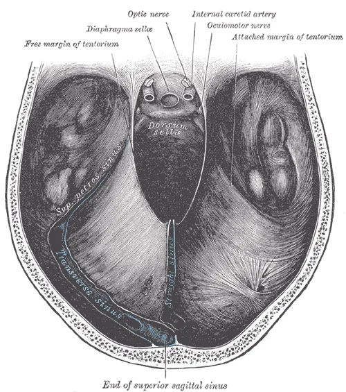
Tentorium cerebelli seen from above. Labelled illustration from Wikimedia Commons, used under its open licence.

Figure 1: Tentorium cerebelli seen from above.
These connections can also be broadly described by their functions[1].
Visual cortex information is also part of the processing that occurs in the cerebrocerebellum[1].
Descending fibers from the cerebrum have branches that connect to neurons in the pons[1]. Those neurons project into the cerebellum, providing a copy of motor commands sent to the spinal cord[1]. Fibers from this nucleus enter the cerebellum and are compared with the descending commands from the cerebrum[1]. [2]
Figure 2Gray705
Long description
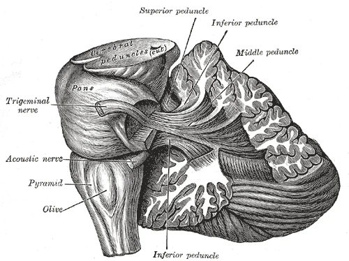
Dissection showing the projection fibers of the cerebellum. (After E. B. Jamieson.)

Figure 2: Dissection showing the projection fibers of the cerebellum (After E.
The primary motor cortex is located in the precentral gyrus of the frontal lobe. A neurosurgeon, Wilder Penfield, described much of the basic understanding of the primary motor cortex by electrically stimulating the surface of the cerebrum. Penfield would probe the surface of the cortex while the patient was only under local anesthesia so that he could observe responses to the stimulation. This led to the belief that the precentral gyrus directly stimulated muscle movement. We now know that the primary motor cortex receives input from several areas that aid in planning movement, and its principal output stimulates spinal cord neurons to stimulate skeletal muscle contraction. [3]
The motor output from the cortex descends into the brain stem and to the spinal cord to control the musculature through motor neurons. Neurons located in the primary motor cortex, named Betz cells, are large cortical neurons that synapse with lower motor neurons in the brain stem or in the spinal cord. The two descending pathways travelled by the axons of Betz cells are the corticobulbar tract and the corticospinal tract, respectively. Both tracts are named for their origin in the cortex and their targets—either the brain stem (the term “bulbar” refers to the brain stem as the bulb, or enlargement, at the top of the spinal cord) or the spinal cord. [3]
The cerebellum, as the name suggests, is the "little brain". It is covered in gyri and sulci like the cerebrum, and looks like a miniature version of that part of the brain. The cerebellum accounts for approximately 10 percent of the mass of the brain. It is attached to the brain stem, specifically the pons, and is considered a separate region of the adult brain. The pons serves as the main connection between the cerebellum and the brain stem. [2] [3] [1]
The cerebellum is largely responsible for comparing information from the cerebrum with sensory feedback from the periphery through the spinal cord. It plays a crucial role in motor coordination by comparing cerebral motor commands with proprioceptive feedback. This comparison allows the cerebellum to send corrective commands when there's a discrepancy between the intended movement and the actual sensory feedback. [3] [2]
The structure of the cerebellum includes the cerebellar cortex, which contains gray matter, and the white matter beneath it.
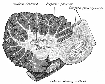
The deep cerebellar nuclei are clusters of gray matter embedded within the white matter of the cerebellum. They serve as the primary output stations for the cerebellar cortex. There are four main pairs of nuclei, arranged from lateral to medial: the dentate, emboliform, globose, and fastigial nuclei. The dentate nucleus is the largest and most lateral, while the fastigial nucleus is the most medial and closest to the midline. These nuclei receive inhibitory input from Purkinje cells in the cortex and excitatory input from climbing and mossy fibers. [3]
Figure 3Gray707
Long description
Sagittal section through right cerebellar hemisphere. The right olive has also been cut sagitally.

Figure 3: Sagittal section through right cerebellar hemisphere.
The cerebellar cortex is composed of three distinct layers, from superficial to deep: the molecular layer, the Purkinje cell layer, and the granular layer. The molecular layer contains the dendrites of Purkinje cells and parallel fibers from granule cells. The Purkinje cell layer consists of a single row of large, flask-shaped Purkinje cells, which are the sole output neurons of the cerebellar cortex. The granular layer is densely packed with small granule cells and Golgi cells. Mossy fibers terminate in glomeruli within the granular layer, where they synapse with granule cell dendrites. Climbing fibers, originating from the inferior olivary nucleus, wrap around the dendrites of Purkinje cells in the molecular layer, providing powerful excitatory input.
The cerebellum is connected to the brainstem by three pairs of fiber tracts known as the cerebellar peduncles. These connections allow the cerebellum to integrate sensory information and motor commands to coordinate movement. [2]
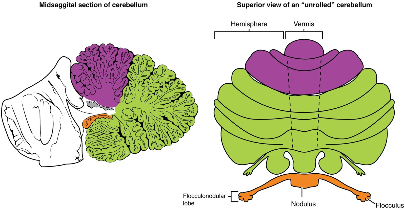
The cerebellum is divided into lobes and functional zones. Anatomically, it is divided into the anterior lobe, posterior lobe, and flocculonodular lobe. Functionally, it is divided into the vestibulocerebellum (archicerebellum), spinocerebellum (paleocerebellum), and cerebrocerebellum (neocerebellum). The vestibulocerebellum, consisting of the flocculonodular lobe, is involved in balance and eye movements. The spinocerebellum, comprising the vermis and intermediate zone, receives proprioceptive input from the spinal cord and is involved in coordinating limb movements. The cerebrocerebellum, consisting of the lateral hemispheres, is involved in planning and timing of complex movements.
Figure 41613 Major Regions of the Cerebellum-02
Long description
The cerebellum can be divided into two basic regions: the midline and the hemispheres. The midline is composed of the vermis and the flocculonodular lobe, and the hemispheres are the lateral regions.

Figure 4: The cerebellum can be divided into two basic regions: the midline and the hemispheres.
Figure 5Gray704
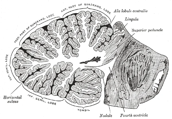
Long description
Sagittal section of the cerebellum, near the junction of the vermis with the hemisphere. (Schäfer.)

Figure 5: Sagittal section of the cerebellum, near the junction of the vermis with the hemisphere (Schäfer.).
Figure 6Gray703
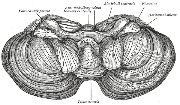
Long description
Under surface of the cerebellum. (Schäfer.)

Figure 6: Under surface of the cerebellum (Schäfer.).
The cerebellum functions as a sophisticated comparator, integrating motor commands with sensory feedback to refine movement. This circuit allows for the coordination of voluntary actions by contrasting intended movement with actual performance. The structural basis for this integration lies in its three pairs of cerebellar peduncles, which serve as the primary afferent and efferent pathways connecting the cerebellum to the brain stem.
Figure 71612 Cerebellar Peduncles-02
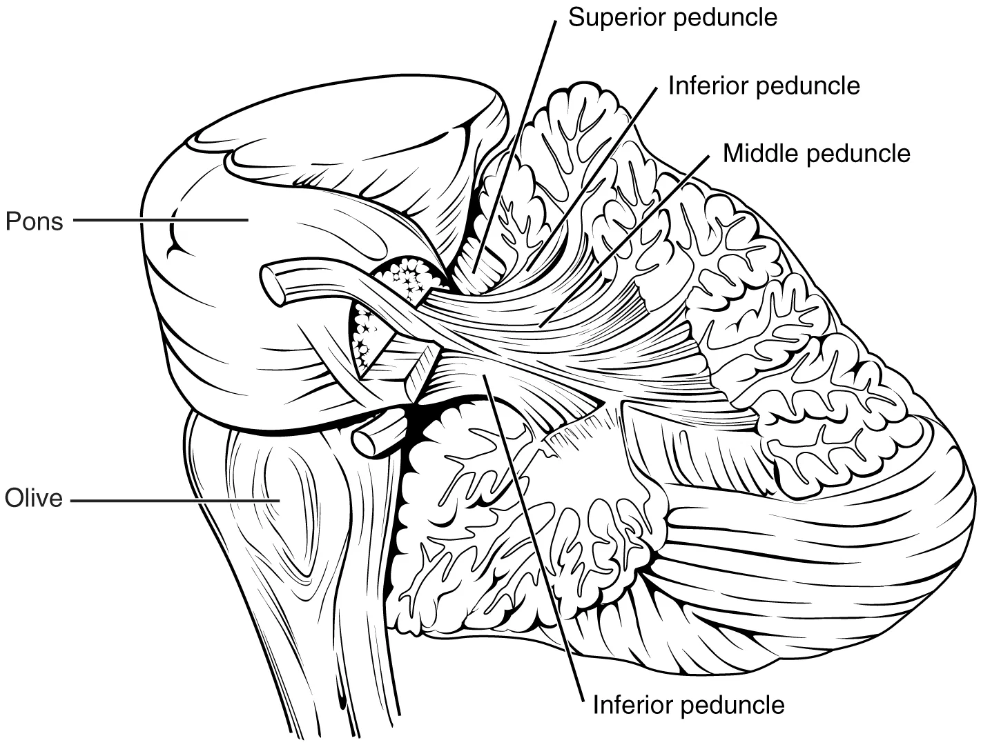
Long description
The connections to the cerebellum are the three cerebellar peduncles, which are close to each other. The ICP arises from the medulla—specifically from the inferior olive, which is visible as a bulge on the ventral surface of the brain stem. The MCP is the ventral surface of the pons. The SCP project

Figure 7: The connections to the cerebellum are the three cerebellar peduncles, which are close to each other.
The superior cerebellar peduncle (SCP) is located most superiorly and laterally, primarily carrying efferent fibres. The middle cerebellar peduncle (MCP) is the largest, connecting laterally to the pons. The inferior cerebellar peduncle (ICP) is situated inferiorly and medially, connecting to the medulla. [1]
Figure 8Gray702
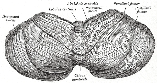
Long description
Upper surface of the cerebellum. (Schäfer.)

Figure 8: Upper surface of the cerebellum (Schäfer.).
The middle cerebellar peduncle serves as the main afferent route from the cerebral cortex. When the cerebral cortex sends a motor command to initiate an action, such as walking, that command is copied by the pons and transmitted into the cerebellum through the MCP. This input allows the cerebellum to receive a 'copy' of the intended movement plan. [1]
This input provides the cerebellum with real-time data on body position and equilibrium. [1]
Ataxia is a movement disorder of the cerebellum, presenting as a loss of coordination in voluntary movements. When the problem is observed in movement, it is ascribed to cerebellar damage. Sensory and vestibular ataxia would likely also present with problems in gait and station. The cerebellum compares motor commands and sensory feedback to generate a new output, making it possible to coordinate movements. [1]
Clinical pearlClinical Pearl
Ataxia indicates cerebellar damage when the deficit is in movement coordination, distinct from sensory ataxia which stems from proprioceptive loss.
| Peduncle | Location | Primary Function | Key Structures |
|---|---|---|---|
| Superior (SCP) | Superior/Lateral | Efferent | Red nucleus, Rubrospinal tract |
| Middle (MCP) | Lateral | Afferent | Pons, Corticopontine fibres |
| Inferior (ICP) | Inferior/Medial | Afferent | Spinal cord, Vestibular nuclei |
The cerebellum is a highly vascular structure, requiring a robust and consistent blood supply to maintain its critical role in motor coordination and balance. Understanding the vascular architecture is essential for localising lesions and interpreting clinical deficits. The blood supply is derived from three major paired arteries that arise from the vertebrobasilar system. These vessels ensure perfusion to the cerebellar hemispheres, the vermis, and the deep cerebellar nuclei.
Figure 9Gray706
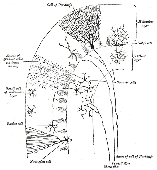
Long description
Transverse section of a cerebellar folium, showing principal cell types and connections

Figure 9: Transverse section of a cerebellar folium, showing principal cell types and connections.
Arising from the vertebral arteries and the basilar artery are the three cerebellar arteries.
DefinitionVertebrobasilar System
The paired vertebral arteries join to form the basilar artery, which supplies the brainstem and cerebellum.
It travels laterally, passing near the facial and vestibulocochlear nerves (CN VII and VIII), and supplies the anterior inferior surface of the cerebellum and the flocculus. It also gives off the labyrinthine artery in most individuals, which supplies the inner ear.
| Artery | Origin | Territory | Key Clinical Deficit |
|---|---|---|---|
| Superior Cerebellar | Basilar Artery | Superior cerebellum, superior peduncle | Contralateral ataxia, dysmetria |
| Anterior Inferior Cerebellar | Basilar Artery | Anterior inferior cerebellum, flocculus | Ipsilateral ataxia, facial palsy, hearing loss |
| Posterior Inferior Cerebellar | Vertebral Artery | Posterior inferior cerebellum, medulla | Lateral medullary syndrome, ipsilateral ataxia |
The cerebellum is integral to the coordination of voluntary movement, balance, and posture. Its functional role is best understood by examining the clinical examination of gait and station, which directly assess cerebellar integrity. Gait is considered a distinct component of the neurological exam or a subtest of the coordination exam that addresses walking and balance.
The hemispheres are lateral to the vermis, coordinating distal limb movements. The flocculonodular lobe is located inferiorly and posteriorly, associated with vestibular input and balance. The spinocerebellum is situated in the vermis and intermediate zone, receiving proprioceptive input. The vestibulocerebellum comprises the flocculonodular lobe, interacting with the vestibular nuclei. The cerebrocerebellum occupies the lateral hemispheres, involved in planning and timing of movements.
The clinical assessment begins with the station subtest, which starts with the patient standing in a normal position to check for the placement of the feet and balance. The patient is asked to hop on one foot to assess the ability to maintain balance and posture during movement. Though the station subtest appears to be similar to the Romberg test, the difference is that the patient’s eyes are open during station. [1]
DefinitionStation Test
A test of balance and posture with eyes open, distinguishing cerebellar ataxia from sensory ataxia.
The cerebellum modulates motor output through three main functional divisions. The spinocerebellum regulates body and limb movements by comparing intended and actual movement. The vestibulocerebellum maintains equilibrium and coordinates eye movements. The cerebrocerebellum plans and initiates voluntary movements. These functions rely on the integration of sensory input from the spinal cord, vestibular system, and cerebral cortex.
| Division | Location | Primary Function |
|---|---|---|
| Spinocerebellum | Vermis and intermediate hemisphere | Coordination of body and limb movements |
| Vestibulocerebellum | Flocculonodular lobe | Equilibrium and eye movement coordination |
| Cerebrocerebellum | Lateral hemispheres | Planning and initiation of voluntary movements |
Disruption in these areas leads to specific clinical signs. Lesions in the vermis cause truncal ataxia, while hemispheric lesions cause limb ataxia.
The integration of these functions allows for complex motor tasks. Walking, reaching, and speaking require precise timing and coordination. The cerebellum ensures that muscle contractions are synchronized. It adjusts muscle tone to maintain posture against gravity. This automatic adjustment is vital for stability.
Cerebellar Function Assessment
- Assess GaitObserve walking and balance
- Question: Abnormal?
Yes
- Check Spinocerebellum
No
- Check Vestibulocerebellum
This input is crucial for maintaining balance. This allows posture, movement, and balance to be modulated on the basis of equilibrium information provided by the vestibular system. [3]
The spinocerebellum receives proprioceptive input from the spinal cord. This input allows for real-time correction of movement. The spinocerebellar tracts carry this information. The cerebellum compares the intended movement with the actual movement. Any discrepancy is corrected by adjusting motor output.
Exam trapCommon Trap
Do not confuse cerebellar ataxia with sensory ataxia. Cerebellar ataxia persists with eyes open.
| Tract | Origin | Termination | Function |
|---|---|---|---|
| Spinocerebellar | Spinal cord | Cerebellum | Proprioceptive input |
| Vestibulocerebellar | Vestibular nuclei | Cerebellum | Balance input |
| Corticopontocerebellar | Cortex | Cerebellum | Motor planning input |
The cerebellum's output is sent to the thalamus and then to the motor cortex. This output modulates the activity of upper motor neurons. The cerebellum does not directly innervate muscles. It influences movement indirectly through the cortex and brain stem. This indirect influence allows for fine-tuning of motor commands.
DefinitionAtaxia
Lack of coordination of muscle movements, often due to cerebellar dysfunction.
However, the primary role remains motor coordination. The cognitive functions are secondary to the motor role.
NoteNote
Cerebellar cognitive affective syndrome is a rare condition involving cognitive and emotional changes.
In summary, the cerebellum is essential for motor coordination, balance, and posture. Its functional divisions work together to ensure smooth and precise movements. Clinical assessment of gait and station is crucial for detecting cerebellar dysfunction. Understanding the cerebellum's role is vital for neurological examination.
Testing for cerebellar function is the basis of the coordination exam. The subtests target appendicular musculature, controlling the limbs, and axial musculature for posture and gait. The assessment of cerebellar function will depend on the normal functioning of other systems addressed in previous sections of the neurological exam. Motor control from the cerebrum, as well as sensory input from somatic, visual, and vestibular senses, are important to cerebellar function. [1]
The midline vermis is central, flanked by the lateral hemispheres. If you are walking along the sidewalk on campus and you hear chirping, the superior colliculus coordinates that information with your awareness of the visual location of the tree right above you. That is the correlation of auditory and visual maps. [2]
DefinitionCheck Reflex
A similar response would be seen if you try to pick up a coffee mug that you believe to be full but turns out to be empty. Without checking the contraction, the mug would be thrown from the overexertion of the muscles expecting to lift a heavier object. [1]
Cerebellar lesions produce distinct signs depending on the location. Lesions of the vermis affect axial musculature, causing truncal ataxia and gait disturbance. Lesions of the hemispheres affect appendicular musculature, causing limb ataxia.
| Sign | Description | Typical Lesion Site |
|---|---|---|
| Dysmetria | Inability to judge distance; overshooting or undershooting a target | Cerebellar hemisphere |
| Dysdiadochokinesia | Impaired ability to perform rapid alternating movements | Cerebellar hemisphere |
| Intention Tremor | Tremor that increases as the limb approaches a target | Cerebellar hemisphere |
| Rebound Phenomenon | Failure to stop movement when resistance is removed | Cerebellar hemisphere |
| Truncal Ataxia | Unsteadiness of the trunk and gait | Vermis |
| Nystagmus | Involuntary rhythmic eye movements | Cerebellar connections |
The tectum is composed of four bumps known as the colliculi (singular = colliculus), which means “little hill” in Latin. If you are walking along the sidewalk on campus and you hear chirping, the superior colliculus coordinates that information with your awareness of the visual location of the tree right above you. That is the correlation of auditory and visual maps. [2]
The cerebellum is often tested via gait analysis, which serves as a primary clinical screen for its function. The examination begins with the patient walking normally away from the examiner and then turning to return. During this normal gait, the examiner observes for abnormal placement of the feet and the movement of the arms relative to the body. Specific variations test distinct aspects of balance and coordination. Tandem gait requires the patient to place the heel of one foot against the toe of the other and walk in a straight line. This specific maneuver tests the ability to maintain a narrow base of support. Walking only on the heels or only on the toes tests additional aspects of balance and motor control. [1]
Exam trapGait Assessment
Do not confuse cerebellar gait with sensory ataxia. Cerebellar lesions cause wide-based, unsteady gait, whereas sensory ataxia worsens significantly with eye closure (positive Romberg).
DefinitionTandem Gait
A test where the patient places the heel of one foot against the toe of the other to walk in a straight line, assessing fine motor coordination and balance.
| Structure | Location | Primary Function |
|---|---|---|
| Cerebrum | Superior, anterior | Higher cognitive function, voluntary movement |
| Diencephalon | Deep, central | Relay station, homeostasis |
| Midbrain | Between thalamus and pons | Visual/auditory reflexes, motor control |
| Cerebellum | Posterior fossa | Coordination, balance, motor learning |
NoteTerminology
Dorsal and ventral are sometimes used interchangeably with posterior and anterior, particularly in reference to nerves and spinal cord structures. [2]
Students frequently confuse the anatomical levels of the spinal cord with the vertebral levels, leading to errors in localising lesions. The spinal cord does not extend the full length of the vertebral column because it ceases significant growth after the first or second year, whereas the skeleton continues to grow. Consequently, the name of a spinal cord region corresponds to the level at which spinal nerves pass through the intervertebral foramina. For example, the sacral spinal cord contains motor neurons for the big toe, yet these nerves must travel a long distance to reach their target muscles. The axon that has to reach all the way to the belly of that muscle may be a meter in length. This anatomical reality creates the cauda equina, a long bundle of nerves that resembles a horse’s tail. [2]
Another frequent error involves the somatic nervous system's control mechanisms. The defining characteristic of the somatic nervous system is that it controls skeletal muscles. Students often assume all somatic movements are voluntary, but the term “voluntary” suggests that there is a conscious decision to make a movement. However, some aspects of the somatic system use voluntary muscles without conscious control. One example is the ability of our breathing to switch to unconscious control while we are focused on another task. Conversely, muscles responsible for basic breathing are also utilized for speech, which is entirely voluntary. [3]
The primary motor cortex is arranged in a similar fashion to the primary somatosensory cortex, in that it has a topographical map of the body, creating a motor homunculus. [3]
Reflex arcs are often oversimplified. In the hot-stove withdrawal reflex, the biceps brachii contracts while the antagonistic triceps brachii needs to relax. Because the neuromuscular junction is strictly excitatory, the biceps will contract when the motor nerve is active. Skeletal muscles do not actively relax. Instead the motor neuron needs to “quiet down,” or be inhibited. This occurs through an interneuron in the spinal cord. The interneuron’s cell body is located in the dorsal horn of the spinal cord. The interneuron receives a synapse from the axon of the sensory neuron that detects that the hand is being burned. In response to this stimulation from the sensory neuron, the interneuron then inhibits the motor neuron that controls the triceps brachii. This is done by releasing a neurotransmitter or other signal that hyperpolarizes the motor neuron connected to the triceps brachii, making it less likely to initiate an action potential. [3]
Exam trapReflex Misconception
Skeletal muscles do not actively relax; they relax when motor neuron inhibition reduces their excitation.
They did not have impairments to understanding speech, only to producing speech sounds, suggesting a damaged or underdeveloped Broca’s area. [3]
In examinations, knowledge of the neuromuscular junction is tested. Motor neuron axons connect to muscle fibers at a neuromuscular junction. This is a specialized synaptic structure at which multiple axon terminals synapse with the muscle fiber sarcolemma. The synaptic end bulbs of the motor neurons secrete acetylcholine, which binds to receptors on the sarcolemma. The binding of acetylcholine opens ligand-gated ion channels, increasing the movement of cations across the sarcolemma. This depolarizes the sarcolemma, initiating muscle contraction. Whereas other synapses result in graded potentials that must reach a threshold in the postsynaptic target, activity at the neuromuscular junction reliably leads to muscle fiber contraction with every nerve impulse received from a motor neuron. However, the strength of contraction and the number of fibers that contract can be affected by the frequency of the motor neuron impulses. [3]
On imaging and in clinical neurology, the distinction between withdrawal reflexes is vital. Another example of a withdrawal reflex occurs when you step on a painful stimulus, like a tack or a sharp rock. The nociceptors that are activated by the painful stimulus activate the motor neurons responsible for contraction of the tibialis anterior muscle. This causes dorsiflexion of the foot. An inhibitory interneuron, activated by a collateral branch of the nociceptor fiber, will inhibit the motor neurons of the gastrocnemius and soleus muscles to cancel plantar flexion. An important difference in this reflex is that plantar flexion is most likely in progress as the foot is pressing down onto the tack. Contraction of the tibialis anterior is not the most important aspect of the reflex, as continuation of plantar flexion will result in further damage from stepping onto the tack. [3]
DefinitionMotor Unit
The motor neuron and all the muscle fibers that it controls make up a motor unit.
Withdrawal Reflex Logic
- Stimulus DetectedPainful stimulus (e.g., tack)
- Question: Foot Pressing Down?
Yes
- Activate Tibialis AnteriorCauses dorsiflexion
- Inhibit Gastrocnemius/SoleusVia inhibitory interneuron
- Cancel Plantar FlexionPrevents further damage
No
- Standard WithdrawalBiceps contract, Triceps inhibit
| Muscle Group | Approximate Fibres per Unit | Control Precision |
|---|---|---|
| Quadriceps | Up to 1000 | Low |
| Extraocular | 10 | High |
The spinal cord regions correspond to the level at which spinal nerves pass through the intervertebral foramina. The spinal cord does not grow significantly longer after the first or second year, but the skeleton continues to grow. [2] The defining characteristic of the somatic nervous system is that it controls skeletal muscles. The primary motor cortex has a topographical map of the body, creating a motor homunculus. The greatest amount of cortical space is given to muscles that perform fine, agile movements. [3] The axons will also branch to innervate multiple muscle fibers. [3] Together, the motor neuron and all the muscle fibers that it controls make up a motor unit. [3] Activity at the neuromuscular junction reliably leads to muscle fiber contraction with every nerve impulse received from a motor neuron.
Answer each one in your head first, then open the answer.
What structure resembles a horse’s tail?
Show answerHide answer
Cauda equina
The neurons responsible for musculature in the feet and lower legs are in the medial wall of the precentral gyrus. [3]
What neurotransmitter is secreted by synaptic end bulbs at the neuromuscular junction?
Show answerHide answer
AcetylcholineWhich muscle is inhibited during the reflex when stepping on a tack?
Show answerHide answer
Gastrocnemius and soleusWhat is the defining characteristic of the somatic nervous system?
Show answerHide answer
It controls skeletal muscles
- Betts JG, Young KA, Wise JA, et al. The Coordination and Gait Exams. OpenStax, Anatomy and Physiology 2e (CC BY-NC-SA 4.0). Accessed .
- Betts JG, Young KA, Wise JA, et al. The Central Nervous System. OpenStax, Anatomy and Physiology 2e (CC BY-NC-SA 4.0). Accessed .
- Betts JG, Young KA, Wise JA, et al. Motor Responses. OpenStax, Anatomy and Physiology 2e (CC BY-NC-SA 4.0). Accessed .
Figure credits
- Figure 1: Henry Vandyke Carter, via Wikimedia Commons, Public domain, Public domain.
- Figure 2: Henry Vandyke Carter, via Wikimedia Commons, Public domain, Public domain.
- Figure 3: Henry Vandyke Carter, via Wikimedia Commons, Public domain, Public domain.
- Figure 4: OpenStax College, via Wikimedia Commons, CC BY 3.0, CC BY 3.0. resized.
- Figure 5: Henry Vandyke Carter, via Wikimedia Commons, Public domain, Public domain.
- Figure 6: Henry Vandyke Carter, via Wikimedia Commons, Public domain, Public domain.
- Figure 7: OpenStax College, via Wikimedia Commons, CC BY 3.0, CC BY 3.0.
- Figure 8: Henry Vandyke Carter, via Wikimedia Commons, Public domain, Public domain.
- Figure 9: Henry Vandyke Carter, via Wikimedia Commons, Public domain, Public domain.
Educational content for medical students and doctors. It is not a substitute for clinical judgement or local guidelines.