Thalamus
The thalamus is the major sensory relay station of the diencephalon, connecting peripheral inputs to the cerebral cortex.
ALIASEs: Thalamic nuclei, Dorsal thalamus, Epithalamus
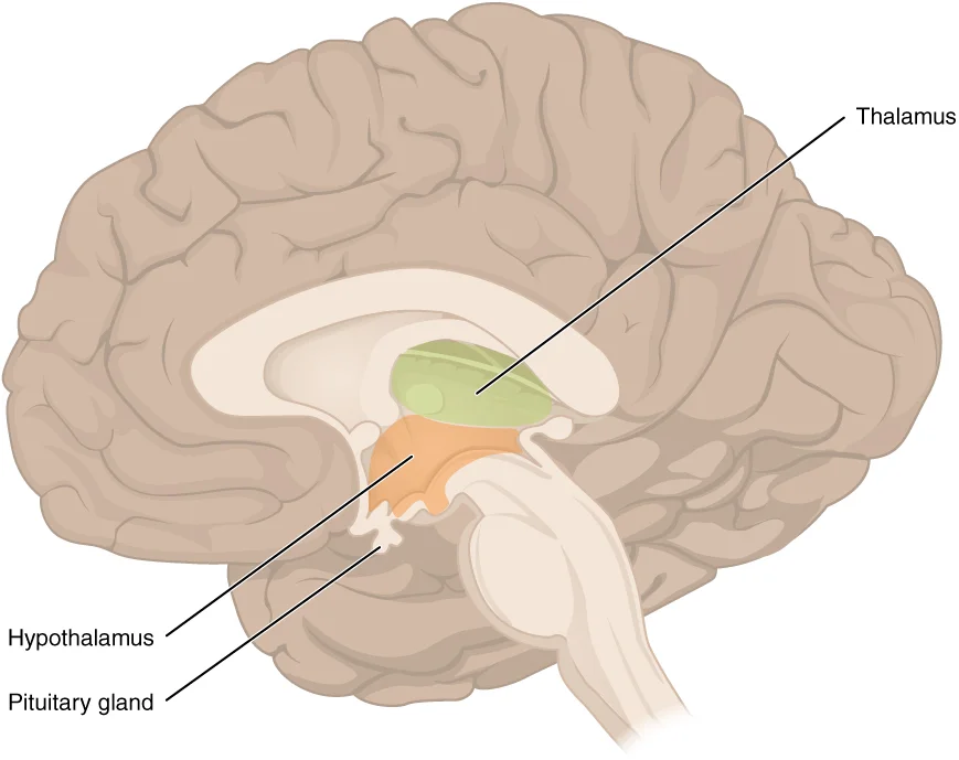
The thalamus is a large, egg-shaped mass of grey matter located deep within the brain, forming the lateral walls of the third ventricle. It sits superior to the midbrain and inferior to the cerebrum, serving as the primary gateway for sensory information entering the cerebral cortex. The diencephalon, which includes the thalamus and hypothalamus, constitutes the central core of the forebrain. [2] [1]
Figure 11310 Diencephalon
Long description
The diencephalon is composed primarily of the thalamus and hypothalamus, which together define the walls of the third ventricle. The thalami are two elongated, ovoid structures on either side of the midline that make contact in the middle. The hypothalamus is inferior and anterior to the thalamus, c

Figure 1: The diencephalon is composed primarily of the thalamus and hypothalamus, which together define the walls of the third ventricle.
Figure 2Gray732
Long description
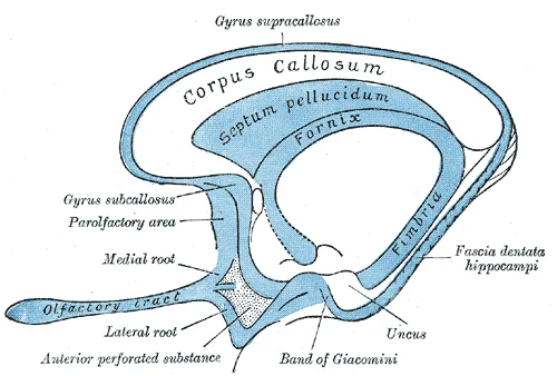
Scheme of rhinencephalon. Labelled illustration from Wikimedia Commons, used under its open licence.

Figure 2: Scheme of rhinencephalon.
DefinitionThalamus
The thalamus is a collection of nuclei that relay information between the cerebral cortex and the periphery, spinal cord, or brain stem. [2]
Structurally, the thalamus is divided into three major regions defined by white matter tracts: the anterior nucleus, the medial nucleus, and the lateral group of nuclei. The anterior nucleus acts as a relay between the hypothalamus and the limbic system, which controls emotions and memory. The medial nuclei relay information from the limbic system and basal ganglia to the cerebral cortex, playing a role in memory creation and alertness. [1]
Superior and posterior to the main thalamic mass lies the epithalamus, a small region that contains the pineal gland. The pineal gland is involved in circadian rhythm regulation. Adjacent to the pineal gland is the habenula, which connects the limbic system to the midbrain. The thalamus also interacts with the basal nuclei; the primary output of the basal nuclei is directed to the thalamus, which then relays motor commands to the cerebral cortex. [2]
| Nucleus Group | Primary Connections | Key Functions |
|---|---|---|
| Anterior | Hypothalamus, Limbic System | Relay for emotion and memory processing |
| Medial | Limbic System, Basal Ganglia | Relay for memory creation and alertness |
| Lateral | Special and Somatic Senses | Relay for sensory information to cortex |
The thalamus is not merely a passive conduit; it actively processes information. For instance, thalamic nuclei influence which visual stimuli receive attention. All sensory information, except for the sense of smell, must pass through the thalamus before cortical processing. The olfactory system is unique, as axons from the olfactory bulb project directly to the cerebral cortex and limbic system without thalamic intervention. [2] [1]
NoteClinical Pearl
Thalamic syndromes, such as Dejerine-Roussy syndrome, can result from thalamic strokes, causing chronic pain and sensory disturbances due to disrupted relay pathways.
The thalamus receives descending input from the cerebrum, which usually communicates motor commands involving interactions with the cerebellum and brain stem nuclei. This bidirectional communication allows the cortex to influence thalamic filtering of sensory data. The thalamus is thus integral to both ascending sensory perception and descending motor control loops. [2]
Figure 3Thalamus3enTRN
Long description
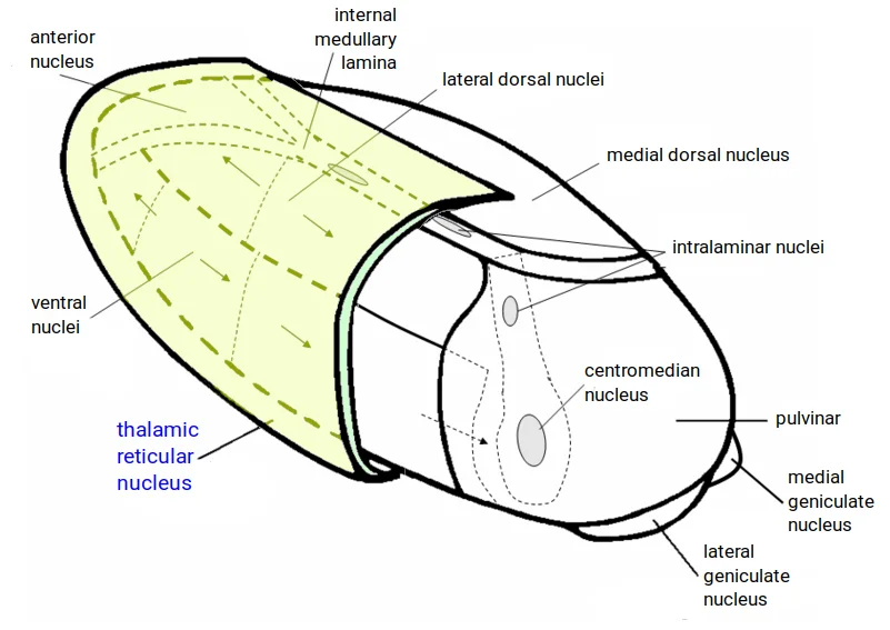
This diagram shows the thalamic reticular nucleus location in three dimensional perspective relative to various other nuclei and nucleus groups of the human left thalamus. It is an English translation from the Dutch-lang

Figure 3: This diagram shows the thalamic reticular nucleus location in three dimensional perspective relative to various other nuclei and nucleus groups of the human left thalamus.
The thalamus is a large, ovoid mass of grey matter situated in the diencephalon, forming the major part of the lateral walls of the third ventricle. It serves as the critical relay station for nearly all sensory information ascending to the cerebral cortex, with the notable exception of the olfactory system which projects directly to the frontal and temporal lobes. Anatomically, it is divided into anterior, medial, and lateral nuclear groups, each with distinct connectivity patterns and functional roles. [1]
Figure 4Gray757
Long description
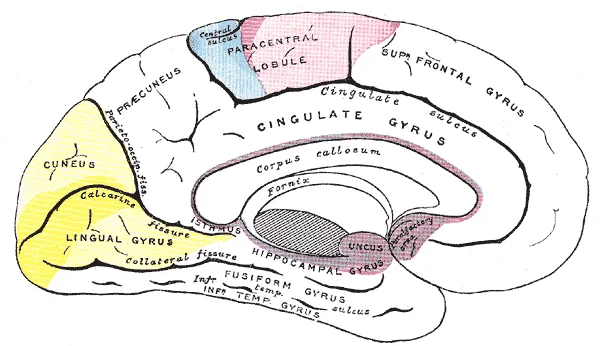
Areas of localization on medial surface of hemisphere. Motor area in red. Area of general sensations in blue. Visual area in yellow. Olfactory area in purple. The psychic portions are in lighter tints.

Figure 4: Areas of localization on medial surface of hemisphere (motor area in red).
Laterally, it is separated from the lentiform nucleus of the basal ganglia by the internal capsule. Medially, the medial surface forms the upper part of the lateral wall of the third ventricle.
The sensory pathways for somatosensation, gustation, and equilibrium all converge on specific nuclei within the thalamus before projecting to the cortex. For somatosensory input from the body, axons from the dorsal column system ascend as the medial lemniscus and terminate in the thalamus, where they synapse with third-order neurons. These third-order neurons then project their axons to the postcentral gyrus of the cerebral cortex, where somatosensory stimuli are initially processed. Similarly, the sensory pathway for gustation travels along the facial and glossopharyngeal cranial nerves, synapsing in the solitary nucleus, with axons then projecting to the ventral posterior nucleus of the thalamus. Finally, the vestibular nuclei project to the thalamus to join the proprioceptive pathway, allowing for the conscious perception of equilibrium. [1]
The visual system also relies heavily on thalamic relay. The optic tract, extending from the optic chiasm, has three major targets, with the majority of connections going to the thalamus—specifically, the lateral geniculate nucleus. Axons from this nucleus then project to the visual cortex of the cerebrum, located in the occipital lobe. This topographical arrangement is maintained throughout the pathway, ensuring that anatomic information is preserved. For instance, axons from the lower body are carried in the fasciculus gracilis, while those from the upper body are in the fasciculus cuneatus, and this relationship is maintained in the medial lemniscus and thalamus. [1]
Figure 5Gray730
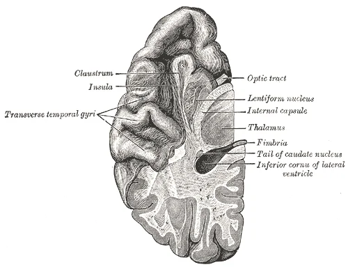
Long description
Section of brain showing upper surface of temporal lobe.

Figure 5: Section of brain showing upper surface of temporal lobe.
DefinitionThalamic Relay
The thalamus is the obligatory relay station for all sensory systems to reach the cerebral cortex, except for the olfactory system.
The thalamus is not merely a passive conduit but plays a role in modulating cortical activity. Subcortical nuclei, such as the basal nuclei, influence the activity of part of the thalamus, which can then increase or decrease cortical activity, often resulting in changes to motor commands. This integration allows for complex processing of sensory and motor information before it reaches the conscious awareness of the cerebral cortex.
The internal medullary lamina is a Y-shaped sheet of white matter that divides the thalamus into anterior, medial, and lateral nuclear groups. Understanding dorsal and ventral terminology is essential, as these terms are often used interchangeably with posterior and anterior in neuroanatomy. [2]
The anterior nuclear group lies anterior to the internal medullary lamina. It receives input from the mammillary bodies via the mammillothalamic tract and projects to the cingulate gyrus, linking it to the limbic system. The medial nuclear group, including the dorsomedial nucleus, is situated medial to the internal medullary lamina. It has extensive connections with the prefrontal cortex and is involved in memory and emotion, functions associated with the limbic cortex. The limbic system is a collection of structures involved in emotion, memory, and behavior.
The lateral nuclear group is the largest and is divided into dorsal and ventral tiers. The dorsal tier includes the dorsolateral and posterior nuclei. The ventral tier contains the ventral anterior, ventral lateral, ventral posterior, and geniculate nuclei. These nuclei serve as relay stations for sensory and motor information. The trigeminal pathway carries somatosensory information from the face, head, mouth, and nasal cavity. Axons from the second neuron in this pathway decussate and ascend to the thalamus along the trigeminothalamic tract.
The ventral posterior nucleus is subdivided into the ventral posteromedial (VPM) and ventral posterolateral (VPL) nuclei. The VPM receives sensory input from the face, while the VPL receives input from the body. The geniculate nuclei are located on the lateral aspect. The medial geniculate nucleus is part of the auditory pathway, while the lateral geniculate nucleus is part of the visual pathway. The sensory pathways of the trigeminal pathway each involve three successive neurons. First, axons from the trigeminal ganglion enter the brain stem at the level of the pons.
The internal medullary lamina separates the medial and lateral nuclear groups. It contains ascending and descending fibers. The pineal gland is located posterior to the thalamus, near the habenula. The habenula connects the basal forebrain to the midbrain and is involved in reward and aversion. Thalamic syndromes can result from vascular lesions affecting these nuclei. The basal nuclei are a set of nuclei in the cerebrum responsible for comparing cortical processing with the general state of activity in the nervous system to influence the likelihood of movement taking place. The basal nuclei are also referred to as the basal ganglia. The major structures of the basal nuclei that control movement are the caudate, putamen, and globus pallidus. The caudate is a long nucleus that follows the basic C-shape of the cerebrum. The putamen is mostly deep in the anterior regions of the frontal and parietal lobes. Together, the caudate and putamen are called the striatum. The globus pallidus is a layered nucleus that lies just medial to the putamen. The globus pallidus has two subdivisions, the external and internal segments. These nuclei are depicted in a frontal section of the brain. The direct pathway is the projection of axons from the striatum to the globus pallidus internal segment (GPi) and the substantia nigra pars reticulata (SNr). The GPi/SNr then projects to the thalamus, which projects back to the cortex. The indirect pathway is the projection of axons from the striatum to the globus pallidus external segment (GPe), then to the subthalamic nucleus (STN), and finally to GPi/SNr. The two streams both target the GPi/SNr, but one has a direct projection and the other goes through a few intervening nuclei.
Figure 6Gray748
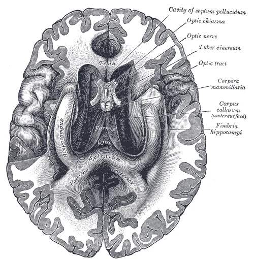
Long description
The fornix and corpus callosum from below. (From a specimen in the Department of Human Anatomy of the University of Oxford.)

Figure 6: The fornix and corpus callosum from below.
Figure 7Gray747
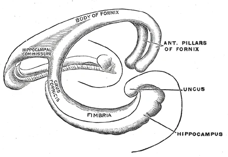
Long description
Diagram of the fornix. (Spitzka.). Labelled illustration from Wikimedia Commons, used under its open licence.

Figure 7: Diagram of the fornix (Spitzka.).
Figure 8Gray744
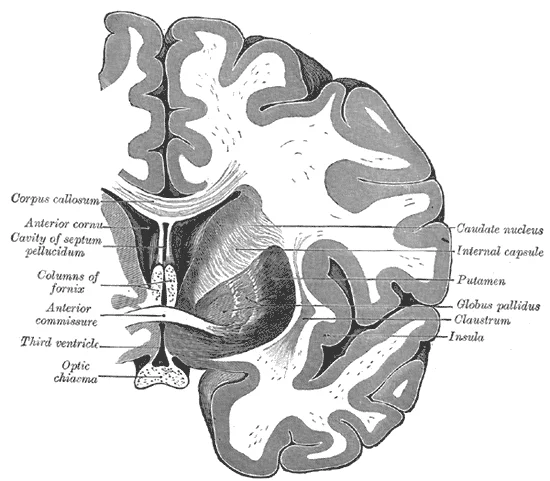
Long description
Coronal section of brain through anterior commissure.

Figure 8: Coronal section of brain through anterior commissure.
Figure 9Gray741
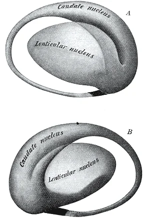
Long description
Two views of a model of the striatum: A, lateral aspect; B, mesal aspect.

Figure 9: Two views of a model of the striatum: A, lateral aspect; B, mesal aspect.
Figure 10Gray733
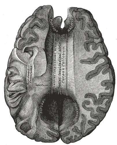
Long description
Corpus callosum from above. Labelled illustration from Wikimedia Commons, used under its open licence.

Figure 10: Corpus callosum from above.
Figure 11Gray731
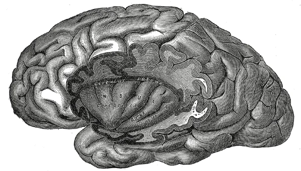
Long description
The insula of the left side, exposed by removing the opercula.

Figure 11: The insula of the left side, exposed by removing the opercula.
Figure 12Gray724
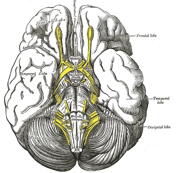
Long description
Base of brain. Labelled illustration from Wikimedia Commons, used under its open licence.

Figure 12: Base of brain.
Figure 13Gray722
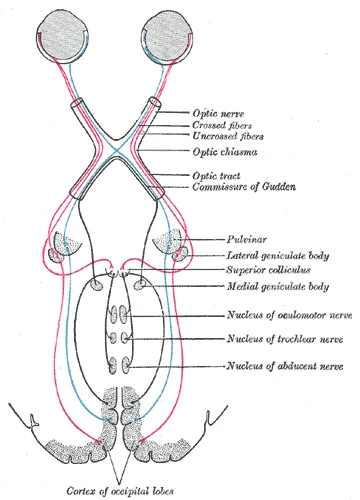
Long description
Scheme showing central connections of the optic nerves and optic tracts.

Figure 13: Scheme showing central connections of the optic nerves and optic tracts.
Figure 14Gray720
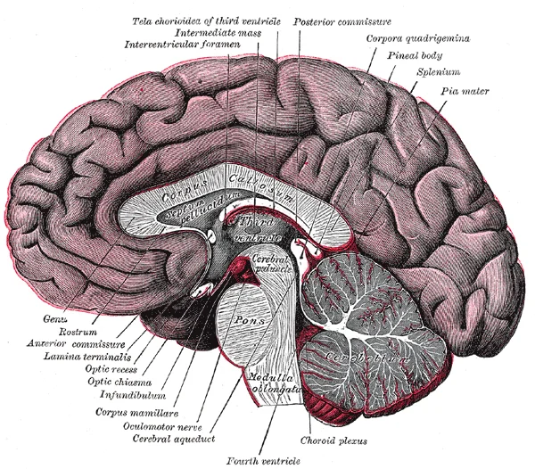
Long description
Median sagittal section of brain. The relations of the pia mater are indicated by the red color.

Figure 14: Median sagittal section of brain.
Figure 15Gray719
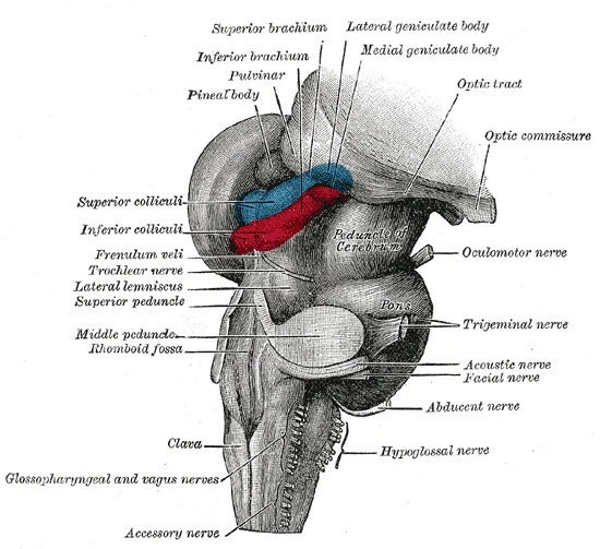
Long description
Hind- and mid-brains; postero-lateral view.

Figure 15: Hind- and mid-brains; postero-lateral view.
Figure 16Gray717
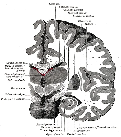
Long description
Coronal section of brain immediately in front of pons.

Figure 16: Coronal section of brain immediately in front of pons.
Figure 17Gray715
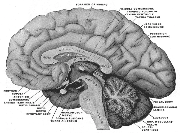
Long description
Mesal aspect of a brain sectioned in the median sagittal plane.

Figure 17: Mesal aspect of a brain sectioned in the median sagittal plane.
Figure 18Gray696
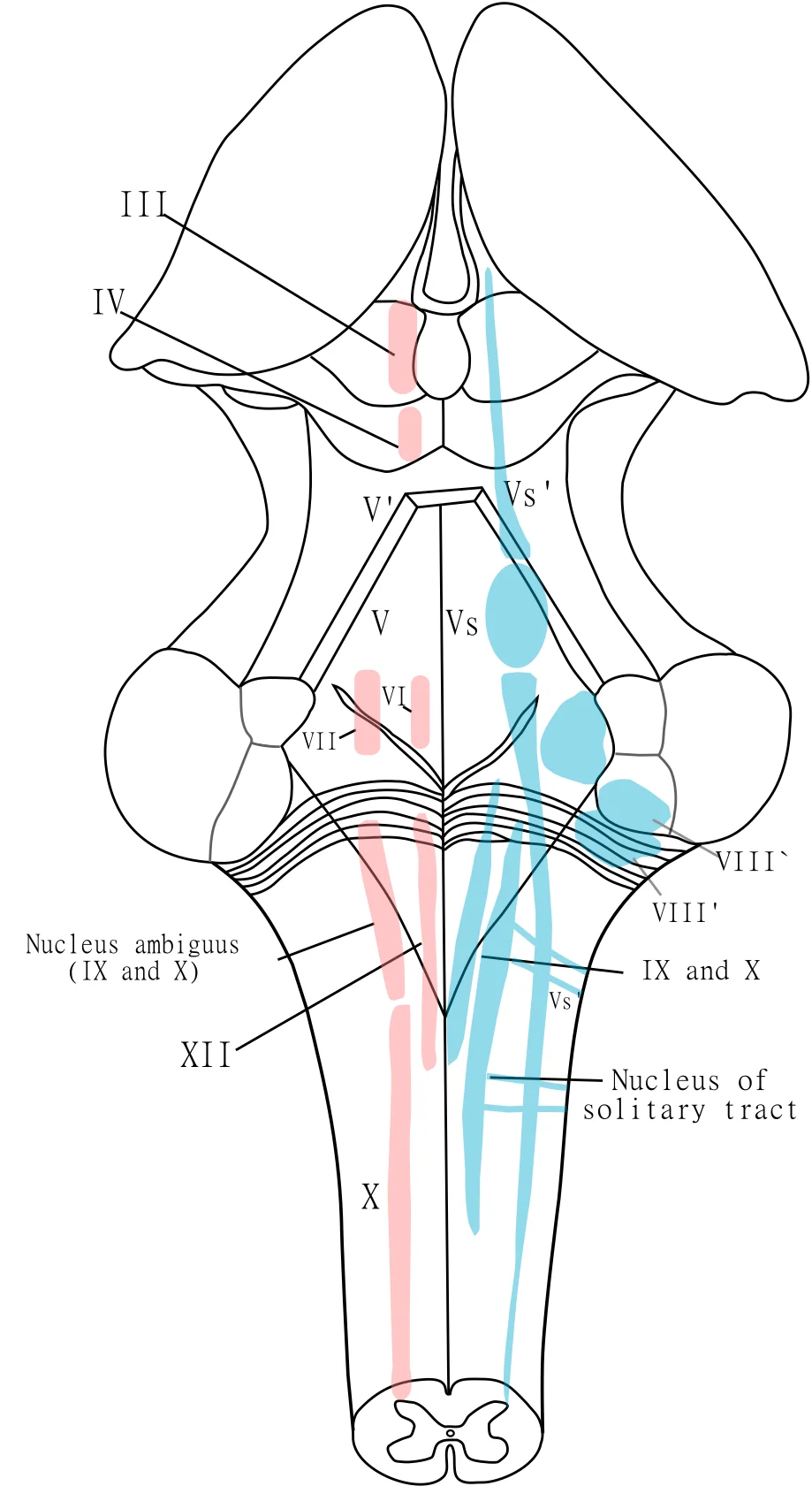
Long description
Vectorization of File:Gray696.png. Depicts a posterior view of the brain with the thalamus dissected out and the cerebellum removed. The 4th ventricle is splayed open in the center of the image. Major motor (red) and sensory (blue) nuclei are depicted.

Figure 18: Vectorization of File:Gray696.png.
The thalamus serves as a major relay station for sensory and motor information throughout the brain. It receives afferent inputs from various brain regions and sends efferent projections to the cerebral cortex. [1]
A key afferent pathway to the thalamus is the dorsal column system, which carries touch sensations and proprioception. In this pathway, second-order neurons decussate in the brainstem before synapsing in the thalamus. Similarly, the spinothalamic tract, responsible for pain and temperature sensations, sends second-order neurons that decussate in the spinal cord at the same level as their entry before reaching the thalamus. In both pathways, the third neurons originate in the thalamus and project to the somatosensory cortex. [1]
The auditory pathway also connects to the thalamus through the inferior colliculus. Neurons of the inferior colliculus project to the thalamus, which then sends auditory information to the cerebrum for the conscious perception of sound. This connection allows us to process and interpret sounds from our environment. [2]
Visual information reaches the thalamus through a more complex pathway. The optic nerve carries visual information where axons from the medial retina decussate at the optic chiasm, while axons from the lateral retina do not cross. This segregation of visual information between the left and right sides of the visual field ultimately projects to the thalamus before reaching the visual cortex. [1]
The superior colliculus, part of the tectum, combines sensory information about visual, auditory, and somatosensory space. It projects to the thalamus and coordinates orienting responses to stimuli, such as turning the eyes toward a sound or touch stimulus. This integration of sensory information helps us navigate and interact with our environment effectively. [2]
The vascular architecture of the thalamus is complex, reflecting its central position in the diencephalon. It receives its blood supply from two distinct circulatory systems: the anterior choroidal artery and the posterior cerebral artery. Understanding this dual supply is critical for localising lesions. This figure displays an axial view of the brain at the level of the thalamus. The anterior choroidal artery is shown arising from the internal carotid artery, coursing posteriorly to supply the anterior limb of the internal capsule and the lateral aspect of the thalamus. The posterior cerebral artery is depicted encircling the midbrain, giving off thalamoperforating and thalamogeniculate branches that penetrate the medial and posterior aspects of the thalamus respectively. The labelled structures include the internal carotid artery, the anterior choroidal artery, the posterior cerebral artery, and the thalamus itself.
This supply is consistent across most individuals. The artery courses posteriorly, passing between the cerebral peduncle and the optic tract. It then enters the lateral ventricle to supply the choroid plexus. In the thalamus, it provides blood to the anterior pole. Occlusion of this artery can lead to infarction of the anterior thalamus, resulting in specific cognitive and motor deficits. The anterior choroidal artery is a critical vessel because it supplies not only the thalamus but also the internal capsule and the optic tract. Therefore, lesions here often present with a triad of hemiparesis, hemianopia, and hemihypesthesia. The anterior choroidal artery arises from the internal carotid artery just before it bifurcates into the middle and anterior cerebral arteries. This anatomical relationship is crucial for surgical planning. [3]
The posterior cerebral artery supplies the majority of the thalamus. It arises from the basilar artery, although in some individuals, it may arise from the internal carotid artery (fetal configuration). The posterior cerebral artery gives off several branches that supply the thalamus. These include the thalamoperforating arteries and the thalamogeniculate arteries. The thalamogeniculate arteries arise from the P2 segment. They supply the lateral thalamus, including the ventral posterior nuclei. The posterior cerebral artery courses around the midbrain, passing between the cerebral peduncle and the tentorium cerebelli. It then enters the quadrigeminal cistern. The branches of the posterior cerebral artery are numerous and variable. The thalamoperforating arteries are particularly important because they supply the paramedian thalamus. Occlusion of these arteries can lead to paramedian thalamic infarction, which presents with altered consciousness and memory deficits. The thalamogeniculate arteries supply the lateral thalamus. Occlusion of these arteries can lead to lateral thalamic syndrome, characterised by contralateral sensory loss and pain.
The dual blood supply of the thalamus means that infarctions can occur in different patterns depending on the affected vessel. Anterior choroidal artery infarctions are rare but significant due to their impact on the internal capsule. Posterior cerebral artery infarctions are more common and can affect various thalamic nuclei. The thalamoperforating arteries are small and susceptible to occlusion by small vessel disease. This can lead to lacunar infarcts in the thalamus. The thalamogeniculate arteries are larger and can be affected by emboli from the heart or proximal arteries. Understanding the vascular anatomy of the thalamus is essential for diagnosing and managing thalamic strokes. The thalamus is a relay station for sensory and motor signals. Therefore, damage to this area can have widespread effects on neurological function. The blood supply to the thalamus is a key area of study in neuroanatomy and neurology.
The thalamus serves as the principal relay station for sensory information ascending to the cerebral cortex. It processes and directs signals from nearly all sensory modalities, with the notable exception of olfaction. This relay function ensures that sensory data is filtered and routed to the appropriate cortical areas for conscious perception. In addition to relay, the thalamus participates in association functions, integrating sensory inputs and modulating cortical activity to facilitate higher-order cognitive processes. [1]
The lateral surface of the thalamus is covered by the internal capsule, a dense band of white matter fibres. The medial surface forms the lateral wall of the third ventricle. The inferior surface rests on the subthalamus.
The relay function involves specific nuclei that transmit discrete sensory signals. For instance, the lateral geniculate nucleus relays visual information from the optic tract to the primary visual cortex in the occipital lobe. The medial geniculate nucleus relays auditory information from the inferior colliculus to the primary auditory cortex in the temporal lobe. The ventral posterior nucleus relays somatosensory information from the spinothalamic tract and medial lemniscus to the primary somatosensory cortex in the parietal lobe. These pathways ensure that sensory data reaches the correct cortical region for initial processing.
Figure 19Gray740
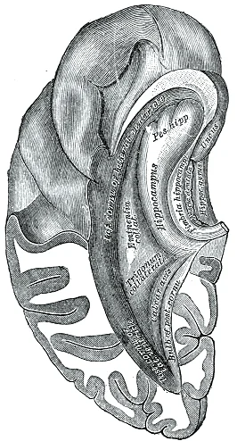
Long description
Inferior and posterior cornua, viewed from above.

Figure 19: Inferior and posterior cornua, viewed from above.
Figure 20Gray729
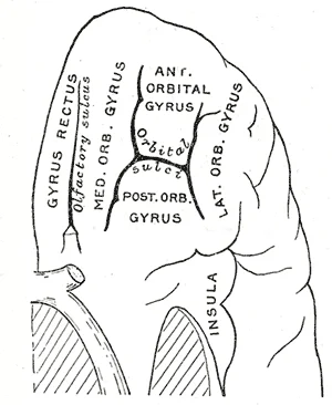
Long description
Orbital surface of left frontal lobe. Labelled illustration from Wikimedia Commons, used under its open licence.

Figure 20: Orbital surface of left frontal lobe.
The association function involves nuclei that project to association cortices rather than primary sensory areas. The dorsomedial nucleus, for example, projects to the prefrontal cortex, playing a role in memory and executive function. The pulvinar is involved in visual attention and integrating visual information with other sensory modalities. These association nuclei help to modulate the flow of information to the cortex, influencing attention, consciousness, and the integration of sensory experiences. The thalamus does not merely pass information; it actively participates in shaping cortical activity, thereby contributing to the complex functions of the nervous system.
The thalamus serves as the critical relay station for sensory information, and its integrity is essential for normal perception. Damage to specific thalamic nuclei or their connecting pathways results in distinct clinical syndromes that help localise the lesion. Understanding these patterns requires correlating anatomical position with functional loss.
Lesions affecting the thalamus can produce a variety of sensory deficits. This condition is distinct from “tunnel vision” because the superior and inferior peripheral fields remain intact. While this specific visual field deficit is often caused by a pituitary gland growth pressing against the optic chiasm, it illustrates how lesions outside the thalamus can mimic thalamic sensory relay failures. Consequently, the patient cannot see objects to their right and left. [1]
Thalamic lesions may also disrupt the integration of sensory data, leading to perceptual abnormalities. However, after a few days of acclimation, their behaviour corrected itself, indicating that the visual system adapts to new sensory inputs. This flexibility suggests that thalamic and cortical pathways can reorganise following injury, although the initial presentation of a thalamic stroke often involves severe sensory loss or pain. [1]
Vascular anomalies near the thalamus can also complicate diagnosis.
| Syndrome | Affected Structure | Clinical Presentation |
|---|---|---|
| Bilateral Hemianopia | Optic Chiasm (mimic) | Loss of lateral peripheral vision; superior and inferior fields intact |
| Thalamic Pain Syndrome | Ventral Posterior Nucleus | Contralateral hemianesthesia, followed by severe burning pain |
| Visual Adaptation Deficit | Visual Cortex/Thalamus | Initial misperception of spatial orientation, correcting over days |
| False Flow Gap | Basilar Artery Fenestration | Mimics occlusion on MRA; requires further diagnostic confirmation |
The thalamus acts as the principal relay station for sensory information, with the exception of olfaction, directing inputs to specific cortical areas. It is located deep within the cerebrum, separated from the contralateral thalamus by the third ventricle. [1]
DefinitionThalamic Relay
The thalamus processes and relays sensory and motor signals to the cerebral cortex, playing a critical role in consciousness, sleep, and alertness.
Sensory pathways converge here before reaching the cortex. The lateral geniculate nucleus relays visual information, while the medial geniculate nucleus handles auditory data. The ventral posterolateral nucleus processes somatosensory input from the body. This segregation ensures that distinct modalities are processed in parallel streams. [1]
| Nucleus | Primary Function | Cortical Target |
|---|---|---|
| Lateral Geniculate | Visual relay | Occipital lobe |
| Medial Geniculate | Auditory relay | Temporal lobe |
| Ventral Posterolateral | Somatosensory relay | Parietal lobe |
| Ventral Anterior | Motor relay | Frontal lobe |
The ventral visual stream connects to structures in the temporal lobe that are important for long-term memory formation. This pathway is crucial for object recognition and identification. Together, these streams influence the activity in the frontal lobe to generate movements of the body in relation to visual information. [1]
Exam trapSensory Exception
Olfactory information bypasses the thalamus initially, projecting directly to the piriform cortex and limbic structures. This is a common exam trap.
Lesions in specific thalamic nuclei produce distinct clinical syndromes. This highlights the thalamus's role not just in relay, but in pain modulation.
The white matter of the corpus callosum provides the major pathway for communication between the two hemispheres of the cerebral cortex. The thalamus connects to both hemispheres via the internal capsule. Damage to the internal capsule often results in dense motor and sensory deficits due to the compression of these ascending and descending tracts. [2]
Clinical pearlClinical Correlation
Thalamic strokes often present with pure sensory loss or sensorimotor stroke. Remember that the thalamus is supplied by small penetrating arteries, making it vulnerable to lacunar infarcts.
Understanding its position relative to the lateral ventricles is key for neuroimaging interpretation. On axial CT scans, the thalami appear as bilateral oval structures flanking the third ventricle.
NoteAnatomical Note
The intralaminar nuclei are involved in arousal and consciousness. Damage here can lead to coma or vegetative states, emphasizing the thalamus's role in global brain function.
Motor circuits also pass through the thalamus. They project to the motor cortex, facilitating smooth and coordinated movement. Disruption of these pathways contributes to movement disorders such as Parkinson’s disease and tremors.
The visual pathway segregates information from the two eyes so that one half of the visual field projects to the other side of the brain. This decussation occurs at the optic chiasm, but the thalamus maintains this segregation in the lateral geniculate nucleus. This organization is preserved in the cortical representation of the visual field.
DefinitionDecussation
Crossing over of nerve fibres from one side of the central nervous system to the other. In the visual system, nasal retinal fibres cross at the chiasm.
Its precise anatomical location and functional specialization make it a critical structure in neuroanatomy. Mastery of its nuclei and connections is essential for understanding neurological deficits.
Students frequently confuse the functional roles of different brainstem nuclei, particularly regarding sensory processing and motor control. A primary error is attributing sound localisation to the thalamus. Sound localisation is a feature of central processing in the auditory nuclei of the brain stem. The brain calculates the interaural time difference and the interaural intensity difference to determine location. The thalamus acts as a relay station for auditory information but does not perform the initial calculation of these differences. [1]
Another common mistake involves the visual pathway. Students often assume the visual cortex processes the visual field in its natural orientation. Light from the superior visual field falls on the inferior retina, and light from the inferior visual field falls on the superior retina. This topography is maintained such that the superior region of the visual cortex processes the inferior visual field and vice versa. Therefore, the visual field information is inverted and reversed as it enters the visual cortex—up is down, and left is right. The thalamus (specifically the lateral geniculate nucleus) relays this inverted information, but the correction to conscious perception occurs in the cortex. [1]
In motor control, students often misidentify the source of dopamine regulation in the basal ganglia. The switch between the two pathways is the substantia nigra pars compacta, which projects to the striatum and releases the neurotransmitter dopamine. Dopamine receptors are either excitatory (D1-type receptors) or inhibitory (D2-type receptors). The direct pathway is activated by dopamine, and the indirect pathway is inhibited by dopamine. The thalamus is the output target of the basal ganglia loops, not the source of the modulatory dopamine signal. [2]
Confusion also arises regarding the anatomical identity of the pons. The word pons comes from the Latin word for bridge. It is visible on the anterior surface of the brain stem as the thick bundle of white matter attached to the cerebellum. The pons is the main connection between the cerebellum and the brain stem. Students may mistake the anterior white matter for the entire structure, ignoring that the gray matter beneath is a continuation of the tegmentum from the midbrain. [2]
Regarding the cerebellum, students often underestimate its mass relative to the whole brain. The cerebellum, as the name suggests, is the “little brain”. It accounts for approximately 10 percent of the mass of the brain. It is largely responsible for comparing information from the cerebrum with sensory feedback from the periphery through the spinal cord. [2]
A frequent error in vascular anatomy is assuming the posterior cerebral artery (PCA) always arises from the basilar artery in a standard configuration. A fetal origin of the PCA is the most frequent variant involving this artery. It has been reported in between 13%-15% of the population. In these cases, the PCA is supplied by the internal carotid artery rather than the basilar artery. [3]
Students also confuse the prevalence of arterial variants. A hypoplastic PCOMM is overall the most common arterial variant reported in the brain. Duplication of the PCOMM artery is a rare event with a prevalence of 1.8% in surgical dissection series. Absence of the PCOMM artery is even rarer (<1.5%). [3]
Finally, students may misattribute the supply of the posterior inferior cerebellar artery (PICA). There have been cases where the HA supplies the PICA artery directly, in which case it is called a "persistent HA variant". This is an anatomical variant, not the standard rule. [3]
Knowledge of thalamic and brainstem anatomy is critical at the bedside for localising neurological deficits. When a patient presents with sensory loss, clinicians must understand that the thalamus relays somatosensory information. Damage to the thalamus can result in contralateral hemisensory loss. Understanding the relay nature of the thalamus helps in distinguishing between peripheral nerve lesions and central nervous system lesions.
In the operating theatre, awareness of the pons is vital. The pons is the main connection between the cerebellum and the brain stem. Surgeons operating in the posterior fossa must navigate the thick bundle of white matter attached to the cerebellum. Injury to the tegmentum region of the pons can disrupt neurons receiving descending input from the forebrain that is sent to the cerebellum. This can lead to significant motor coordination deficits. [2]
On imaging, radiologists use the topographic relationships of the visual pathway. The topographic relationship is evident in that information from the foveal region of the retina is processed in the center of the primary visual cortex. Information from the peripheral regions of the retina are correspondingly processed toward the edges of the visual cortex. Lesions in specific areas of the thalamus or optic radiations can produce characteristic visual field defects, such as quadrantanopias, based on this inverted and reversed mapping.
In examinations, students are tested on the embryological development of cerebral arteries. At the 11-12 mm embryological stage (35 days), the development of the MCA is first identified as small buds originating proximal to the ACA on the anterior division of the primitive ICA. Although the MCA is still plexiform and not a true artery, it is the major blood source for the cerebral hemispheres at this stage. At the16-18 mm stage, the MCA becomes more prominent, the plexi fuse into a single artery and further branches pierce the cerebral hemisphere. [3]
Clinical practice also involves recognising vascular variants. A fetal origin of the PCA is the most frequent variant involving this artery. It has been reported in between 13%-15% of the population. Recognising this variant is crucial during endovascular procedures or when interpreting angiograms, as the PCA territory is supplied by the carotid system rather than the vertebrobasilar system.
The cerebellum's role in motor coordination is assessed clinically. The cerebellum is largely responsible for comparing information from the cerebrum with sensory feedback from the periphery through the spinal cord. It can compare this with sensory feedback input through the medulla and send output through the midbrain that can correct motor commands for coordination. Tests such as the finger-to-nose test assess this function. [2]
Understanding the substantia nigra's role is essential in movement disorders. When the substantia nigra pars compacta is firing, it signals to the basal nuclei that the body is in an active state, and movement will be more likely. When the substantia nigra pars compacta is silent, the body is in a passive state, and movement is inhibited. This knowledge underpins the pathophysiology of Parkinson's disease, where dopamine production is lost. [2]
In the context of atherosclerosis, the geometry of the carotid bifurcation is relevant. In the carotid bifurcation, intima thickening and atheromas rarely occur in the flow divider but are more common in the outer wall of the bifurcation, where low or oscillating wall shear stress is found. This knowledge informs surgical approaches to carotid endarterectomy. [3]
Visual Pathway Topography
- Light enters eyeSuperior field hits inferior retina
- Question: Where does info go?
Yes
- Optic nerve to LGN
No
- Inferior field hits superior retina
Sound localisation is calculated in the brain stem auditory nuclei, not the thalamus. Visual field information is inverted and reversed as it enters the visual cortex. The substantia nigra pars compacta releases dopamine to regulate the direct and indirect pathways. The pons is the main connection between the cerebellum and the brain stem. The cerebellum accounts for approximately 10 percent of the mass of the brain. A fetal origin of the PCA occurs in between 13%-15% of the population. A hypoplastic PCOMM is the most common arterial variant reported in the brain. At the 11-12 mm embryological stage, the MCA is identified as small buds.
Answer each one in your head first, then open the answer.
What calculates the interaural time difference?
Show answerHide answer
Auditory nuclei of the brain stemWhat percentage of brain mass does the cerebellum account for?
Show answerHide answer
10 percentWhat is the most common arterial variant in the brain?
Show answerHide answer
Hypoplastic PCOMMAt what embryological stage does the MCA become a single artery?
Show answerHide answer
16-18 mm stageWhat happens to visual field information in the visual cortex?
Show answerHide answer
It is inverted and reversed
- Betts JG, Young KA, Wise JA, et al. Central Processing. OpenStax, Anatomy and Physiology 2e (CC BY-NC-SA 4.0). Accessed .
- Betts JG, Young KA, Wise JA, et al. The Central Nervous System. OpenStax, Anatomy and Physiology 2e (CC BY-NC-SA 4.0). Accessed .
- Menshawi K, Mohr JP, Gutierrez J. A Functional Perspective on the Embryology and Anatomy of the Cerebral Blood Supply. Journal of stroke 2015;17(2):144-158. doi:10.5853/jos.2015.17.2.144. Accessed .
Figure credits
- Figure 1: OpenStax, via Wikimedia Commons, CC BY 4.0, CC BY 4.0.
- Figure 2: Henry Vandyke Carter, via Wikimedia Commons, Public domain, Public domain.
- Figure 3: Alan U. Kennington, via Wikimedia Commons, CC0, CC0.
- Figure 4: Henry Vandyke Carter, via Wikimedia Commons, Public domain, Public domain.
- Figure 5: Henry Vandyke Carter, via Wikimedia Commons, Public domain, Public domain.
- Figure 6: Henry Vandyke Carter, via Wikimedia Commons, Public domain, Public domain.
- Figure 7: Henry Vandyke Carter, via Wikimedia Commons, Public domain, Public domain.
- Figure 8: Henry Vandyke Carter, via Wikimedia Commons, Public domain, Public domain.
- Figure 9: Henry Vandyke Carter, via Wikimedia Commons, Public domain, Public domain.
- Figure 10: Henry Vandyke Carter, via Wikimedia Commons, Public domain, Public domain.
- Figure 11: Henry Vandyke Carter, via Wikimedia Commons, Public domain, Public domain.
- Figure 12: Henry Vandyke Carter, via Wikimedia Commons, Public domain, Public domain.
- Figure 13: Henry Vandyke Carter, via Wikimedia Commons, Public domain, Public domain.
- Figure 14: Henry Vandyke Carter, via Wikimedia Commons, Public domain, Public domain.
- Figure 15: Henry Vandyke Carter, via Wikimedia Commons, Public domain, Public domain.
- Figure 16: Henry Vandyke Carter, via Wikimedia Commons, Public domain, Public domain.
- Figure 17: Henry Vandyke Carter, via Wikimedia Commons, Public domain, Public domain.
- Figure 18: Gray696.png: User:mcstrother derivative work: Mcstrother, via Wikimedia Commons, Public domain, Public domain.
- Figure 19: Henry Vandyke Carter, via Wikimedia Commons, Public domain, Public domain.
- Figure 20: Henry Vandyke Carter, via Wikimedia Commons, Public domain, Public domain.
Educational content for medical students and doctors. It is not a substitute for clinical judgement or local guidelines.