Hypothalamus
The hypothalamus is a diencephalic structure regulating endocrine and autonomic functions via the pituitary gland.
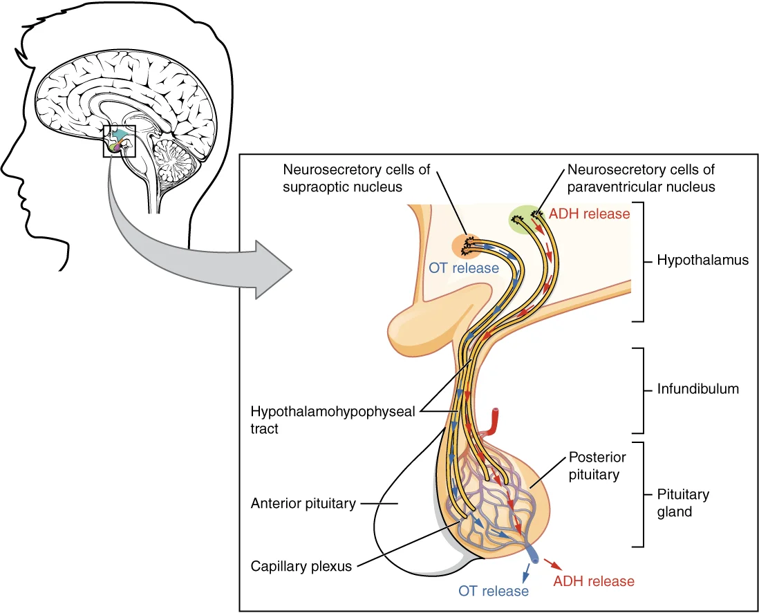
Figure 11807 The Posterior Pituitary Complex
Long description
Neurosecretory cells in the hypothalamus release oxytocin (OT) or ADH into the posterior lobe of the pituitary gland. These hormones are stored or released into the blood via the capillary plexus.

Figure 1: Neurosecretory cells in the hypothalamus release oxytocin (OT) or ADH into the posterior lobe of the pituitary gland.
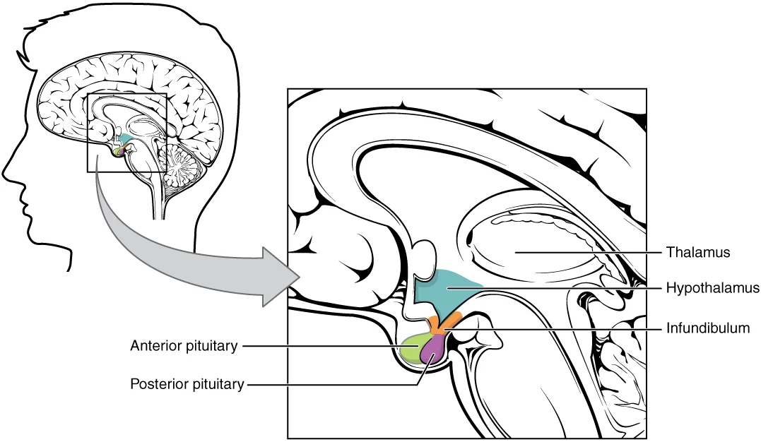
It has both neural and endocrine functions, producing and secreting many hormones[1]. In addition, the hypothalamus is anatomically and functionally related to the pituitary gland (or hypophysis), a bean-sized organ suspended from it by a stem called the infundibulum (or pituitary stalk)[1]. The pituitary gland is cradled within the sellaturcica of the sphenoid bone of the skull[1].
DefinitionHypothalamus
A diencephalic structure anterior and inferior to the thalamus, linking the nervous and endocrine systems via the pituitary gland.
The pituitary gland consists of two lobes that arise from distinct parts of embryonic tissue: the posterior pituitary (neurohypophysis) is neural tissue, whereas the anterior pituitary (also known as the adenohypophysis) is glandular tissue that develops from the primitive digestive tract[1].
The hypothalamus–pituitary complex is located in the diencephalon of the brain[1]. The hypothalamus and the pituitary gland are connected by a structure called the infundibulum, which contains vasculature and nerve axons[1]. The posterior pituitary is actually an extension of the neurons of the paraventricular and supraoptic nuclei of the hypothalamus[1].
The posterior pituitary gland does not produce hormones, but rather stores and secretes hormones produced by the hypothalamus[1]. These hormones travel along the axons into storage sites in the axon terminals of the posterior pituitary[1]. In response to signals from the same hypothalamic neurons, the hormones are released from the axon terminals into the bloodstream[1].
Figure 21806 The Hypothalamus-Pituitary Complex
Long description
The hypothalamus region lies inferior and anterior to the thalamus. It connects to the pituitary gland by the stalk-like infundibulum. The pituitary gland consists of an anterior and posterior lobe, with each lobe secreting different hormones in response to signals from the hypothalamus.

Figure 2: The hypothalamus region lies inferior and anterior to the thalamus.
NotePituitary Origins
The posterior pituitary is neural tissue extending from the hypothalamus, while the anterior pituitary is glandular tissue derived from the primitive digestive tract.
The lateral nucleus is part of the lateral hypothalamic area. Lesions here can affect feeding behaviour.
The ventromedial nucleus is involved in satiety signalling. Damage may lead to hyperphagia.
The anterior nucleus contributes to thermoregulation and parasympathetic output.
The hypothalamus forms the floor and lower lateral walls of the third ventricle. Superiorly, it is bounded by the hypothalamic sulcus, which separates it from the thalamus above. Anteriorly, it extends to the lamina terminalis, a thin membrane that marks the anterior limit of the third ventricle. Inferiorly, the hypothalamus gives rise to the infundibulum, or pituitary stalk, which connects to the pituitary gland. This anatomical arrangement positions the hypothalamus as the primary interface between the nervous system and the endocrine system. [3]
The hypothalamus acts as the command center for the endocrine system, coordinating messages between neural and hormonal signals. It secretes hormones that directly produce responses in target tissues or regulate other glands. A stimulus received by the nervous system often passes through this complex to be translated into hormonal responses. This structural integration allows the hypothalamus to maintain homeostasis through precise control of physiological processes. [1]
DefinitionHypothalamus
The inferior part of the diencephalon, forming the floor of the third ventricle and connecting to the pituitary gland via the infundibulum.
The hypothalamus contains specialized cells known as osmoreceptors, which monitor the solute concentration of the blood, or blood osmolarity. These cells are particularly sensitive to the concentration of sodium ions and other solutes. Blood osmolarity changes in response to food and fluid consumption, disease, injury, or medications. The hypothalamus constantly monitors these changes to regulate fluid balance and thirst. [1]
| Function | Mechanism | Key Hormones/Neurotransmitters |
|---|---|---|
| Growth Regulation | Anabolic effects on protein synthesis | GHRH, GHIH (Somatostatin) |
| Thyroid Regulation | Negative feedback loop | TRH, TSH |
| Stress Response | Regulation of ACTH release | CRH |
| Prolactin Control | Inhibition via neurotransmitter | Dopamine (PIH) |
| Reward and Arousal | Orexin production | Orexin (Hypocretin) |
The endocrine system regulates growth, protein synthesis, and cellular replication. Growth hormone (GH), or somatotropin, is produced by the anterior pituitary gland and promotes protein synthesis. Its levels are controlled by the release of GHRH and GHIH from the hypothalamus. Similarly, thyroid activity is regulated by TSH, released from the anterior pituitary in response to TRH from the hypothalamus. Elevated thyroid hormones trigger a drop in TRH and TSH production via negative feedback. [1]
The release of ACTH is regulated by CRH from the hypothalamus in response to physiologic rhythms and stressors. In non-pregnant females, prolactin secretion is inhibited by dopamine, released from hypothalamic neurons. During pregnancy, prolactin levels rise due to PRH from the hypothalamus. [1]
Orexin, also known as hypocretin, is a peptide produced only in the hypothalamus. It is implicated in hunger and arousal. Orexin neurons are located in the perifornical and mid-tuberal regions of the lateral hypothalamus. These neurons extend to the dorsomedial hypothalamus, influencing arousal, sleep/wake cycles, and attention. Orexin has reciprocal connections with the aminergic and cholinergic systems and stimulates CRH neurons. It shows diurnal fluctuations, which are absent in depression. [1] [3]
The hypothalamus is a complex structure with distinct nuclei, laminae, and tracts that can be observed in cross-section.
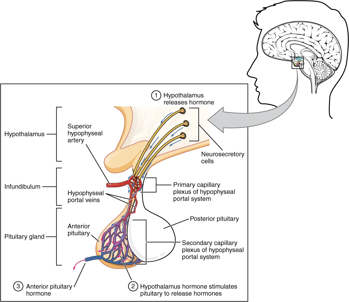
This portal system allows hypothalamic releasing hormones to directly regulate anterior pituitary hormone secretion without entering the systemic circulation first. The supraoptic-paraventricular tract connects the hypothalamic nuclei to the posterior pituitary, where oxytocin and vasopressin are stored and released.
Lesions to specific hypothalamic nuclei can produce distinct clinical syndromes. Posterior lesions in the ventral pallidium are particularly effective at inducing "disgust" reactions, while anterior lesions or lesions in other areas like the lateral hypothalamus or lateral preoptic area do not produce the same effect.
The hypothalamus acts as the primary integration centre for the autonomic nervous system and the endocrine system. It receives input from and sends output to numerous brain regions, forming a complex network that regulates homeostasis. Understanding these afferent and efferent pathways is essential for localising lesions and understanding clinical syndromes.
Afferent inputs arrive via several major tracts. The ventral tegmental area projects dopaminergic fibres to the hypothalamus, influencing reward and motivation. Additionally, direct neural inputs from the brainstem convey visceral sensory data regarding blood pressure, pH, and nutrient status.
Efferent outputs are divided into neural and hormonal pathways. Neural efferents project to the brainstem and spinal cord to regulate autonomic function.
Hormonal efferents originate from neurosecretory cells in the hypothalamus. These hormones travel to the anterior pituitary to regulate the release of trophic hormones. These hormones travel down axons to the posterior pituitary for release into the systemic circulation.
In the centre, the hypothalamus lies inferior to the thalamus and lateral to the third ventricle. Superiorly, the lamina terminalis forms the anterior boundary.
The regulation of childbirth involves a specific positive feedback loop. When fetal development is complete, the peptide-derived hormone oxytocin stimulates uterine contractions and dilation of the cervix. Throughout most of pregnancy, oxytocin hormone receptors are not expressed at high levels in the uterus. Toward the end of pregnancy, the synthesis of oxytocin receptors in the uterus increases, and the smooth muscle cells of the uterus become more sensitive to its effects. Oxytocin is continually released throughout childbirth through a positive feedback mechanism. As noted earlier, oxytocin prompts uterine contractions that push the fetal head toward the cervix. In response, cervical stretching stimulates additional oxytocin to be synthesized by the hypothalamus and released from the pituitary. This increases the intensity and effectiveness of uterine contractions and prompts additional dilation of the cervix. [1] [3]
Although the high blood levels of oxytocin begin to decrease immediately following birth, oxytocin continues to play a role in female and newborn health. First, oxytocin is necessary for the milk ejection reflex (commonly referred to as “let-down”) in breastfeeding people. As the newborn begins suckling, sensory receptors in the nipples transmit signals to the hypothalamus. In response, oxytocin is secreted and released into the bloodstream. Within seconds, cells in the milk ducts contract, ejecting milk into the infant’s mouth. Secondly, in all people, oxytocin is thought to contribute to parent–newborn bonding, known as attachment. Oxytocin is also thought to be involved in feelings of love and closeness, as well as in the sexual response. [1]
The hypothalamus receives its arterial blood supply from a rich network of branches derived primarily from the circle of Willis. This vascular arrangement ensures that the hypothalamus, despite its small size, has high metabolic demands met by consistent perfusion. The primary sources are the anterior cerebral artery, the middle cerebral artery, and the internal carotid artery. This plexus is the first part of the hypophyseal portal system, allowing hypothalamic releasing and inhibiting hormones to travel directly to the anterior pituitary without entering the general systemic circulation first.
Figure 31808 The Anterior Pituitary Complex
Long description
The anterior pituitary manufactures seven hormones. The hypothalamus produces separate hormones that stimulate or inhibit hormone production in the anterior pituitary. Hormones from the hypothalamus reach the anterior pituitary via the hypophyseal portal system.

Figure 3: The anterior pituitary manufactures seven hormones.
These vessels are distinct from the portal system and provide direct arterial blood to the neurohypophysis. This unique vascular anatomy is critical for the endocrine function of the hypothalamus, as it allows for precise, rapid regulation of pituitary hormone secretion.
Disruption of this blood supply, such as through aneurysm compression or surgical injury, can lead to significant endocrine dysfunction. For instance, damage to the superior hypophyseal arteries may impair the delivery of hypothalamic hormones to the anterior pituitary, leading to hypopituitarism. Similarly, injury to the inferior hypophyseal arteries can affect the posterior pituitary, potentially causing diabetes insipidus due to impaired ADH storage and release. These areas lack a tight blood-brain barrier, allowing them to sense changes in blood osmolarity and hormone levels, which is essential for the regulation of thirst and ADH release. [3]
DefinitionHypophyseal Portal System
A unique vascular connection between the hypothalamus and the anterior pituitary that allows direct transport of releasing and inhibiting hormones.
Key structures within the hypothalamus include the optic chiasm, the tuber cinereum, the infundibulum, and the mammillary bodies.
| Structure | Location | Relation |
|---|---|---|
| Optic Chiasm | Anterior | Superior to the infundibulum |
| Tuber Cinereum | Central | Between the optic chiasm and mammillary bodies |
| Infundibulum | Central | Connects hypothalamus to pituitary |
| Mammillary Bodies | Posterior | Posterior to the tuber cinereum |
The hypothalamus serves as the primary integrative centre for maintaining homeostasis, coordinating autonomic, endocrine, and behavioural responses. Its functions are organised around specific nuclei that regulate vital physiological drives and environmental rhythms.
Thermoregulation is managed by the preoptic area of the anterior hypothalamus, which acts as the body’s thermostat. This region contains warm-sensitive neurons that detect increases in blood temperature. When activated, these neurons initiate heat-loss mechanisms, including vasodilation of cutaneous blood vessels and sweating. Conversely, the posterior hypothalamus is critical for heat conservation and generation. It integrates signals from peripheral thermoreceptors and, when cold is detected, triggers shivering thermogenesis and vasoconstriction to preserve core body temperature. Damage to the anterior hypothalamus can result in poikilothermia, where the body temperature fluctuates with the environment, while lesions to the posterior area may cause hyperthermia due to an inability to dissipate heat.
Appetite control involves a complex interplay between the lateral hypothalamus and the arcuate nucleus. The lateral hypothalamic area functions as the 'hunger centre'; stimulation here induces eating, whereas destruction leads to aphagia and starvation. In contrast, the ventromedial hypothalamus acts as the 'satiety centre'. Stimulation of this region suppresses food intake, while lesions result in hyperphagia and obesity. These centres receive hormonal signals such as leptin and ghrelin, which inform the brain about energy stores and gastric emptying, respectively. The integration of these signals ensures that energy intake matches metabolic demand. [2]
Thirst is primarily regulated by the organum vasculosum of the lamina terminalis (OVLT) and the subfornical organ, which are circumventricular organs lacking a blood-brain barrier. These structures detect increases in plasma osmolality and decreases in blood volume. When osmolality rises, osmoreceptors in the anterior hypothalamus are stimulated, triggering the sensation of thirst and the release of antidiuretic hormone (ADH) from the posterior pituitary. ADH acts on the renal collecting ducts to increase water reabsorption, thereby diluting the plasma and restoring osmotic balance. This mechanism is crucial for preventing dehydration and maintaining blood pressure.
The suprachiasmatic nucleus (SCN) of the hypothalamus is the master circadian pacemaker. It receives direct photic input from the retina via the retinohypothalamic tract, allowing it to synchronise the body’s internal clock with the external light-dark cycle. The SCN regulates the secretion of melatonin from the pineal gland and influences cortisol release through the hypothalamic-pituitary-adrenal axis. Disruptions in SCN function can lead to sleep-wake disorders and mood disturbances. The SCN also modulates autonomic functions, ensuring that physiological processes such as body temperature and heart rate follow a daily rhythm.
The hypothalamus is situated inferior to the thalamus and forms the floor and lower lateral walls of the third ventricle. The preoptic area is located anteriorly and superiorly within the hypothalamic region. The lateral hypothalamic area lies laterally, adjacent to the internal capsule. The ventromedial nucleus is positioned medially, near the third ventricle. The arcuate nucleus is found at the base of the hypothalamus, just above the pituitary stalk. The suprachiasmatic nucleus is located dorsally, just above the optic chiasm. The organum vasculosum of the lamina terminalis is situated at the anterior wall of the third ventricle.
The hypothalamus is a small but critical structure located at the base of the brain, forming the floor and part of the walls of the third ventricle. It serves as the primary integration centre for the autonomic nervous system and the endocrine system. To understand its clinical significance, one must appreciate its anatomical position and the functional zones it contains.
The hypothalamus is functionally divided into three longitudinal zones: the periventricular zone, the medial zone, and the lateral zone. The periventricular zone contains nuclei that project to the posterior pituitary (neurohypophysis) via the hypothalamo-hypophyseal tract. These include the supraoptic and paraventricular nuclei, which produce vasopressin and oxytocin. The medial zone contains major regulatory nuclei such as the anterior, ventromedial, dorsomedial, and arcuate nuclei. These nuclei regulate hunger, satiety, temperature, and circadian rhythms. The lateral zone contains the lateral hypothalamic area, which is crucial for feeding behaviour, and the tuberomammillary nucleus, which regulates wakefulness.
Lesions of the hypothalamus produce distinct syndromes depending on the specific nucleus or zone affected. The following table summarises key lesion syndromes.
| Structure | Lesion Effect | Clinical Syndrome |
|---|---|---|
| Supraoptic/Paraventricular Nuclei | Loss of ADH/Oxytocin production | Diabetes Insipidus (polyuria, polydipsia) |
| Ventromedial Nucleus | Loss of satiety signal | Hyperphagia, severe obesity |
| Lateral Hypothalamic Area | Loss of hunger signal | Aphagia, weight loss |
| Anterior Hypothalamus | Loss of parasympathetic cooling | Hyperthermia, anhidrosis |
| Posterior Hypothalamus | Loss of sympathetic heating | Hypothermia, inability to shiver |
| Mammillary Bodies | Damage to Papez circuit | Amnesia, confabulation (Korsakoff syndrome) |
The pituitary stalk (infundibulum) descends from the median eminence to the posterior pituitary. Lesion sites are marked on the ventromedial nucleus and the lateral hypothalamic area. The ventromedial nucleus is located medial to the arcuate nucleus and lateral to the third ventricle. The lateral hypothalamic area is situated lateral to the ventromedial nucleus and medial to the internal capsule. Understanding these relations helps localise deficits. For example, a lesion in the ventromedial nucleus disrupts the satiety centre, leading to uncontrolled eating. A lesion in the lateral area removes the hunger drive, causing refusal to eat. [2]
The hypothalamus also regulates the hypothalamic-pituitary-adrenal (HPA) axis. Chronic stress or lesions affecting this axis can lead to mood disorders. Abnormalities in the stress system are documented in depression. Long-term activation of the stress system may increase the risk of depression.
Clinical pearlClinical Pearl
Lesions of the mammillary bodies are classically associated with Korsakoff syndrome, characterised by anterograde amnesia and confabulation, often due to thiamine deficiency in alcoholism.
Exam trapCommon Trap
Do not confuse the anterior hypothalamus (cooling) with the posterior hypothalamus (heating). Anterior lesions cause hyperthermia; posterior lesions cause hypothermia.
The hypothalamus integrates autonomic, endocrine, and behavioural outputs via distinct nuclear groups. Understanding the hypothalamic-pituitary axes is central to clinical neuroendocrinology.
The hypothalamus secretes gonadotropin-releasing hormone (GnRH) in a pulsatile manner to regulate the anterior pituitary. This axis controls reproductive function through two key glycoprotein hormones. Follicle-stimulating hormone (FSH) stimulates the production and maturation of sex cells, or gametes, including ova and sperm. FSH also promotes follicular growth; these follicles then release estrogens in ovaries. Luteinizing hormone (LH) triggers ovulation, as well as the production of estrogens and progesterone by the ovaries. LH stimulates production of testosterone by the testes. Disruption of this pulsatility leads to hypogonadism. [1]
The paraventricular nucleus (PVN) is the primary site for corticotropin-releasing hormone (CRH) synthesis. Under basal conditions, the HPA axis is constrained by two major inhibitory mechanisms. These are the corticosteroid feedback and the inhibition of the PVN by the neurotransmitter GABA. These two mechanisms work together and influence each other. [3]
While the hypothalamus regulates homeostatic hunger, the nucleus accumbens (NAc) mediates the hedonic aspects of feeding. The NAc contains an opioid-mediated hedonic hotspot where opioid stimulation can increase “liking” reactions to sucrose taste. This distinguishes "liking" from "wanting," as LH and VP mediate separable “wanting” vs. “liking” aspects of food reward. [2]
GABAergic and glutamatergic inputs to the NAc modulate motivated behaviour. GABA inhibition of neurons in a rostral strip of medial shell increases hedonic “liking” reactions to a bittersweet sucrose/quinine solution. By contrast, GABAergic agonist microinjections at sites in the caudal half of shell decrease “liking” reactions and increase negative “disgust” reactions. This rostrocaudal gradient allows for a spectrum of motivational states. Progressive shifts along the rostrocaudal axis produces many different quantitative mixtures of appetitive/defensive responses. The sounds change progressively in pitch as the brick moves in one direction, just as elicited mixtures of desire/dread behaviors change progressively in valence as NAc microinjection sites are moved along the anterior-posterior gradient of medial shell. [2]
Exam trapCommon Trap
Do not confuse the hypothalamic regulation of homeostatic hunger with the NAc-mediated hedonic "liking" of food. The PVN regulates the HPA axis via GABA, while the NAc medial shell regulates reward via opioids and dopamine.
| Structure | Primary Neurotransmitter/Peptide | Key Function |
|---|---|---|
| PVN | CRH, GABA | HPA axis activation; stress response |
| NAc Medial Shell | Opioids, Dopamine | Hedonic "liking" of rewards |
| Anterior Pituitary | FSH, LH | Gonadal steroid and gamete production |
Students frequently confuse the anatomical location and functional output of the hypothalamus with that of the pituitary gland. A primary error is attributing the secretion of melanocyte-stimulating hormone (MSH) directly to the hypothalamic neurons themselves. MSH is actually formed by the cleavage of the pro-opiomelanocortin (POMC) precursor protein within the cells located in the zone between the pituitary lobes. While the hypothalamus regulates the pituitary via releasing and inhibiting hormones, it does not secrete MSH into the systemic circulation in the same direct manner. This distinction is critical when interpreting hormonal axes. [1]
Another frequent mistake involves the role of dopamine in stress and mood disorders. Students often assume that stress uniformly suppresses dopaminergic activity. In reality, stress exposure and elevated glucocorticoid levels enhance dopamine release in the nucleus accumbens. Furthermore, dopamine via D-1 receptors may regulate the amount of circulating corticosterone, creating a complex feedback loop rather than a simple suppression. Misunderstanding this bidirectional relationship leads to incorrect predictions about behavioural responses to chronic stress. [3]
A third pitfall is the oversimplification of the neurobiology of reward within the nucleus accumbens. Many students believe that neuronal excitation is the sole driver of appetitive motivation. However, a popular hypothesis suggests that inhibitory hyperpolarization of medium spiny neurons in the nucleus accumbens is the primary mechanism for generating appetitive motivation. This inhibition releases downstream neurons from chronic GABAergic suppression, disinhibiting target structures. Yet, contradictory evidence shows that approximately 30% of nucleus accumbens core and shell neurons increase firing in response to sweet rewards. Another study reported that nearly 75% of shell and core neurons showed increased firing elicited by sucrose rewards. The truth lies in the nuance: while inhibition is a chief mechanism, excitation of at least some neurons also supports appetitive motivation. [2]
Students also misinterpret the hormonal changes in posttraumatic stress disorder (PTSD). It is commonly assumed that PTSD is characterised by high cortisol levels due to chronic stress. However, a meta-analysis showed that plasma or serum cortisol levels in PTSD did not differ from controls. When controls not exposed to trauma were compared to PTSD patients, cortisol levels in the PTSD group were even lower. This apparent paradox is explained by enhanced sensitivity for cortisol feedback, partly due to genetic polymorphisms in the glucocorticoid receptor gene or FKBP5 gene. Assuming high cortisol in PTSD leads to incorrect diagnostic reasoning. [3]
Finally, the transition from acute to chronic stress mechanisms is often overlooked. Students may assume corticotrophin-releasing hormone (CRH) remains the primary driver of adrenocorticotropic hormone (ACTH) release throughout the course of stress. Animal experiments show that in chronic stress there is a switch from CRH to arginine vasopressin (AVP) stimulation of ACTH release. Since depression is a chronic disorder, AVP-driven hypothalamic-pituitary-adrenal axis hyperactivity receives significant attention. Failing to recognise this switch limits understanding of the pathophysiology of recurrent major depressive disorder. [3]
At the bedside, understanding the hypothalamic-pituitary axis is essential for managing endocrine disorders. For instance, when evaluating skin pigmentation changes in pregnancy, clinicians must recognise that increased MSH production, in combination with estrogens, leads to darker skin pigmentation, especially on the areolas and labia minora. This is a physiological response, not a pathological one, and distinguishing it from Addison’s disease is vital. Similarly, in patients with PTSD, recognising that cortisol levels may be lower rather than higher prevents misdiagnosis and guides appropriate therapeutic interventions focusing on receptor sensitivity rather than hormone replacement. [1]
In the operating theatre, knowledge of neuroanatomy and neurochemistry informs anaesthetic and surgical planning. While the hypothalamus itself is not directly operated on in routine procedures, understanding the hedonic hotspots in the ventral pallidum is relevant for deep brain stimulation research. Microinjection mapping studies have identified a site in the posterior half of the ventral pallidum, roughly 0.8 mm3 in volume, where mu opioid receptor stimulation increases hedonic “liking” reactions. Although this is primarily research data, it underscores the importance of precise anatomical localisation when dealing with deep brain structures. [2]
On imaging, radiologists must differentiate between hypothalamic and pituitary pathologies. Lesions in the zone between the pituitary lobes can affect MSH production, leading to unusual pigmentation patterns. Recognising that MSH is derived from POMC cleavage helps in understanding associated syndromes involving pro-opiomelanocortin processing. Furthermore, in cases of suspected hypothalamic dysfunction affecting stress responses, imaging may reveal structural abnormalities, but functional assessment requires understanding the genetic basis of cortisol hypersensitivity, such as SNPs in the GR or CRH-R-1 genes. [2]
In examinations, students are tested on the specific neurochemical mechanisms of reward and stress. Questions may ask about the relative roles of neuronal excitation and inhibition in the nucleus accumbens. The correct answer often involves acknowledging that while inhibition of medium spiny neurons is a primary mechanism for appetitive motivation, excitation of a subset of neurons also plays a role. Similarly, questions on chronic stress require identifying the switch from CRH to AVP stimulation of ACTH release. Mastery of these specific details, rather than general principles, is required to answer examination questions correctly.
MSH is secreted by cells between the pituitary lobes, not the hypothalamus. Pregnancy increases MSH and estrogens, causing darker pigmentation of areolas. Stress enhances dopamine release in the nucleus accumbens. Dopamine via D-1 receptors may regulate circulating corticosterone. Inhibition of nucleus accumbens neurons is a primary mechanism for appetitive motivation. Approximately 30% to 75% of nucleus accumbens neurons increase firing with rewards. PTSD patients may have lower cortisol levels than non-trauma controls. Chronic stress switches ACTH stimulation from CRH to AVP.
Answer each one in your head first, then open the answer.
Where is melanocyte-stimulating hormone primarily secreted from in the context of the pituitary axis?
Show answerHide answer
Cells in the zone between the pituitary lobesWhat combination of hormones causes darker skin pigmentation on the areolas during pregnancy?
Show answerHide answer
Increased MSH production and estrogensWhat is the primary mechanism for generating appetitive motivation in the nucleus accumbens according to the hyperpolarization hypothesis?
Show answerHide answer
Inhibitory hyperpolarization of medium spiny neuronsHow do cortisol levels in PTSD patients compare to non-trauma exposed controls?
Show answerHide answer
Cortisol levels are even lower in the PTSD groupWhat switch occurs in ACTH stimulation during chronic stress?
Show answerHide answer
A switch from CRH to AVP stimulation of ACTH release
- Betts JG, Young KA, Wise JA, et al. The Pituitary Gland and Hypothalamus. OpenStax, Anatomy and Physiology 2e (CC BY-NC-SA 4.0). Accessed .
- Castro DC, Cole SL, Berridge KC. Lateral hypothalamus, nucleus accumbens, and ventral pallidum roles in eating and hunger: interactions between homeostatic and reward circuitry. Frontiers in systems neuroscience 2015;9:90. doi:10.3389/fnsys.2015.00090. Accessed .
- Bao AM, Swaab DF. The human hypothalamus in mood disorders: The HPA axis in the center. IBRO reports 2019;6:45-53. doi:10.1016/j.ibror.2018.11.008. Accessed .
Figure credits
Educational content for medical students and doctors. It is not a substitute for clinical judgement or local guidelines.