Basal ganglia
The basal ganglia are subcortical nuclei that regulate voluntary movement by modulating cortical activity through direct and indirect pathways.
Beneath the cerebral cortex lie sets of nuclei known as subcortical nuclei that augment cortical processes. The basal nuclei are a set of nuclei in the cerebrum responsible for comparing cortical processing with the general state of activity in the nervous system to influence the likelihood of movement taking place. For example, while a student is sitting in a classroom listening to a lecture, the basal nuclei will keep the urge to jump up and scream from actually happening. The basal nuclei are [1]
The major structures of the basal nuclei that control movement are the caudate, putamen, and globus pallidus, which are located deep in the cerebrum. The caudate is a long nucleus that follows the basic C-shape of the cerebrum from the frontal lobe, through the parietal and occipital lobes, into the temporal lobe. The putamen is mostly deep in the anterior regions of the frontal and parietal lobes. Together, the caudate and putamen are called the striatum. The globus pallidus is a layered nucleus that lies just medial to the putamen; they are called the lenticular nuclei because they look like curved pieces fitting together like lenses. The globus pallidus has two subdivisions, the external and internal segments, which are lateral and medial, respectively. [1]
The basal nuclei in the cerebrum are connected with a few more nuclei in the brain stem that together act as a functional group that forms a motor pathway. The primary output of the basal nuclei is to the thalamus, which relays that output to the cerebral cortex. The diencephalon includes the thalamus and the hypothalamus, along with some other structures. The thalamus is a relay between the cerebrum and the rest of the nervous system. The subthalamus includes the subthalamic nucleus that is par [1]
DefinitionBasal Nuclei
The basal nuclei are a set of nuclei in the cerebrum responsible for comparing cortical processing with the general state of activity in the nervous system to influence the likelihood of movement taking place.
The caudate and putamen form the striatum, the main input station for the basal ganglia. All input to the basal nuclei is from the cortex into the striatum. The caudate follows the C-shape of the cerebrum, while the putamen lies deep in the anterior frontal and parietal lobes. [1]
The globus pallidus is medial to the putamen and consists of external and internal segments. It serves as a key relay in the output pathways of the basal ganglia.
The subthalamic nucleus is part of the subthalamus and is included within the basal nuclei functional group. It receives input from the globus pallidus external segment in the indirect pathway.
The substantia nigra is located in the brain stem and connects with the basal nuclei. It contains the pars compacta, which releases dopamine to modulate the direct and indirect pathways.
Two streams of information processing take place in the basal nuclei. The direct pathway is the projection of axons from the striatum to the globus pallidus internal segment (GPi) and the substantia nigra pars reticulata (SNr). The GPi/SNr then projects to the thalamus, which projects back to the cortex. The direct pathway causes the disinhibition of the thalamus (inhibition of one cell on a target cell that then inhibits the first cell). The thalamus then can either excite the cortex (as a resu [1]
The indirect pathway is the projection of axons from the striatum to the globus pallidus external segment (GPe), then to the subthalamic nucleus (STN), and finally to GPi/SNr. The indirect pathway causes, or reinforces, the normal inhibition of the thalamus. The thalamus then can either excite the cortex (as a result of the direct pathway) or fail to excite the cortex (as a result of the indirect pathway). [1]
The switch between the two pathways is the substantia nigra pars compacta, which projects to the striatum and releases the neurotransmitter dopamine. Dopamine receptors are either excitatory (D1-type receptors) or inhibitory (D2-type receptors). The direct pathway is activated by dopamine, and the indirect pathway is inhibited by dopamine. When the substantia nigra pars compacta is firing, it signals to the basal nuclei that the body is in an active state, and movement will be more likely. When the substantia nigra pars compacta is silent, the body is in a passive state, and movement is inhibited. To illustrate this situation, while a student is sitting listening to a lecture, the substantia nigra pars compacta would be silent and the student less likely to get up and walk around. [1]
Clinical pearlDopamine Effect
Dopamine activates the direct pathway and inhibits the indirect pathway, promoting movement initiation.
The nigrostriatal system involves the substantia nigra pars compacta projecting to the striatum. This dopaminergic pathway is crucial for regulating the balance between the direct and indirect pathways.
The direct pathway facilitates movement by disinhibiting the thalamus. It projects from the striatum to the GPi/SNr and then to the thalamus.
The indirect pathway suppresses unwanted movement by reinforcing thalamic inhibition. It involves the striatum, GPe, STN, and GPi/SNr.
The cerebrum is the most complex part of the brain, comprising outer gray matter known as the cortex and several deep nuclei. These deep nuclei are grouped into three important functional categories. The basal nuclei, often referred to as the basal ganglia, are primarily responsible for cognitive processing, with their most critical function being the planning of movements. Other deep structures include the basal forebrain nuclei, which are vital for learning and memory, and the limbic cortex, which forms part of the limbic system involved in emotion and behaviour.
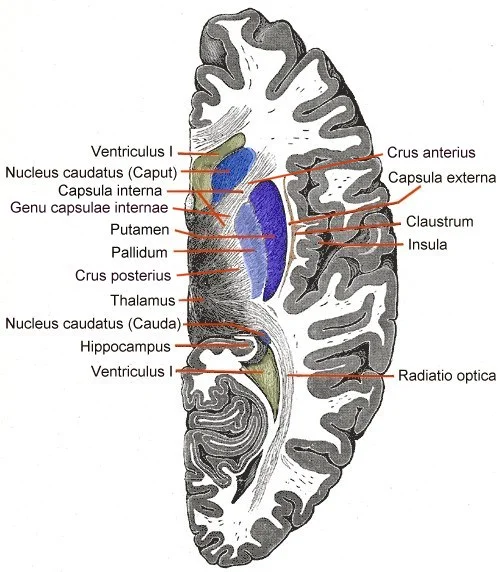
Figure 1Telencephalon-Horiconatal
Long description
Horizontal section through Telencephalon. Basal ganglia blue.

Figure 1: Horizontal section through Telencephalon (basal ganglia blue).
Lateral to the lateral ventricles lies the caudate nucleus, a C-shaped structure that follows the contour of the ventricle. Its head bulges into the anterior horn of the lateral ventricle. Posterior to the head, the body of the caudate nucleus runs alongside the lateral ventricle.
Medial to the caudate nucleus and forming the lateral wall of the third ventricle is the thalamus. Below the thalamus lies the lentiform nucleus, which is divided into two parts. The outer part is the putamen, and the inner part is the globus pallidus. The putamen and the caudate nucleus together form the striatum. The globus pallidus is separated from the thalamus by the internal medullary lamina. Lateral to the lentiform nucleus is the internal capsule, a dense band of white matter fibres that separates the lentiform nucleus from the caudate and thalamus. These fibres carry motor and sensory information between the cortex and the brainstem.
The amygdala is located in the temporal lobe, anterior to the hippocampus. It is part of the limbic system and is involved in emotional processing. The basal forebrain nuclei are situated inferior to the anterior commissure and are involved in cholinergic projection to the cortex.
DefinitionBasal Nuclei
The basal nuclei are responsible for cognitive processing, the most important function being that associated with planning movements. [1]
The structural arrangement allows for complex integration of motor commands. The striatum receives input from the cortex. The output from the basal nuclei influences the thalamus, which then projects back to the cortex. This loop is essential for smooth movement execution. The proximity of the internal capsule to these nuclei means that lesions here can cause significant motor deficits. The distinction between the putamen and globus pallidus is anatomically clear in this view, separated by the medial medullary lamina. The caudate nucleus head is particularly prominent in this coronal section, highlighting its role in connecting with the frontal lobes via the internal capsule.
| Structure | Location | Primary Function |
|---|---|---|
| Caudate Nucleus | Lateral to lateral ventricle | Cognitive processing, movement planning |
| Putamen | Lateral to globus pallidus | Motor control, part of striatum |
| Globus Pallidus | Medial to putamen | Output of basal ganglia circuit |
| Thalamus | Medial to lentiform nucleus | Relay station for motor signals |
| Amygdala | Temporal lobe | Emotional processing |
The cerebral cortex surrounds these deep structures. The complexity of the cerebrum varies across species, but in mammals, it includes the cortex and these deep nuclei. The basal forebrain nuclei are crucial for learning and memory, distinct from the motor planning role of the basal nuclei. The limbic cortex is part of the limbic system, handling emotion and behaviour. Understanding these gross features provides the foundation for studying the functional pathways of the basal ganglia.
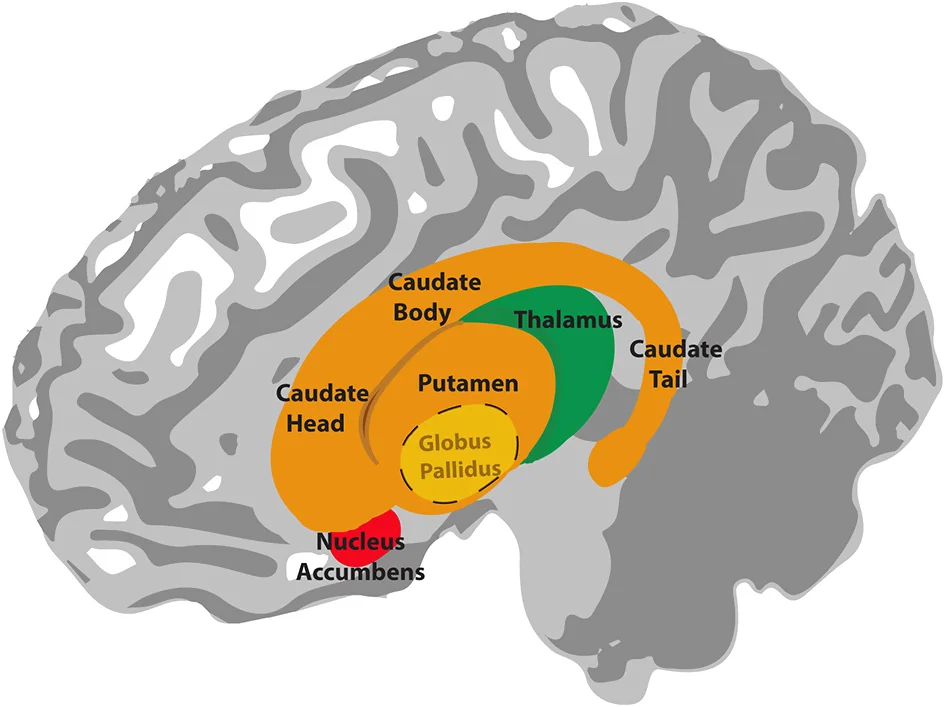
Figure 2Anatomy of the basal ganglia
Long description
Illustration of the anatomy of the basal ganglia. The globus pallidus lies inside the putamen. The thalamus is located underneath the basal ganglia, in the medial position of the brain.

Figure 2: Illustration of the anatomy of the basal ganglia.
The basal ganglia are a set of nuclei in the cerebrum[1]. They are buried within the white matter of each cerebral hemisphere, beneath the cerebral cortex[1]. In a cross-section of the brain, they appear as islands of grey matter interspersed among the white matter fibre tracts. The internal capsule, a dense band of projection fibres carrying motor and sensory information to and from t
The principal nuclei of the basal ganglia are the caudate nucleus, the putamen, and the globus pallidus. The caudate nucleus is a large C-shaped nucleus that curves around the lateral ventricle, with a head, body, and tail. The putamen is a rounded, lens-shaped nucleus lying lateral to the globus pallidus. Together, the caudate and putamen are often referred to as the striatum, as they are connected anteriorly by grey matter bridges. The globus pallidus lies medial to the putamen and is divided into an external and an internal segment by a thin layer of white matter, the medial medullary lamina.
Two further structures are functionally part of the basal ganglia: the subthalamic nucleus, located in the diencephalon just below the thalamus, and the substantia nigra, located in the midbrain[1]. The substantia nigra is divided into a dorsal pars compacta, which contains dopaminergic neurons, and a ventral pars reticulata, which contains GABAergic neurons. The subthalamic nucleus is a small, lens-shaped nucleus that lies just medial to the internal capsule.
The internal structure of the basal ganglia is defined by its laminae and fibre tracts. The external medullary lamina is a thin sheet of white matter that separates the putamen from the globus pallidus. The internal medullary lamina divides the globus pallidus into its external and internal segments. The internal capsule, the major white matter tract, passes between the lentiform nucleus (putamen and globus pallidus) medially and the caudate nucleus laterally. The anterior limb of the internal capsule lies between the head of the caudate and the putamen; the posterior limb lies between the thalamus and the globus pallidus.
The putamen appears as a large, oval grey mass lateral to the globus pallidus ограничењима. The globus pallidus, with its two segments separated by the internal medullary lamina, lies medial to the putamen.
The basal ganglia receive their main input from the cerebral cortex and the substantia nigra pars compacta, and their output projects via the globus pallidus internus and substantia nigra pars reticulata to the thalamus, which in turn projects back to the cortex. This circuitry modulates cortical activity, particularly for the initiation and control of voluntary movement.
The basal ganglia are a group of subcortical nuclei that form the core of the extrapyramidal motor system. They do not initiate movement directly but modulate the intensity and scale of motor commands generated by the cerebral cortex. To understand their function, one must trace the complex loops of afferent (incoming) and efferent (outgoing) connections that link them to the thalamus and the brainstem.
The caption states this is a schematic of the direct and indirect pathways.
DefinitionStriatum
The striatum comprises the caudate nucleus and the putamen. It is the main receiving centre for cortical input within the basal ganglia. [2]
The afferent connections originate primarily from the cerebral cortex. Almost all areas of the cortex send glutamatergic excitatory projections to the striatum. This widespread cortical input allows the basal ganglia to integrate sensory, motor, and cognitive information. The striatum also receives dopaminergic input from the substantia nigra pars compacta, which modulates the activity of the striatal neurons. This dopaminergic modulation is critical for the balance between movement initiation and suppression.
The efferent connections follow a more restricted pathway. The output nuclei of the basal ganglia are the internal segment of the globus pallidus (GPi) and the substantia nigra pars reticulata (SNr). These structures send inhibitory GABAergic projections to the thalamus. By inhibiting the thalamus, the basal ganglia control the flow of motor signals from the thalamus to the motor cortex. This inhibition is tonically active; therefore, disinhibition of the thalamus is required to facilitate movement.
| Structure | Primary Input | Primary Output | Neurotransmitter |
|---|---|---|---|
| Striatum | Cerebral Cortex | Globus Pallidus / Substantia Nigra | GABA |
| Globus Pallidus Internus | Striatum / Subthalamic Nucleus | Thalamus | GABA |
| Subthalamic Nucleus | Globus Pallidus Externus | Globus Pallidus Internus | Glutamate |
| Substantia Nigra Pars Compacta | - | Striatum | Dopamine |
The circuitry is often described in terms of direct and indirect pathways. The direct pathway facilitates movement by inhibiting the inhibitory output nuclei, thereby disinhibiting the thalamus. The indirect pathway suppresses unwanted movements by increasing the inhibition of the thalamus. The subthalamic nucleus plays a pivotal role in the indirect pathway, providing excitatory drive to the output nuclei. This delicate balance ensures smooth, coordinated motor control. Disruption of these connections leads to the hyperkinetic or hypokinetic movement disorders seen in clinical practice.
The basal ganglia receive their blood supply from branches of the anterior and posterior cerebral circulations. The anterior circulation is primarily supplied by the internal carotid artery (ICA), which divides into anterior and posterior divisions. The anterior division gives rise to the anterior cerebral artery (ACA), middle cerebral artery (MCA), and anterior choroidal artery (AChA). The MCA is the primary supplier of the basal ganglia, particularly the caudate nucleus, putamen, and globus pa [2]
The posterior circulation is supplied by the vertebral arteries (VA) and basilar artery (BA). The VA typically supplies the more inferior aspects of the cerebellum and the lower brain stem. The BA forms during the 5-8 mm embryonic stage from the consolidation of neural arteries. The posterior division of the ICA produces the posterior cerebral artery (PCA) and posterior choroidal artery (PChA). The PCA contributes to the blood supply of the basal ganglia, particularly the globus pallidus and parts of the thalamus.
Collateral circulation plays a crucial role in maintaining blood flow to the basal ganglia in cases of vascular occlusion. The circle of Willis, formed by the anterior communicating artery (ACOMM) and posterior communicating arteries (PCOMM), provides important connections between the anterior and posterior circulations. Individuals with decreased collateral routes, such as lack of PCOMM or ACOMM, may be at higher risk of developing atherosclerosis in areas with chronic low or oscillating blood flow. The persistence of carotid-vertebrobasilar anastomoses may indicate complete occlusion or agenesis of anterior circulation arteries or vertebral arteries, potentially compromising the ability to provide backup blood supply.
The ACA contributes to the blood supply of the head of the caudate nucleus. The PCA and posterior choroidal arteries supply the medial aspects of the basal ganglia, particularly the globus pallidus and thalamus.
The basal ganglia are a group of subcortical nuclei that play a critical role in the regulation of voluntary motor control, procedural learning, and habit formation. [1]
The caudate nucleus and the putamen form the striatum, the primary input station for the basal ganglia. The globus pallidus lies medial to the putamen, divided into internal and external segments. The subthalamic nucleus is located inferior to the thalamus and lateral to the cerebral peduncle. Understanding this spatial arrangement is crucial for localizing lesions that cause movement disorders. [1]
The cerebral cortex, which surrounds these subcortical structures, is responsible for higher functions. The cortex is divided into lobes, each with specific roles. The frontal lobe, anterior to the central sulcus, is primarily associated with motor functions. Within this lobe, the precentral gyrus serves as the primary motor cortex, where upper motor neurons originate to instruct spinal cord cells. Anterior to this, the premotor area plans movements, while the frontal eye fields control eye movements. Broca’s area, typically on the left side, controls speech production. The most anterior portion, the prefrontal cortex, influences personality and decision-making. [1]
Other lobes handle sensory processing. The parietal lobe processes somatosensation. The occipital lobe is the site where visual processing begins. The temporal lobe contains areas for auditory processing and memory formation. The basal ganglia interact with these cortical areas, particularly the motor and premotor cortices, to refine motor output. For instance, the motor cortex sends excitatory signals to the striatum, which then modulates the activity of the globus pallidus and substantia nigra. This modulation ultimately affects the thalamus, which provides feedback to the cortex. Disruption in this loop can lead to hypokinetic disorders, such as Parkinson’s disease, characterized by bradykinesia and rigidity, or hyperkinetic disorders, such as Huntington’s disease, characterized by chorea. [1]
DefinitionBasal Ganglia Circuit
A neural loop involving the cortex, striatum, globus pallidus, subthalamic nucleus, and substantia nigra that regulates voluntary movement by facilitating desired actions and inhibiting unwanted ones.
The integration of these subcortical nuclei with the cerebral cortex ensures that motor commands are not just initiated but are also appropriately scaled and timed. This system allows for the execution of complex, multi-joint movements and the learning of new motor skills. The basal ganglia are thus integral to the seamless transition from intention to action, working in concert with the cerebral cortex to maintain motor homeostasis.
The basal ganglia are a group of subcortical nuclei critical for the regulation of voluntary movement, posture, and tone. Damage to these structures results in distinct movement disorders, broadly categorised into hypokinetic syndromes (reduced movement) and hyperkinetic syndromes (excessive movement). Understanding the specific anatomical location of a lesion allows for precise clinical correlation. [1]
The caudate nucleus lies lateral to the lateral ventricle, with its head forming the lateral wall of the anterior horn. The lentiform nucleus is situated lateral to the internal capsule and medial to the claustrum. The thalamus forms the major part of the lateral wall of the third ventricle. The internal capsule is a white matter tract separating the caudate and thalamus medially from the lentiform nucleus laterally.
Parkinson disease is a progressive neurodegenerative disorder characterised by the loss of dopaminergic neurons in the substantia nigra pars compacta. This loss disrupts the direct and indirect pathways of the basal ganglia circuitry, leading to a hypokinetic state. The clinical triad consists of resting tremor, rigidity, and bradykinesia. Postural instability typically occurs in later stages. The pathology involves the accumulation of alpha-synuclein aggregates known as Lewy bodies.
Huntington disease is an autosomal dominant neurodegenerative disorder caused by a CAG trinucleotide repeat expansion in the huntingtin gene. It leads to selective atrophy of the caudate nucleus and putamen. Clinically, it presents with chorea (involuntary, jerky movements), psychiatric disturbances, and cognitive decline. The loss of GABAergic neurons in the striatum reduces inhibition of the thalamus, resulting in excessive movement.
Hemiballismus is a rare hyperkinetic movement disorder characterised by violent, flinging movements of the limbs on one side of the body. It is typically caused by a lesion in the contralateral subthalamic nucleus of Luys. The subthalamic nucleus normally excites the globus pallidus internus, which inhibits the thalamus. Damage to this nucleus removes this excitation, leading to disinhibition of the thalamus and excessive motor output.
| Structure Lesioned | Primary Effect | Clinical Syndrome |
|---|---|---|
| Substantia Nigra | Loss of dopamine | Parkinson disease (Hypokinetic) |
| Caudate/Putamen | Loss of GABAergic output | Huntington disease (Hyperkinetic) |
| Subthalamic Nucleus | Loss of excitation to GPi | Hemiballismus (Hyperkinetic) |
The basal nuclei receive input from cortical areas and compare it with the general state of the individual through the activity of a dopamine-releasing nucleus. The output influences the activity of part of the thalamus that can then increase or decrease cortical activity that often results in changes to motor commands. [1]
Parkinson’s disease is associated with the loss of dopaminergic neurons in the substantia nigra pars compacta. This leads to reduced dopamine release, causing the indirect pathway to dominate and the direct pathway to be underactive. The result is excessive inhibition of the thalamus, leading to bradykinesia and rigidity.
Huntington’s disease involves degeneration of the striatum, particularly the neurons involved in the indirect pathway. This leads to reduced inhibition of the thalamus, resulting in chorea and hyperkinesia.
Hemiballismus is caused by a lesion in the subthalamic nucleus. This disrupts the indirect pathway, leading to violent, flinging movements on the contralateral side of the body.
Exam trapBasal Ganglia vs. Ganglia
The term basal ganglia is potentially confusing because the term ganglia is typically used for peripheral structures. [1]
Parkinson’s disease results from degeneration of the substantia nigra pars compacta. This reduces dopamine, impairing the direct pathway and enhancing the indirect pathway.
Huntington’s disease involves degeneration of the striatum. This disrupts the indirect pathway, leading to excessive movement.
Hemiballismus is caused by lesions in the subthalamic nucleus. This disrupts the indirect pathway, causing violent movements.
The diencephalon serves as the primary relay station for the central nervous system. It is the only adult brain region retaining its embryological name, translating to “through brain”. All sensory and motor pathways between the cerebrum and the rest of the nervous system, including the spinal cord and peripheral nervous system, must pass through this structure. The sole exception is the olfactory system, which connects directly to the cerebrum without diencephalic intervention. In early vertebrates, the cerebrum consisted mainly of olfactory bulbs receiving chemical environmental data. [1]
The midbrain is a small region situated between the thalamus and the pons. It is anatomically divided into the tectum and the tegmentum, derived from Latin for “roof” and “floor” respectively. The cerebral aqueduct runs through the centre of the midbrain, defining these regions as its roof and floor. [1]
Terminology in neuroanatomy requires precision. The terms dorsal and ventral are often used interchangeably with posterior and anterior, particularly regarding nerves and spinal cord structures. Students must be comfortable using both sets of directional terms. [1]
Arterial variants are common and clinically significant. Fenestrations involve the splitting of an arterial lumen into two channels that later fuse, restoring the primary vessel. This is distinct from duplication, which involves two distinct arteries with separate origins that fuse distally. Fenestrations are the second most common arterial variant after hypoplastic arteries and are more frequent in the anterior circulation. The most commonly duplicated or fenestrated artery is the ACOMM [2].
The Circle of Willis exhibits significant anatomical variation. A “normal” Circle, containing all typical arteries, is found in only 52.3% of normal brains. The most common anomaly is a hypoplastic artery with a patent lumen, typically the posterior communicating artery (PCOMM) or ACOMM. Hypoplastic arteries are defined as smaller than expected, such as a PCOMM that is one half the size of the posterior cerebral artery. Complete absence of an artery is rarer, occurring in up to 2.0% of cases, usually involving the ACOMM. [2]
Specific variants have defined prevalence rates. The ACOMM has a duplication prevalence of 18% and fenestrations in up to 21%. Definitive absence of the ACOMM is found in 5% of surgical dissections. Basilar artery fenestration is a common embryological variant, with prevalence ranging from 3.9% in autopsy reports to 1.1% in angiographic reports. These usually occur proximally, near the vertebrobasilar junction. [2]
The hypoplastic PCOMM is the most common arterial variant reported in the brain. Duplication of the PCOMM is rare, with a prevalence of 1.8% in surgical dissection series. Absence of the PCOMM is even rarer, affecting less than 1.5% of individuals. A fetal posterior cerebral artery is defined by the agenesis of the ipsilateral PCOMM, technically representing a failure of the P1 segment to form. [2]
Persistent proatlantal arteries are classified by their origin. Type 1 arises from the internal carotid artery and joins the V4 segment of the vertebral artery. Type 2 arises from the external carotid artery and joins the V3 segment. These differ from hypoglossal arteries, which enter through the hypoglossal canal, whereas proatlantal arteries enter via the foramen magnum. [2]
Exam trapConfusing basal ganglia with cerebellum
The basal ganglia are distinct from the cerebellum, despite both being involved in motor control. The cerebellum accounts for approximately 10 percent of the mass of the brain, while the basal ganglia are deep nuclei within the cerebral hemispheres. Students often mistakenly attribute cerebellar functions, such as comparing information from the cerebrum with sensory feedback, to the basal ganglia. [1]
Exam trapMisunderstanding basal ganglia connections
A common error is confusing the connections between basal ganglia nuclei. The basal ganglia receive input from the entire cerebral cortex (not just the motor cortex) and project back to it via the thalamus, forming parallel loops. This differs from the cerebellum, which receives a copy of descending input from the cerebrum to the spinal cord primarily at the pons. [1]
Exam trapIncorrectly attributing all movement disorders to basal ganglia
While basal ganglia dysfunction causes disorders like Parkinson's and Huntington's disease, not all movement disorders originate here. For example, cerebellar lesions cause ataxia, which presents differently from basal ganglia disorders. The cerebellum compares motor commands with sensory feedback, a function distinct from basal ganglia's role in motor planning and selection.
Exam trapOverlooking non-motor functions
Students often focus solely on motor functions of basal ganglia while neglecting their roles in cognition, emotion, and habit formation. The basal ganglia participate in multiple parallel circuits beyond the motor loop, including oculomotor, prefrontal, and limbic circuits.
Exam trapConfusing direct and indirect pathways
A fundamental misunderstanding involves the direct (facilitatory) and indirect (inhibitory) pathways of basal ganglia. The direct pathway promotes desired movements by disinhibiting the thalamus, while the indirect pathway suppresses unwanted movements. Imbalance between these pathways underlies many movement disorders.
Exam trapMisidentifying basal ganglia components
The basal ganglia include the striatum (caudate and putamen), globus pallidus (internal and external segments), substantia nigra (compact and reticular parts), and subthalamic nucleus. Students often incorrectly include structures like the thalamus or red nucleus, which are part of the basal ganglia circuits but not technically part of the basal ganglia themselves.
NoteClinical examination
Basal ganglia function is assessed clinically through examination of motor control, including rigidity, bradykinesia, tremor, and gait. Parkinson's disease, characterized by degeneration of dopaminergic neurons in the substantia nigra pars compacta, presents with these cardinal features. The basal ganglia's role in motor planning is evident when these patients have difficulty initiating voluntary movements despite preserved muscle strength.
NoteNeuroimaging
Basal ganglia structures are routinely evaluated on MRI scans for various pathologies. The basal ganglia appear as well-defined gray matter structures on axial T2-weighted images. Common pathologies include basal ganglia infarcts (typically in the territory of the middle cerebral artery), hemorrhages, and degenerative changes. [2]
NoteMovement disorder clinics
Neurologists specializing in movement disorders use their knowledge of basal ganglia anatomy and circuitry to diagnose and treat conditions. For example, in Huntington's disease, degeneration of the striatum leads to choreiform movements. Deep brain stimulation targets specific basal ganglia nuclei (subthalamic nucleus or globus pallidus interna) in Parkinson's disease and essential tremor to modulate pathological activity in these circuits.
NoteNeurosurgery
In the operating theatre, neurosurgeons must have precise knowledge of basal ganglia anatomy when performing procedures like stereotactic surgery for tumor resection or placement of deep brain stimulation electrodes. The subthalamic nucleus is a key basal ganglia nucleus and requires precise targeting. Surgeons must avoid damaging adjacent structures like the internal capsule, which carries important motor and sensory fibers. [2]
NoteNeuropathology
Basal ganglia pathology is examined in post-mortem studies and biopsies. The substantia nigra pars compacta shows loss of neuromelanin-containing neurons in Parkinson's disease. In Wilson's disease, copper accumulation leads to characteristic changes in the basal ganglia. These pathological findings correlate with clinical manifestations and help confirm diagnoses.
Basal Ganglia Motor Circuit
- Striatum (Direct Pathway)
- Striatum (Indirect Pathway)
Clinical pearlExamination tips
When examining a patient for basal ganglia disorders, observe for hypokinetic (Parkinson's) or hyperkinetic (Huntington's, hemiballismus) movement disorders. Test for rigidity by passively moving the limbs, look for cogwheeling. Assess bradykinesia by having the patient perform rapid alternating movements. The basal ganglia's role in automatic movements is revealed when patients have difficulty with repetitive actions but can perform the same movement when prompted.
- The basal ganglia are distinct from the cerebellum, with different anatomical locations and functions
- Basal ganglia include the striatum, globus pallidus, substantia nigra, and subthalamic nucleus
- They participate in direct (facilitatory) and indirect (inhibitory) pathways that modulate motor output
- Basal ganglia disorders present with either hypokinetic (Parkinson's) or hyperkinetic (Huntington's) movement disorders
- The basal ganglia receive blood supply from branches of the middle cerebral artery
- Deep brain stimulation targets specific basal ganglia nuclei to treat movement disorders
- Basal ganglia function extends beyond motor control to include cognition and emotion
Answer each one in your head first, then open the answer.
Which structure is NOT part of the basal ganglia?
Show answerHide answer
ThalamusWhat is the primary function of the direct pathway in basal ganglia?
Show answerHide answer
Facilitate desired movementsWhich artery primarily supplies the basal ganglia?
Show answerHide answer
Middle cerebral arteryWhat is the effect of degeneration in the substantia nigra pars compacta?
Show answerHide answer
Parkinson's diseaseWhich movement disorder is associated with basal ganglia dysfunction and presents with chorea?
Show answerHide answer
Huntington's disease
- Betts JG, Young KA, Wise JA, et al. The Central Nervous System. OpenStax, Anatomy and Physiology 2e (CC BY-NC-SA 4.0). Accessed .
- Menshawi K, Mohr JP, Gutierrez J. A Functional Perspective on the Embryology and Anatomy of the Cerebral Blood Supply. Journal of stroke 2015;17(2):144-158. doi:10.5853/jos.2015.17.2.144. Accessed .
Figure credits
Educational content for medical students and doctors. It is not a substitute for clinical judgement or local guidelines.