Cerebral cortex
The cerebral cortex is the outer layer of the cerebrum responsible for higher-order brain functions.
Figure 1Gray754
Long description
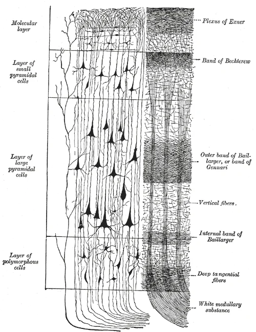
Cerebral cortex. (Poirier.) To the left, the groups of cells; to the right, the systems of fibers. Quite to the left of the figure a sensory nerve fiber is shown. Cell body layers are labeled on the left, and fiber layers are labeled on the right.

Figure 1: Cerebral cortex.
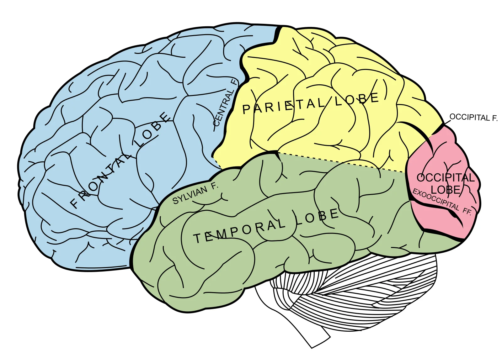
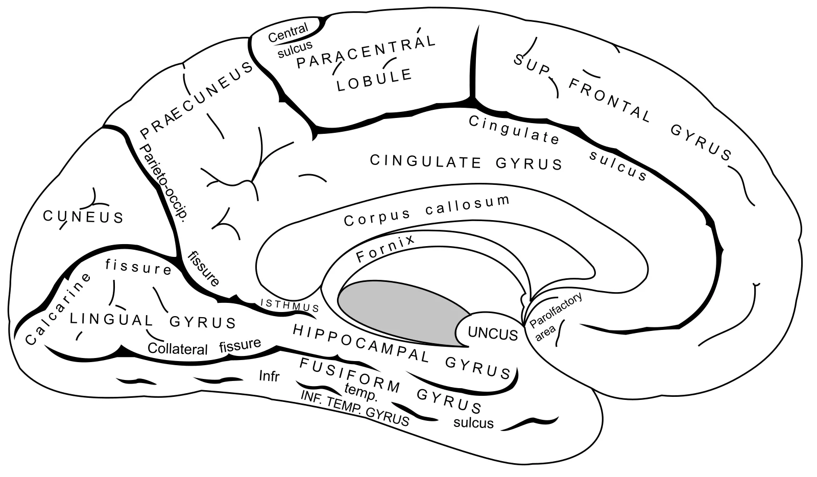
The cerebral cortex is the highly folded outer layer of the cerebrum, composed of grey matter. Its surface features are defined by sulci (grooves) and gyri (ridges), which increase the surface area for neuronal cell bodies. The cerebrum is divided into four major lobes: frontal, parietal, temporal, and occipital. These lobes are separated by prominent anatomical landmarks. The lateral sulcus (Sylvian fissure) separates the temporal lobe from the frontal and parietal lobes. The central sulcus separates the frontal lobe from the parietal lobe. From the medial surface, the parieto-occipital sulcus separates the parietal and occipital lobes. [2] [1]
Figure 2Gray725
Long description
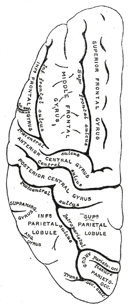
Lateral surface of left cerebral hemisphere, viewed from above.

Figure 2: Lateral surface of left cerebral hemisphere, viewed from above.
Figure 3Gray728
Long description
Principal fissures and lobes of the cerebrum viewed laterally.

Figure 3: Principal fissures and lobes of the cerebrum viewed laterally.
Figure 4Gray727
Long description
Medial surface of left cerebral hemisphere.

Figure 4: Medial surface of left cerebral hemisphere.
Figure 5Gray726
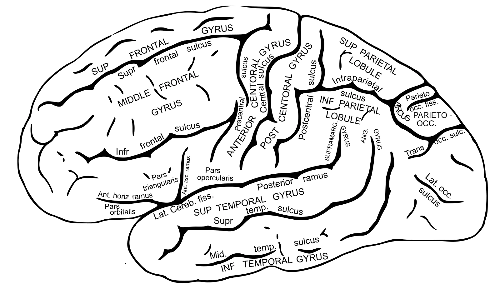
Long description
Lateral surface of left cerebral hemisphere, viewed from the side.

Figure 5: Lateral surface of left cerebral hemisphere, viewed from the side.
The functional organization of the cortex is classically described using Brodmann’s areas, a system developed by Korbinian Brodmann based on cytoarchitecture. This system divides the cortex into 52 distinct regions. Primary cortical areas are where sensory information is initially received from the thalamus for conscious perception, or where descending commands are sent down to the brain stem or spinal cord to execute movements. [2] [1]
Figure 61604 Types of Cortical Areas-02
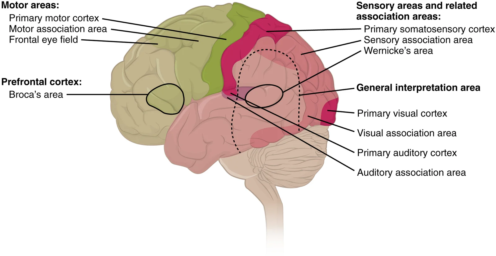
Long description
The cerebral cortex can be described as containing three types of processing regions: primary, association, and integration areas. The primary cortical areas are where sensory information is initially processed, or where motor commands emerge to go to the brain stem or spinal cord. Association areas

Figure 6: The cerebral cortex can be described as containing three types of processing regions: primary, association, and integration areas.
The primary motor cortex is located in the precentral gyrus of the frontal lobe and corresponds to Brodmann area 4. Adjacent to it is the premotor cortex (area 6). The primary somatosensory cortex is located in the postcentral gyrus of the parietal lobe and comprises Brodmann areas 1, 2, and 3. The primary visual cortex is located in the occipital lobe and corresponds to area 17. Adjacent areas 18 and 19 constitute subsequent regions of visual processing. [1][2]
DefinitionLocalization of Function
Different regions of the cerebral cortex can be associated with particular functions, a concept known as localization of function. [2]
The regions adjoining the primary areas are referred to as association areas. These areas process information in these modalities more complexly. A number of other regions, which extend beyond these primary or association areas of the cortex, are referred to as integrative areas. These areas are found in the spaces between the domains for particular sensory or motor functions, and they integrate multisensory information, or process sensory or motor information in more complex ways.
Language function is mediated by specific integrative areas. Wernicke’s area is located adjacent to the auditory association cortex, at the end of the lateral sulcus just anterior to the visual cortex. Broca’s area is located in the lateral aspect of the frontal lobe, just anterior to the region of the motor cortex associated with the head and neck. Lesions in Broca’s area cause expressive aphasia, characterized by non-fluent, halting speech with compromised grammar. Lesions in Wernicke’s area cause receptive aphasia, affecting speech comprehension.
Figure 71605 Brocas and Wernickes Areas-02
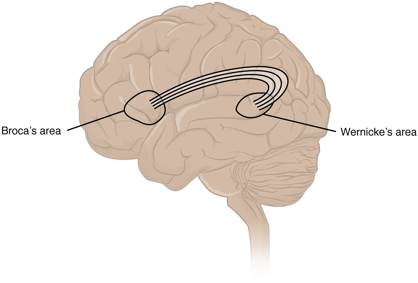
Long description
Two important integration areas of the cerebral cortex associated with language function are Broca’s and Wernicke’s areas. The two areas are connected through the deep white matter running from the posterior temporal lobe to the frontal lobe.

Figure 7: Two important integration areas of the cerebral cortex associated with language function are Broca’s and Wernicke’s areas.
Figure 8Gray756
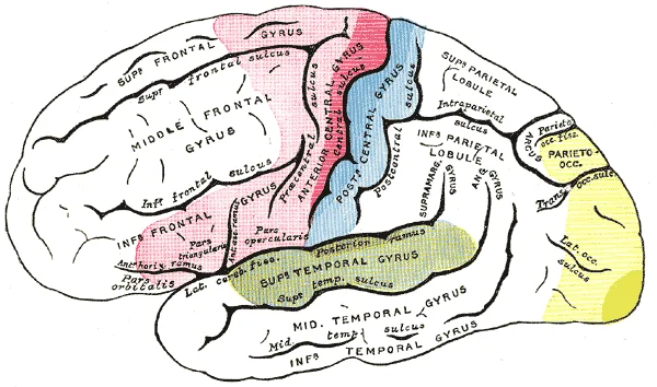
Long description
Areas of localization on lateral surface of hemisphere. Motor area in red. Area of general sensations in blue. Auditory area in green. Visula area in yellow. The psychic portions are in lighter tints.

Figure 8: Areas of localization on lateral surface of hemisphere (motor area in red).
The sensorium refers to those parts of the brain involved in the reception and interpretation of sensory stimuli. The cerebral cortex is the seat of conscious sensory perception. The prefrontal cortex, located anterior to the premotor cortex, is responsible for judgment, abstract reasoning, and working memory. The somatotopic organization of the motor and sensory cortices is represented by the homunculus, where body parts are mapped according to their sensory or motor representation density. Agnosia, aphasia, and neglect are clinical syndromes resulting from damage to these association and integrative areas.
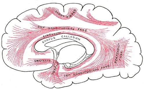
Figure 9Gray751
Long description
Diagram showing principal systems of association fibers in the cerebrum.

Figure 9: Diagram showing principal systems of association fibers in the cerebrum.
The cerebral cortex is the thin layer of gray matter covering the outside of the cerebrum, derived from the Latin word for "bark of a tree"[1]. On average, this layer is approximately 2.55 mm thick. It is highly folded to fit within the limited space of the cranial vault, a constraint imposed by the size of the birth canal[1]. This extensive folding allows more gray matter to fit into the limited space of the skull[1]. The wrinkled portion of the cerebrum is the cerebral cortex, while the rest of the structure lies beneath this outer covering[1].
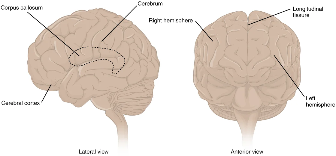
A gyrus is the ridge of one of the wrinkles, and a sulcus is the groove between two gyri[1]. The pattern of these folds indicates specific regions of the cerebral cortex[1]. A large separation between the two sides of the cerebrum is called the longitudinal fissure[1]. This fissure separates the cerebrum into two distinct halves, the right and left cerebral hemispheres[1]. Deep within the cerebrum, the white matter of the corpus callosum provides the major pathway for communication between the two hemispheres of the cerebral cortex[1]. [2]
Figure 101305 CerebrumN
Long description
The cerebrum is a large component of the CNS in humans, and the most obvious aspect of it is the folded surface called the cerebral cortex.

Figure 10: The cerebrum is a large component of the CNS in humans, and the most obvious aspect of it is the folded surface called the cerebral cortex.
The ridges are identified as gyri, and the grooves between them are sulci[1]. The corpus callosum lies deep within the structure, connecting the hemispheres[1]. The cortex appears as the outer wrinkled mantle, distinct from the underlying white matter[1].
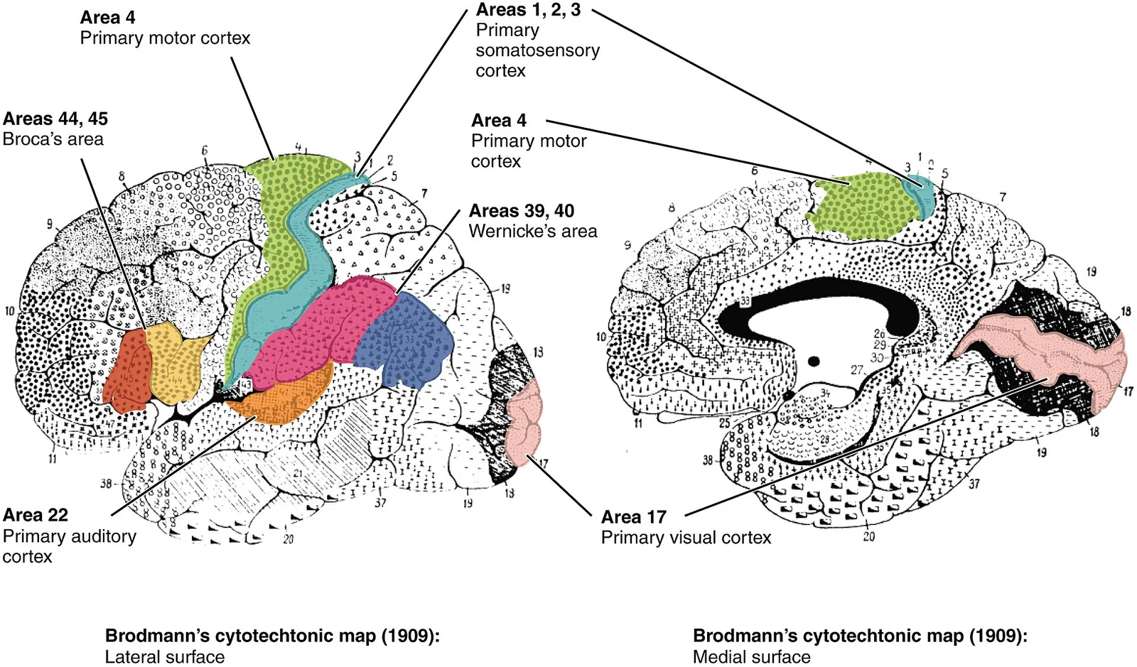
The basis for parceling out areas of the cortex and attributing them to various functions has its root in pure anatomical underpinnings. The German neurologist Korbinian Brodmann described approximately 50 regions of the cortex that differed enough from each other to be considered separate areas. He compared the size, shape, and number of neurons to find anatomical differences in the various parts of the cerebral cortex. Continued investigation into these anatomical areas over the subsequent 100 or more years has demonstrated a strong correlation between the structures and the functions attributed to those structures. [1]
Figure 111603 Brodmann Areas-02
Long description
On the basis of cytoarchitecture, the anatomist Korbinian Brodmann described the extensive array of cortical regions, as illustrated in this figure. Subsequent investigations found that these areas corresponded very well to functional differences in the cerebral cortex. (credit: modification of work

Figure 11: On the basis of cytoarchitecture, the anatomist Korbinian Brodmann described the extensive array of cortical regions, as illustrated in this figure.
The cerebrum is the seat of many of the higher mental functions, such as memory and learning, language, and conscious perception. These higher functions are distributed across various regions of the cortex. Specific locations can be said to be responsible for particular functions. For example, there is a limited set of regions involved in language function, which can be subdivided on the basis of the particular part of language function that each governs. The prefrontal cortex may be related to personality. A clinical situation suggesting this link is the story of Phineas Gage, a railroad worker who had a metal spike impale his prefrontal cortex. The injury led to changes in his personality, transforming a quiet, dependable worker into a raucous, irritable individual. [1] [2]
Beneath the cerebral cortex are sets of nuclei known as subcortical nuclei that augment cortical processes[1]. The nuclei of the basal forebrain serve as the primary location for acetylcholine production, which modulates the overall activity of the cortex[1]. The hippocampus and amygdala are medial-lobe structures involved in long-term memory formation and emotional responses[1]. The basal nuclei are responsible for comparing cortical processing with the general state of activity in the nervous system to influence the likelihood of movement taking place[1]. The primary output of the basal nuclei is to the thalamus, which relays that output to the cerebral cortex[1]. The cortex also sends information to the thalamus that will then influence the effects of the basal nuclei[1]. [2]
The adult brain is separated into four major regions: the cerebrum, the diencephalon, the brain stem, and the cerebellum. The cerebrum is the largest portion and contains the cerebral cortex and subcortical nuclei. It is divided into two halves by the longitudinal fissure. The folding of the cortex maximizes the amount of gray matter in the cranial cavity. During embryonic development, as the telencephalon expands within the skull, the brain goes through a regular course of growth that results in everyone’s brain having a similar pattern of folds. The surface of the brain can be mapped on the basis of the locations of large gyri and sulci. Using these landmarks, the cortex can be separated into four major regions, or lobes. [2]
Figure 12Gray745
Long description
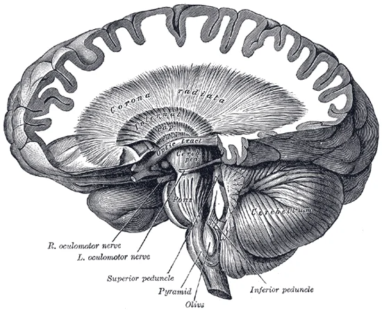
Dissection showing the course of the cerebrospinal fibers. (E. B. Jamieson.)

Figure 12: Dissection showing the course of the cerebrospinal fibers (E.
Anterior to the central sulcus is the frontal lobe, which is primarily associated with motor functions. The precentral gyrus is the primary motor cortex. Cells from this region of the cerebral cortex are the upper motor neurons that instruct cells in the spinal cord to move skeletal muscles. Anterior to this region are a few areas that are associated with planned movements. The premotor area is responsible for thinking of a movement to be made. The frontal eye fields are important in eliciting eye movements and in attending to visual stimuli. Broca’s area is responsible for the production of language, or controlling movements responsible for speech; in the vast majority of people, it is located only on the left side. The prefrontal cortex contains structures important for planning, judgment, reasoning, and working memory. Damage to these areas can result in changes to personality, mood, and behavior. [2] [1]
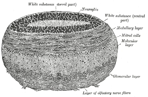
Figure 13Gray755
Long description
Coronal section of olfactory bulb. (Schwalbe.)

Figure 13: Coronal section of olfactory bulb (Schwalbe.).
DefinitionBasal Nuclei
The major structures of the basal nuclei that control movement are the caudate, putamen, and globus pallidus, which are located deep in the cerebrum. [2]
The caudate is a long nucleus that follows the basic C-shape of the cerebrum from the frontal lobe, through the parietal and occipital lobes, into the temporal lobe. The putamen is mostly deep in the anterior regions of the frontal and parietal lobes. Together, the caudate and putamen are called the striatum. The globus pallidus is a layered nucleus that lies just medial to the putamen; they are called the lenticular nuclei because they look like curved pieces fitting together like lenses. The globus pallidus has two subdivisions, the external and internal segments, which are lateral and medial, respectively. [2]
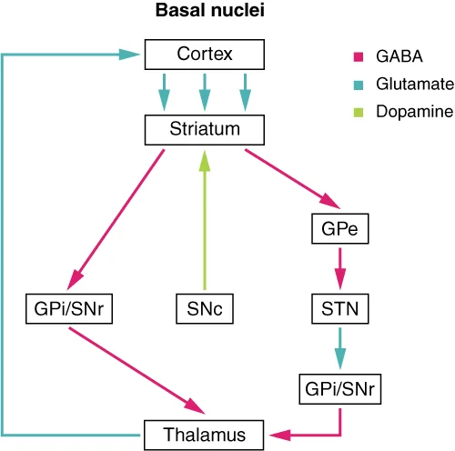
The basal nuclei in the cerebrum are connected with a few more nuclei in the brain stem that together act as a functional group that forms a motor pathway. Two streams of information processing take place in the basal nuclei. All input to the basal nuclei is from the cortex into the striatum. The direct pathway is the projection of axons from the striatum to the globus pallidus internal segment (GPi) and the substantia nigra pars reticulata (SNr). The GPi/SNr then projects to the thalamus, which projects back to the cortex. [2]
The direct pathway causes the disinhibition of the thalamus (inhibition of one cell on a target cell that then inhibits the first cell), whereas the indirect pathway causes, or reinforces, the normal inhibition of the thalamus. The thalamus then can either excite the cortex (as a result of the direct pathway) or fail to excite the cortex (as a result of the indirect pathway). [2]
NoteThalamic Relay
The thalamus is a collection of nuclei that relay information between the cerebral cortex and the periphery, spinal cord, or brain stem. All sensory information, except for the sense of smell, passes through the thalamus before processing by the cortex. Axons from the peripheral sensory organs, or intermediate nuclei, synapse in the thalamus, and thalamic neurons project directly to the cerebrum. It is a requisite synapse in any sensory pathway, except for olfaction. The thalamus does not just pass the information on, it also processes that information. For example, the portion of the thalamus that receives visual information will influence what visual stimuli are important, or what receives attention. [2]
Ascending tracts of nervous system fibers in these columns carry sensory information up to the brain, whereas descending tracts carry motor commands from the brain. Looking at the spinal cord longitudinally, the columns extend along its length as continuous bands of white matter. Between the two posterior horns of gray matter are the posterior columns. Between the two anterior horns, and bounded by the axons of motor neurons emerging from that gray matter area, are the anterior columns. The white matter on either side of the spinal cord, between the posterior horn and the axons of the anterior horn neurons, are the lateral columns. The posterior columns are composed of axons of ascending tracts. The anterior and lateral columns are composed of many different groups of axons of both ascending and descending tracts—the latter carrying motor commands down from the brain to the spinal cord to control output to the periphery. [2]
Figure 141309 Basal Nuclei Connections
Long description
Input to the basal nuclei is from the cerebral cortex, which is an excitatory connection releasing glutamate as a neurotransmitter. This input is to the striatum, or the caudate and putamen. In the direct pathway, the striatum projects to the internal segment of the globus pallidus and the substanti

Figure 14: Input to the basal nuclei is from the cerebral cortex, which is an excitatory connection releasing glutamate as a neurotransmitter.
The cerebral cortex forms the outer layer of gray matter. Deep to the cortex lies the white matter, containing tracts. Within the white matter are the basal nuclei, including the caudate, putamen, and globus pallidus. The lateral ventricles are present, bordered by the caudate nucleus.
The cerebral cortex has both afferent and efferent connections that enable communication between different brain regions and between the brain and the rest of the body. Afferent connections bring sensory information to the cortex, while efferent connections transmit motor commands and other signals from the cortex to other areas. [1]
The frontal lobe contains the prefrontal cortex, which has extensive connections responsible for planning, decision-making, and organizing information. These connections allow the prefrontal cortex to manage working memory and cognitive functions. The prefrontal cortex also connects with other brain regions, and historically, severing these connections through prefrontal lobotomy was performed to treat various psychiatric disorders before the advent of antipsychotic drugs. [1]
The temporal lobe, particularly the medial temporal structures including the hippocampus and amygdala, has connections essential for memory formation and storage. These structures connect with multimodal integration areas of the cerebral cortex to facilitate the consolidation of short-term memories into long-term memories. [1]
Language and speech functions depend on connections within the left hemisphere, specifically involving Broca's and Wernicke's areas and the white matter tracts connecting them. Damage to these connections or areas results in different types of aphasia.
The parietal lobe contains the postcentral gyrus (Brodmann's areas 1, 2, and 3), which processes somatosensory information. This area receives afferent connections carrying tactile sensations including touch, pressure, pain, vibration, as well as proprioception and kinesthesia. [2]
The temporal lobe connections, particularly in the medial temporal region, link to the hippocampus and amygdala. The parietal lobe's postcentral gyrus is positioned posterior to the central sulcus, receiving sensory inputs from the body.
The cerebral cortex relies on a dense and highly organized vascular network to support its high metabolic demand. Understanding this supply is fundamental to localizing neurological deficits. The blood supply is primarily derived from three paired arteries: the anterior cerebral artery, the middle cerebral artery, and the posterior cerebral artery. These vessels arise from the circle of Willis, a polygonal anastomotic ring at the base of the brain that ensures collateral circulation.
Posteriorly, the posterior cerebral artery curves around the midbrain to supply the occipital lobe and the inferior temporal lobe.
DefinitionAnterior Cerebral Artery Territory
Supplies the medial surface of the frontal and parietal lobes, including the paracentral lobule which controls lower limb movement and sensation.
DefinitionMiddle Cerebral Artery Territory
Supplies the lateral surface of the hemisphere, encompassing the primary motor and sensory cortices for the face and upper extremities, as well as language areas in the dominant hemisphere.
DefinitionPosterior Cerebral Artery Territory
Supplies the occipital lobe and the inferior temporal lobe, critical for visual processing and object recognition.
The middle cerebral artery is the most common site of occlusion in stroke due to its large caliber and direct continuation from the internal carotid artery. Occlusion leads to contralateral hemiparesis and hemisensory loss affecting the face and arm more than the leg. It also supplies the insula and deep structures via lenticulostriate branches. The anterior cerebral artery supplies the medial frontal lobe; infarction here causes contralateral leg weakness and sensory loss, often with abulia or urinary incontinence. The posterior cerebral artery supplies the visual cortex; infarction results in contralateral homonymous hemianopia with macular sparing.
| Artery | Origin | Primary Cortical Territory | Key Clinical Deficit if Occluded |
|---|---|---|---|
| Anterior Cerebral | Internal Carotid | Medial frontal/parietal lobes | Contralateral leg weakness/sensory loss |
| Middle Cerebral | Internal Carotid | Lateral frontal/parietal/temporal lobes | Contralateral face/arm weakness/sensory loss; Aphasia |
| Posterior Cerebral | Basilar Artery | Occipital/Inferior temporal lobes | Contralateral homonymous hemianopia |
This vascular arrangement explains why specific cortical functions are lost in predictable patterns. For instance, language deficits such as aphasia often localize to the lateral frontal and temporal lobes supplied by the middle cerebral artery. The precise mapping of these territories allows clinicians to correlate clinical signs with vascular lesions, forming the basis of neuroanatomical diagnosis.
The cerebral cortex is the outer layer of the cerebrum and serves as the primary centre for higher-order brain functions. Its organisation follows a fundamental developmental principle derived from the spinal cord. The spinal cord is divided into the basal plate and the alar plate. The basal plate is closest to the ventral midline of the neural tube, which will become the anterior face of the spinal cord and gives rise to motor neurons. [2]
Figure 151306 Lobes of Cerebral CortexN
Long description
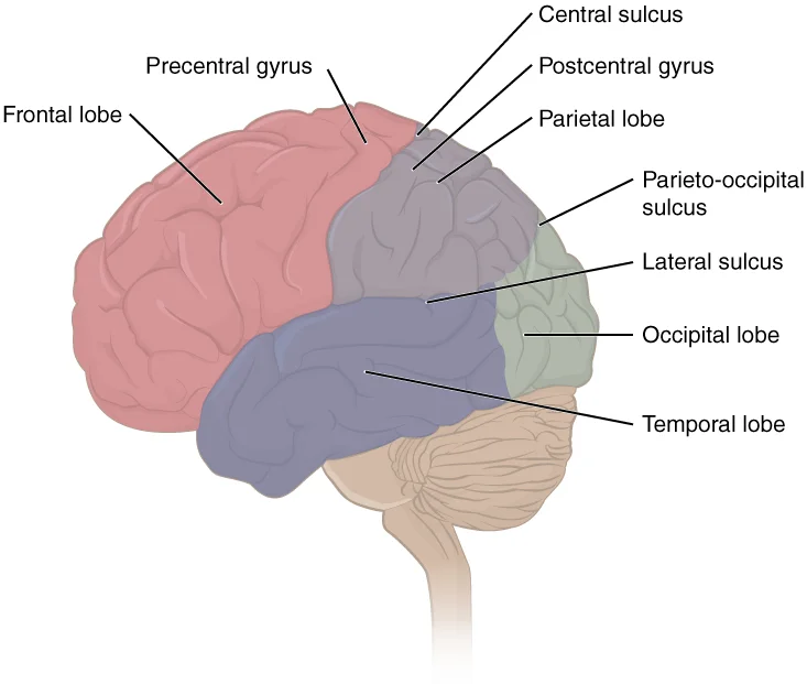
The cerebral cortex is divided into four lobes. Extensive folding increases the surface area available for cerebral functions.

Figure 15: The cerebral cortex is divided into four lobes.
The frontal lobe occupies the anterior portion, situated behind the forehead. It is separated from the parietal lobe by the central sulcus. The parietal lobe lies posterior to the frontal lobe and superior to the temporal lobe. The occipital lobe is located at the posterior pole of the brain. The temporal lobe is positioned inferior to the lateral sulcus. Each lobe contains specific cortical areas responsible for distinct physiological processes.
DefinitionCortical Organisation
The cerebral cortex is functionally organised such that anterior regions handle motor output while posterior regions process sensory input, reflecting the embryological division of the neural tube.
The frontal lobe is primarily associated with motor functions. It contains the primary motor cortex, which initiates voluntary movement. The parietal lobe is where somatosensation is processed. This includes the perception of touch, pressure, pain, and temperature. The occipital lobe is where visual processing begins, although the other parts of the brain can contribute to visual function. This lobe is dedicated to interpreting visual information received from the eyes. The temporal lobe contains the cortical area for auditory processing, but also has regions crucial for memory formation. It is involved in hearing and the encoding of long-term memories. [2]
Figure 16Gray752
Long description
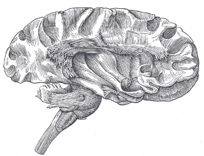
Dissection of cortex and brain-stem showing association fibers and island of Reil after removal of its superficial gray substance.

Figure 16: Dissection of cortex and brain-stem showing association fibers and island of Reil after removal of its superficial gray substance.
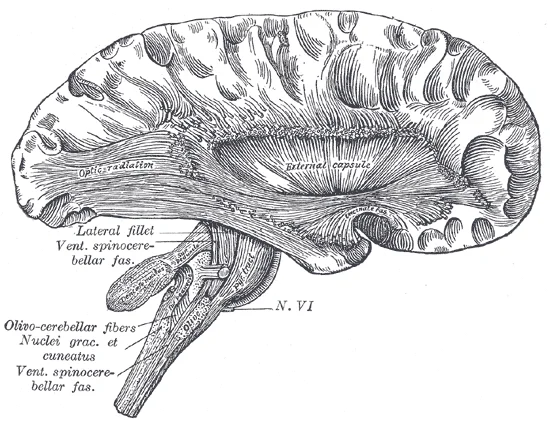
Figure 17Gray753
Long description
Deep dissection of cortex and brain-stem.

Figure 17: Deep dissection of cortex and brain-stem.
| Lobe | Primary Function | Key Structures |
|---|---|---|
| Frontal | Motor control | Primary motor cortex |
| Parietal | Somatosensation | Somatosensory cortex |
| Occipital | Visual processing | Visual cortex |
| Temporal | Auditory processing | Auditory cortex, Memory regions |
The diencephalon includes the thalamus and the hypothalamus, along with some other structures. The thalamus is a relay between the cerebrum and the rest of the nervous system. It directs sensory information to the appropriate cortical areas. The hypothalamus coordinates homeostatic functions through the autonomic and endocrine systems. It regulates body temperature, hunger, and thirst. [2]
The midbrain and hindbrain (composed of the pons and the medulla) are collectively referred to as the brain stem. The structure emerges from the ventral surface of the forebrain as a tapering cone that connects the brain to the spinal cord. Attached to the brain stem, but considered a separate region of an adult brain, is the cerebellum. [2]
Two subtests assess specific functions of these cortical areas. These assessments help localise lesions within the cortex. Understanding the precise location of function allows clinicians to predict deficits based on anatomical damage. For example, damage to the occipital lobe results in visual field defects. Damage to the frontal lobe may cause motor weakness or personality changes. The tegmentum is continuous with the gray matter of the rest of the brain stem. Throughout the midbrain, pons, and medulla, the tegmentum contains the nuclei that receive and send information through the cranial nerves, as well as regions that regulate important functions such as those of the cardiovascular and respiratory systems. [2] [1]
The cerebral cortex is the outer layer of the cerebrum, responsible for higher-order functions such as perception, cognition, and voluntary movement. Understanding its clinical significance requires linking specific anatomical regions to their functional deficits when damaged. Lesions in the cortex often present with distinct syndromes that help localise the injury.
DefinitionCortical Lesion
A focal area of damage to the cerebral cortex, resulting in loss of function corresponding to the affected region.
This image displays the clinical anatomy of the cerebral cortex, showing lesion sites, injury patterns, and imaging appearance. The view is a lateral perspective of the left hemisphere, oriented with the frontal lobe to the left and the occipital lobe to the right. The image highlights key sulci and gyri, including the central sulcus, which separates the frontal and parietal lobes. The precentral gyrus, located anterior to the central sulcus, is the primary motor cortex. The postcentral gyrus, posterior to the central sulcus, is the primary somatosensory cortex. The lateral sulcus (Sylvian fissure) separates the temporal lobe from the frontal and parietal lobes. The superior temporal gyrus is visible within the temporal lobe. The angular gyrus is located at the junction of the parietal, temporal, and occipital lobes. The cuneus is part of the occipital lobe, involved in visual processing. Each structure is clearly demarcated, allowing for precise localisation of deficits based on the lesion's position.
| Structure | Function | Deficit on Lesion |
|---|---|---|
| Precentral Gyrus | Voluntary motor control | Contralateral hemiparesis or hemiplegia |
| Postcentral Gyrus | Somatosensation | Contralateral hemianesthesia or neglect |
| Superior Temporal Gyrus | Auditory processing | Cortical deafness or auditory agnosia |
| Angular Gyrus | Language integration, reading | Gerstmann syndrome, alexia |
| Cuneus | Visual processing | Contralateral homonymous hemianopia |
| Column | Column |
|---|---|
| cell | cell |
Lesions in the precentral gyrus result in contralateral motor deficits. The severity depends on the extent of the damage. Small lesions may cause weakness in specific muscle groups, while large lesions can lead to complete paralysis. The postcentral gyrus lesion causes sensory loss on the opposite side of the body. Patients may report numbness or tingling. The superior temporal gyrus is crucial for auditory processing. Damage here can lead to difficulty understanding speech or recognizing sounds. The angular gyrus is involved in language and reading. Lesions here can cause Gerstmann syndrome, characterized by agraphia, acalculia, finger agnosia, and left-right disorientation. The cuneus is part of the visual cortex. Lesions here cause visual field defects, specifically contralateral homonymous hemianopia.
Clinical pearlLocalisation
Always correlate the clinical deficit with the anatomical location to identify the lesion site.
Neurons of the inferior colliculus project to the thalamus, which then sends auditory information to the cerebrum for the conscious perception of sound. Activity in the superior colliculus is related to orienting the eyes to a sound or touch stimulus. If you are walking along the sidewalk on campus and you hear chirping, the superior colliculus coordinates that information with your awareness of the visual location of the tree right above you. That is the correlation of auditory and visual maps. [2]
NoteIntegration
The superior colliculus integrates multiple sensory modalities to guide orienting responses.
Lesions in the superior colliculus may impair the ability to orient towards stimuli. Patients might have difficulty turning their eyes or head towards a sudden sound or touch. This highlights the importance of the superior colliculus in multisensory integration. Understanding these correlations is essential for diagnosing and managing patients with cortical lesions.
DefinitionLanguage testing has two parts
The neurological exam uses two specific subtests for language: one measures comprehension of language, the is fluency and coherency. [1]
Clinical pearlComprehension is tested by action
Comprehension is tested by asking the patient to follow a set of instructions to perform an action, such as "touch your right finger to your left elbow and then to your right knee". [1]
Clinical pearlFluency is tested by production
Fluency is tested by having the patient generate descriptions of objects or scenes depicted in drawings, and by reciting sentences or explaining a written passage. [1]
Clinical pearlMemory testing avoids overlearned sequences
Memory is tested by reciting the months of the year in reverse order to avoid the overlearned sequence, by spelling common words backwards, or by reciting a list of numbers back. [1]
Exam trapThree-word recall is a memory test
The three-word recall test gives words such as book, clock, and shovel, then asks for them after a short interval filled with other parts of the interview. [1]
DefinitionGnosis is the second sensory subtest
The second subtest for sensory perception is gnosis, which involves stereognosis and graphesthesia. [1]
Clinical pearlStereognosis uses common objects
Stereognosis involves naming objects strictly on the basis of the somatosensory information that comes from manipulating them, with the eyes closed, such as a coin. [1]
Clinical pearlCoin details give it away
A patient should distinguish a dime from a penny, or a nickel from a quarter, using size, thickness, weight, or the smooth or corrugated edge of the coin. [1]
Clinical pearlGraphesthesia uses the palm
Graphesthesia is the recognition of numbers or letters written on the palm of the hand with a dull pointer, such as a pen cap. [1]
Exam trapVerbs and nouns localise differently
Damage at the junction of the frontal and temporal lobes, including the insula, is associated with verb (V) impairment; damage to the middle and inferior temporal lobe is associated with noun (N) impairment. [1]
Clinical pearlConduction aphasia is a disconnection
Conduction aphasia results from damage to the white matter tracts connecting the posterior temporal lobe and the lateral aspect of the frontal lobe, and presents as an inability to faithfully repeat spoken language. [1]
Clinical pearlDopamine gates the basal nuclei
The substantia nigra pars compacta projects to the striatum and releases dopamine, which activates the direct pathway and inhibits the indirect pathway. [2]
Exam trapSilence means stillness
When the substantia nigra pars compacta is silent, the body is in a passive state and movement is inhibited, as in a student sitting through a lecture. [2]
Clinical pearlSpinal grey matter is an H
The gray matter of the spinal cord looks like a bulbous capital "H" in cross-section, subdivided into the posterior horn (sensory), anterior horn (motor) and lateral horn.
Clinical pearlSome axons are a metre long
The motor neuron that causes contraction of the big toe is located in the sacral spinal cord, and its axon may be a meter in length. [2]
Clinical pearlBig axon, big cell body
The neuronal cell body maintaining such a long fibre must be large, possibly several hundred micrometers in diameter, making it one of the largest cells in the body. [2]
The cortical examination is built from these subtests: comprehension and fluency for language, gnosis for sensory perception, and recall and sequencing tasks for memory. Localising a lesion then follows from the pattern: a repetition deficit points to the arcuate connection between posterior temporal and lateral frontal cortex, verb loss to the frontal-temporal junction and insula, and noun loss to the middle and inferior temporal lobe. Deep to the cortex, the substantia nigra pars compacta gates movement through dopamine acting on the direct and indirect pathways, while the diencephalon channels nearly all traffic between the cerebrum and the rest of the nervous system. [2]
Students frequently conflate the structural anatomy of the brain with its functional output, leading to diagnostic errors. A primary error is assuming that any confusion in a patient’s response indicates a global cognitive decline. It is crucial to distinguish between a deficit in understanding reality and a deficit in speech production. For instance, if a patient answers “Santa Claus” to a simple question, this suggests a problem understanding reality, whereas a confused stare may indicate a problem understanding or producing speech[1]. This distinction is vital for localising lesions to specific cortical areas rather than attributing symptoms to general encephalopathy.
Another significant pitfall involves the definition of orientation. Students often treat orientation as a single binary state of being "awake" or "asleep." In clinical practice, orientation is a multi-component awareness of immediate circumstances. It requires awareness of time, specifically the date and current events, rather than just the clock time[1]. It also demands awareness of place, meaning the patient must know where they are and why they are there[1]. Finally, it includes awareness of self, recognising personal identity and relating it to the examiner[1]. Failing to test all three components can lead to an underestimation of cognitive impairment.
Students also frequently misunderstand the role of the temporal lobe in memory. It is a common mistake to believe that the temporal lobe stores long-term memories permanently. While structures in the temporal lobe are responsible for establishing long-term memory, the ultimate location of those memories is usually in the region in which the sensory perception was processed. Memory is essentially a sensory function; memories are recalled sensations such as the smell of Mom’s baking or the sound of a barking dog. Therefore, damage to the temporal lobe affects the encoding and retrieval pathways, not necessarily the storage site of the memory trace itself. [2]
A further error relates to the cerebellum’s role in motor control. Many students view the cerebellum as the origin of motor commands. In reality, the cerebellum compares information from the cerebrum with sensory feedback from the periphery through the spinal cord. It accounts for approximately 10 percent of the mass of the brain. If walking is not coordinated, the cerebellum sends out a corrective command to compensate for the difference between the original cortical command and the sensory feedback. The output of the cerebellum is into the midbrain, which then sends a descending input to the spinal cord to correct the messages going to skeletal muscles. Confusing the cerebellum with the primary motor cortex leads to incorrect localisation of ataxia. [2]
Finally, students often overlook the anatomical relationship between the spinal cord and the vertebral column. It is incorrect to assume that the spinal cord segments align perfectly with the vertebral levels throughout the entire column. The spinal cord is not the full length of the vertebral column because the spinal cord does not grow significantly longer after the first or second year, but the skeleton continues to grow. Misunderstanding this growth disparity leads to errors in interpreting spinal imaging and localising neurological deficits. [2]
This knowledge is applied at the bedside during the initial mental state examination. The initial tests of orientation are based on the questions, “Do you know what the date is?” or “Do you know where you are?” or “What is your name?”[1]. These questions verify that the patient is aware, oriented, and capable of interacting with another person[1]. Further understanding of a patient’s awareness of orientation can come from questions that address remote memory, such as “Who is the President of the United States?”, or asking what happened on a specific date[1]. This structured approach allows clinicians to rapidly assess cortical function and detect subtle deficits in language or reality testing.
In the operating theatre and neurosurgical contexts, understanding the brain stem is critical. The pons is the main connection between the cerebellum and the brain stem. The bridge-like white matter is only the anterior surface of the pons; the gray matter beneath that is a continuation of the tegmentum from the midbrain. Gray matter in the tegmentum region of the pons contains neurons receiving descending input from the forebrain that is sent to the cerebellum. Surgeons must respect these pathways to avoid disrupting motor coordination. Similarly, the medulla is the region known as the myelencephalon in the embryonic brain. The tegmentum of the midbrain and pons continues into the medulla because this gray matter is responsible for processing cranial nerve information. Awareness of the reticular formation, a diffuse region of gray matter throughout the brain stem, is related to sleep and wakefulness, such as general brain activity and attention. [2]
On imaging, radiologists use anatomical landmarks to identify structures. The cerebellum is largely responsible for comparing information from the cerebrum with sensory feedback from the periphery through the spinal cord. The cerebellum is connected to the brain stem, primarily at the pons, where it receives a copy of the descending input from the cerebrum to the spinal cord. It can compare this with sensory feedback input through the medulla and send output through the midbrain that can correct motor commands for coordination. Identifying these connections helps in diagnosing conditions like cerebellar ataxia or brain stem strokes. The inferior and slightly anterior to the thalamus is the hypothalamus, the other major region of the diencephalon. The hypothalamus is a collection of nuclei that are largely involved in regulating homeostasis. The hypothalamus is the executive region in charge of the autonomic nervous system and the endocrine system through its regulation of the anterior pituitary gland. Other parts of the hypothalamus are involved in memory and emotion as part of the limbic system. [2]
In examinations, students must demonstrate precise anatomical knowledge. The length of the spinal cord is divided into regions that correspond to the regions of the vertebral column. The name of a spinal cord region corresponds to the level at which spinal nerves pass through the intervertebral foramina. Immediately adjacent to the brain stem is the cervical region, followed by the thoracic, then the lumbar, and finally the sacral region. The nerves that emerge from the spinal cord pass through the intervertebral foramina at the respective levels. This precise terminology is tested to ensure students can localise lesions accurately. [2]
The temporal lobe is associated with primary auditory sensation, known as Brodmann’s areas 41 and 42 in the superior temporal lobe. Because regions of the temporal lobe are part of the limbic system, memory is an important function associated with that lobe. Understanding this association helps in interpreting seizures or memory loss. Language is, arguably, a very human aspect of neurological function[1]. Formulating an understanding of yourself is largely describing who you are to yourself[1]. Language is certainly at the core of what it means to be self-aware[1]. This conceptual understanding is often tested in ethics and communication modules alongside anatomy. [2]
The prefrontal lobotomy is an outdated mode of treatment for personality disorders (psychiatric conditions) that profoundly affected the personality of the patient. While historical, this is often cited in examinations to contrast modern psychiatric care with past surgical interventions. The word pons comes from the Latin word for bridge. It is visible on the anterior surface of the brain stem as the thick bundle of white matter attached to the cerebellum. This etymological knowledge aids in remembering its function as a connector. [2]
Clinical pearlOrientation Triad
Orientation requires testing time, place, and person. A patient may know the date but not where they are, indicating a specific deficit rather than global confusion.
DefinitionCerebellar Function
The cerebellum compares cortical motor commands with sensory feedback to correct movement. It does not initiate movement but refines it via the midbrain and spinal cord.
Assessing Orientation
- Ask 'What is your name?'Verify identity and speech production
- Question: Is the answer appropriate?
Yes
- Ask 'Where are you?'
No
- Note confusion or reality distortion
- Ask 'What is the date?'Assess awareness of time
- Question: Is the date correct?
Yes
- Ask remote memory question
No
- Note disorientation to time
Orientation involves awareness of time, place, and person. The temporal lobe establishes long-term memory but stores it in sensory regions. The cerebellum corrects motor commands using sensory feedback. The pons connects the cerebellum to the brain stem. The spinal cord is shorter than the vertebral column. The hypothalamus regulates homeostasis and the autonomic nervous system. Language is core to self-awareness and human experience. The medulla processes cranial nerve information via the tegmentum.
Answer each one in your head first, then open the answer.
What does a patient's answer of 'Santa Claus' to 'What is your name?' suggest?
Show answerHide answer
A problem understanding realityWhich areas of the temporal lobe are associated with primary auditory sensation?
Show answerHide answer
Brodmann’s areas 41 and 42What percentage of the brain's mass does the cerebellum account for?
Show answerHide answer
Approximately 10 percentWhat is the name of the bundle of nerves resembling a horse's tail?
Show answerHide answer
Cauda equinaWhat is the primary function of the hypothalamus?
Show answerHide answer
Regulating homeostasis
- Betts JG, Young KA, Wise JA, et al. The Mental Status Exam. OpenStax, Anatomy and Physiology 2e (CC BY-NC-SA 4.0). Accessed .
- Betts JG, Young KA, Wise JA, et al. The Central Nervous System. OpenStax, Anatomy and Physiology 2e (CC BY-NC-SA 4.0). Accessed .
Figure credits
- Figure 1: Henry Vandyke Carter, via Wikimedia Commons, Public domain, Public domain.
- Figure 2: Henry Vandyke Carter, via Wikimedia Commons, Public domain, Public domain.
- Figure 3: Mysid, via Wikimedia Commons, Public domain, Public domain.
- Figure 4: Henry Vandyke Carter, via Wikimedia Commons, Public domain, Public domain.
- Figure 5: Henry Vandyke Carter, via Wikimedia Commons, Public domain, Public domain.
- Figure 6: OpenStax College, via Wikimedia Commons, CC BY 3.0, CC BY 3.0. resized.
- Figure 7: OpenStax College, via Wikimedia Commons, CC BY 3.0, CC BY 3.0.
- Figure 8: Henry Vandyke Carter, via Wikimedia Commons, Public domain, Public domain.
- Figure 9: Henry Vandyke Carter, via Wikimedia Commons, Public domain, Public domain.
- Figure 10: OpenStax, via Wikimedia Commons, CC BY 4.0, CC BY 4.0.
- Figure 11: OpenStax College, via Wikimedia Commons, CC BY 3.0, CC BY 3.0. resized.
- Figure 12: Henry Vandyke Carter, via Wikimedia Commons, Public domain, Public domain.
- Figure 13: Henry Vandyke Carter, via Wikimedia Commons, Public domain, Public domain.
- Figure 14: OpenStax, via Wikimedia Commons, CC BY 4.0, CC BY 4.0.
- Figure 15: OpenStax, via Wikimedia Commons, CC BY 4.0, CC BY 4.0.
- Figure 16: Henry Vandyke Carter, via Wikimedia Commons, Public domain, Public domain.
- Figure 17: Henry Vandyke Carter, via Wikimedia Commons, Public domain, Public domain.
Educational content for medical students and doctors. It is not a substitute for clinical judgement or local guidelines.