Limbic system
Limbic system integrates emotion, memory and olfaction in brain border structures.
The limbic system is a collection of cortical and subcortical structures that form a border around the brainstem[1]. The term "limbic" comes from the Latin word for border, first introduced by Thomas Willis to designate cortical border structures encircling the brainstem[1]. Broca initially proposed that olfactory structures were the main components of the limbic system in mammalian brains[1].
The limbic system includes several key structures: the hippocampus, amygdala, cingulate gyrus, parahippocampal gyrus, fornix, and mammillary bodies[1]. The hippocampus is the largest gray matter nucleus of the limbic system, residing in the mesial temporal lobe[1]. The amygdala is connected to the hippocampus anatomically, with their heads nearly contacting each other[1].
The Papez circuit, first proposed by James Papez, is a major pathway of the limbic system believed to be involved in controlling both memory and emotion[1]. This circuit connects the hippocampus, hypothalamus (mammillary bodies), thalamus, cingulate gyrus, and parahippocampal gyrus[1].
The limbic system has several important functions, including learning, memory, olfaction, adjusting visceral functions, emotional processing, attention, and cognition[1]. Paul MacLean integrated the Papez and Yakovlev models as part of the limbic system and further modified the Papez circuit to include the prefrontal cortex, septum, and amygdalae[1].
Yakovlev introduced the basolateral circuit, which includes thalamo-amygdaloid connections and their connectivity with the orbitofrontal gyrus, associating this circuit with emotions and motivations[1]. The limbic system was later extended to include the midbrain[1]. Mishkin demonstrated that stimulation of higher-order sensory areas activates a cortico-limbo-thalamo-cortical circuit involved in memory function, proposing two parallel loops - one involving the amygdala and dorsomedial nucleus of the thalamus, and the other involving the hippocampus and anterior nuclei[1].
Figure 1The Limbic System and Nearby Structures - John Taylor
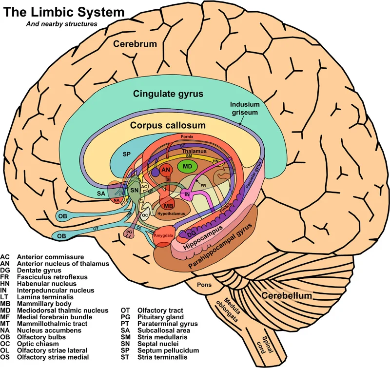
Long description
Cutout anatomy diagram of the human limbic system and some nearby structures.

Figure 1: Cutout anatomy diagram of the human limbic system and some nearby structures.
The limbic system is not a single anatomical structure but a functionally integrated network of cortical and subcortical elements. MacLean’s model established that the limbic cortex and subcortical limbic structures operate as a unified system, interconnected by both short and long-range fiber bundles. This integration allows for the complex processing of emotion, memory, and autonomic regulation. [1]
Below the corpus callosum lies the hippocampal formation, curving into the temporal lobe. The hypothalamus forms the floor of the third ventricle, situated below the thalamus. The thalamus itself is centrally located, acting as a relay station. The stria terminalis runs parallel to the fornix along the medial margin of the lateral ventricles. These structures are arranged to facilitate rapid communication between the cerebral cortex and deeper subcortical nuclei.
It has reciprocal connections with many structures, including the cerebral cortex, basal ganglia, cerebellum, brainstem, and spinal cord. The thalamus plays a critical role in modulating limbic functions through many connections with major bodies of the limbic system including the hippocampus, amygdala, hypothalamic and septal nuclei. In high spatial resolution diffusion-weighted imaging, the thalamus is divided into three segments by three major colors indicating different orientation of the thalamic fiber tracts. [1]
It is a collection of nuclei located lateral to the third ventricle and superior and posterior to the optic chiasm. Similar to the amygdala, the hypothalamic and septal nuclei are among the most connected structures of the limbic system. The hypothalamus is directly connected to wide areas of the cerebral cortex, including the frontal, parietal, occipital, and temporal lobes, along with the cerebellum and mesencephalon. [1]
The stria terminalis (ST) is a major amygdalo-septal/hypothalamic connectivity of the limbic system and is believed to be one of the major output pathways of the amygdala. The ST courses side by side with the fornix along the medial margin of the lateral ventricles. The ST follows a C-shaped course laterally and parallel to the fornix, descends through the caudothalamic groove, and reaches the hypothalamic and septal nuclei and the bed nucleus of the ST. [1]
Studies demonstrated direct engagement of the prefrontal cortex with the limbic system in primates, including the human brain. The entire cerebral cortex is also indirectly connected to the anterior limbic nuclei such as the amygdalae, hypothalamic and septal nuclei mediated by the thalamus via extensive cortico-thalamic connectivity. These newly described fiber tracts incorporate multiple additional limbic circuits to the already known complex limbic network. The purpose of this review is to assess the anatomy of the limbic structures and to elaborate on the anatomical connectivity of the limbic circuits as an update to the original Papez circuit. [1]
The hypothalamus is at the center of multiple loops and circuits due to its central location and crucial role in many limbic functions. Some of the circuits involving the hypothalamic and septal nuclei are detailed in the hypothalamus section and illustrated in Figs. –. The cortico-basal ganglia-thalamo-cortical pathway is another loop including the thalamus, limbic and basal ganglia. The cortico-basal ganglia-thalamo-cortical loops are thought to have consisted of parallel circuits that individually process limbic, associative, and sensorimotor information. Efferent pathways have also been described projecting from the NAC into several limbic structures, including the stria terminalis, preoptic and lateral hypothalamic nuclei, amygdala, thalamus, and cingulum. Given the connections between the NAC with both the limbic system and motor nuclei of the basal ganglia, the NAC may play a role in communication between limbic and motor systems. [1]
The primary focus is the septal region, situated deep within the medial cerebral hemisphere. The septal nuclei appear as a distinct band of gray matter. They are located rostral to the anterior commissure and anterior to the lamina terminalis. Inferiorly, they lie beneath the rostrum of the corpus callosum. The nuclei extend laterally alongside the lamina terminalis towards the optic chiasm. This positioning places them in close proximity to the hypothalamus. Consequently, most pathways connecting to the hypothalamic nuclei also exhibit connectivity with the septal nuclei. [1]
The internal architecture of the limbic system relies on complex networks of nuclei, laminae, and tracts. A key component is the prefronto-caudo-thalamic tract (PFCT). This pathway was first described in 2010. It includes the caudate nucleus and lentiform nuclei. Eventually, it projects into the prefrontal cortex. The tract contains cortico-striatal pathways between the putamina and caudate nuclei. It directly connects the thalamus with the bed nucleus of the stria terminalis (BNST) and septal nuclei. The PFCT communicates with multiple limbic nuclei, including the hypothalamic, septal, BNST, and nucleus accumbens. [1]
At its thalamic origin, the PFCT arises from the dorsomedial nucleus of the thalamus. This segment exhibits convexity toward the midline and concavity toward the lateral thalamus. After arising, the PFCT projects into the BNST. It receives projections from the septal nuclei and lentiform nuclei. Projections join the PFCT superiorly from the caudate body. Laterally, fibers join from the lenticular nuclei, comprising the putamen and globus pallidus. Inferiorly, connections arrive from the hypothalamic nuclei. The PFCT connects all these structures with the caudate head, nucleus accumbens, and prefrontal cortex. [1]
Indirectly, it connects to the cerebral cortex through the thalamus via the amygdalothalamic tract. The amygdala connects to the prefrontal cortex indirectly through the hypothalamic and septal nuclei. This occurs via the amygdalofugal tract, which connects the amygdala to the hypothalamic and septal nuclei. The PFCT then connects these nuclei to the prefrontal cortex. [1]
The septal nuclei send projections mainly to the habenular nuclei and the medial dorsal nucleus of the thalamus via the stria medullaris. They are connected to the hippocampus and amygdala via the fornix and stria terminalis. Direct connectivity of the septal nuclei to the amygdala is described via the amygdalofugal tract. This tract connects the amygdala to the septal nuclei, hypothalamic nuclei, and BNST. The thalamus is connected to the septal nuclei via the stria medullaris and PFCT. A direct thalamo-septal connection is also reported via fibers running side by side with the dorsomedial thalamic tract. [1]
DefinitionSeptal Nuclei
A band of gray matter nuclei located rostral to the anterior commissure, anterior to the lamina terminalis, and inferior to the rostrum of the corpus callosum.
| Structure | Primary Connection | Target/Pathway |
|---|---|---|
| PFCT | Thalamus (DM nucleus) | Prefrontal cortex, BNST, Septal nuclei |
| Amygdalofugal Tract | Amygdala | Hypothalamic/Septal nuclei, BNST |
| Stria Medullaris | Septal nuclei | Habenular nuclei, Medial dorsal thalamus |
The limbic system has extensive afferent and efferent connections that integrate it with other brain regions. The thalamus serves as a major hub for limbic connections, directly linking to multiple limbic nuclei including the hypothalamic/septal nuclei, hippocampus, and amygdala. Key tracts include the mammillothalamic tract (MTT), which connects the ventral thalamus to the mammillary bodies of the hypothalamus, and the dorsal thalamo-hypothalamic (DTH) tract, which connects the pulvinar, latero [1]
The thalamus also connects to widespread cortical areas through three major radiation systems: the anterior thalamic radiation (ATR), superior thalamic radiation (STR), and posterior thalamic radiation (PTR). The ATR mainly connects the thalamus to the prefrontal cortices, containing reciprocal fibers that run between the thalamus and prefrontal cortex along the medial aspect of the anterior limbs of the internal capsules. The STR connects the ventral nuclear group of the thalamus to the pre and post-central gyrus, including thalamo-cortical projections of the spinothalamic tract to the parietal cortex. The PTR serves as a connection between the occipital and parietal lobes and the thalamus by passing through the retro lenticular limb of the internal capsule. [1]
The uncinate fasciculus (UF) is the most laterally located limbic white matter pathway, curving upward into the extreme and external capsule and medial to the insular cortex, with connections to the lenticular nucleus, internal capsule, and medial insular cortex, then continuing into the orbitofrontal cortex. The ventral amygdalofugal tract consists of crucial amygdalo-diencephalic connections, including the amygdalo-hypothalamic, amygdalo-septal, amygdalo-thalamic, and amygdalo-mesencephalic tracts. [1]
The olfactory bulb receives input from the nasal epithelium and projects directly to the piriform cortex and amygdala through the olfactory tract. The amygdala then connects to the hypothalamus via the ventral amygdalofugal tract, forming a pathway by which olfactory information can influence emotional and autonomic responses. The mammillary bodies in the hypothalamus connect back to the thalamus via the mammillothalamic tract, completing the Papez circuit. This circuit demonstrates how olfactory information can be integrated with limbic structures to influence emotional processing and memory formation.
DefinitionLimbic Circuit
The limbic system forms interconnected circuits that integrate emotion, memory, and autonomic function, with the thalamus serving as a major relay station.
The nucleus basalis, located in the substantia innominate of the basal forebrain, represents another important limbic connection point. As the largest collection of cholinergic neurons in the brain, it has widespread cholinergic cortical projections that modulate activity in limbic areas and connects with structures such as the lateral hypothalamus, amygdala, and midbrain. These connections highlight the limbic system's extensive network that allows for integration of sensory information, emotional processing, and memory formation. [1]
The limbic system is a complex network of structures that relies on a rich and redundant vascular supply to maintain its high metabolic demands. This perspective allows for the clear identification of the major arterial contributions to the medial and temporal structures that form the core of the limbic circuitry. Understanding this vascular anatomy is critical, as lesions in these specific territories can disrupt the intricate loops that govern emotion, memory, and behaviour.
The anterior cerebral artery (ACA) plays a pivotal role in supplying the medial aspects of the cerebral hemispheres. Its cortical branches extend to supply the cingulate gyrus, a key component of the limbic lobe, and the medial frontal cortex. The ACA also contributes to the blood supply of the hippocampal formation via its pericallosal branches. The fornix, which is the only limbic pathway that generates a complete loop on its own, receives its vascular support from branches of the anterior cerebral artery and the anterior choroidal artery. This loop, a hypothalamo-septo-hippocampo-hypothalamic circuit via the forniceal columns, body, crura, and ventral arms, is essential for memory consolidation and emotional processing. [1]
The middle cerebral artery (MCA) supplies the lateral aspects of the temporal lobe, including the amygdala and the insular cortex. The amygdala is a central hub in the limbic system, involved in the processing of emotions such as fear and aggression. It receives blood from the temporal branches of the MCA and the anterior choroidal artery. The insular cortex, which is involved in the integration of sensory and emotional information, is also supplied by the MCA. The MCA’s deep branches, including the lenticulostriate arteries, supply the basal ganglia, which have extensive connections with the limbic system through cortico-thalamo-striata-cortical circuits.
The posterior cerebral artery (PCA) is crucial for the supply of the medial temporal lobe structures. It provides blood to the hippocampus, parahippocampal gyrus, and parts of the thalamus that connect to limbic structures. The hippocampus is vital for the formation of new memories and is supplied by the hippocampal branches of the PCA. The thalamus, particularly the mediodorsal nucleus, receives blood from the PCA and the posterior communicating artery. The mediodorsal thalamus is a key relay station in the limbic system, connecting the amygdala and the prefrontal cortex.
The hypothalamus receives its blood supply primarily from the superior hypophyseal artery, a branch of the internal carotid artery, and from branches of the posterior cerebral artery. The mammillary bodies, part of the hypothalamus, are supplied by the posterior communicating artery. The circle of Willis, formed by anastomoses between the internal carotid and vertebral-basilar systems, ensures collateral circulation to limbic structures in cases of occlusion. This redundancy is vital for maintaining perfusion to these critical areas.
The basal nucleus of Meynert, located in the substantia innominata, receives blood from the anterior choroidal artery and the lenticulostriate arteries. This nucleus is a major source of cholinergic input to the cerebral cortex and is involved in attention and memory. The bed nucleus of the stria terminalis (BNST), which may participate in many limbic circuits involving the hypothalamic and septal nuclei, receives blood from the anterior choroidal artery and the lenticulostriate arteries.
DefinitionLimbic vascular supply
The limbic system is supplied by the anterior, middle, and posterior cerebral arteries, with the fornix forming a complete hypothalamo-septo-hippocampo-hypothalamic loop.
The limbic system is a complex network of structures and pathways that underpins emotional processing, memory formation, and motivational behaviour. Its function is not isolated to a single nucleus but emerges from the intricate connectivity between the cortex, thalamus, and hypothalamus. Modern neuroanatomy, supported by diffusion-weighted imaging and primate dissection studies, reveals a dense web of fiber tracts that facilitate this integration. These pathways ensure that sensory input is evaluated for emotional significance and that memory traces are consolidated with affective context. [1]
The amygdala is situated within the temporal lobe, anterior to the hippocampus. It connects to the hypothalamus via the amygdalofugal tract and to the thalamus via the amygdalothalamic tract. The stria terminalis also runs along the lateral ventricle, connecting the amygdala to the septal nuclei and hypothalamus. Understanding these spatial relationships is vital for localising lesions and interpreting clinical syndromes. [1]
The functional output of these structures is mediated through specific neural circuits. The prefronto-hypothalamic/septal connecting arm, carried by the MFB, integrates cognitive appraisal with autonomic and endocrine responses. Simultaneously, the amygdalo-thalamo-amygdalo-amygdala loop, facilitated by the AC, ensures bilateral consistency in emotional processing. The POHT provides a conduit for visual and sensory data to reach the nucleus basalis and anterior hypothalamic nuclei, linking perception directly to visceral states. These connections demonstrate that the limbic system is not merely a seat of emotion but a central hub for integrating sensory, cognitive, and homeostatic information. The development of complex behaviours, such as endurance hunting, likely drove the evolution of these mnemonic and regulatory networks, requiring retention of geographical areas and precise neuro-hormonal control. Thus, the limbic system’s function is defined by its connectivity, enabling the brain to assign value to experiences and coordinate appropriate behavioural responses. [1]
The limbic system is not a single anatomical entity but a functional network involving the hippocampus, amygdala, cingulate gyrus, and associated white matter tracts. Damage to these structures produces distinct clinical syndromes that reveal their normal roles in memory, emotion, and behaviour. Understanding the anatomical basis of these lesions allows for precise localisation of pathology in clinical practice.
Adjacent to it lies the amygdala, a almond-shaped nucleus located anteriorly.
In contrast, Kluver-Bucy syndrome results from bilateral lesions of the amygdala and anterior temporal lobes. This syndrome is characterised by hyperorality, hypersexuality, and visual agnosia. Patients exhibit a lack of fear and anger responses. The amygdala is critical for processing emotional significance. Its destruction removes the emotional filter from sensory perception. The hippocampus, also often involved, leads to severe anterograde amnesia. The inability to form new memories is a hallmark of this condition.
| Structure Lesioned | Primary Clinical Deficit | Associated Syndrome |
|---|---|---|
| Bilateral Amygdala | Loss of fear, hyperorality, hypersexuality | Kluver-Bucy Syndrome |
| Hippocampus / Medial Temporal Lobe | Anterograde amnesia, memory consolidation failure | Alzheimer’s Disease (early stage) |
| Cingulate Gyrus | Apathy, abulia, emotional blunting | Anterior Cingulate Syndrome |
| Nucleus Basalis of Meynert | Cognitive decline, attention deficits | Alzheimer’s Disease (progressive) |
Alzheimer’s disease represents a progressive neurodegenerative condition. It begins with subtle memory impairment due to hippocampal involvement. As the disease advances, atrophy spreads to the parietal and temporal lobes. This widespread cortical damage leads to global cognitive decline. Their degeneration disrupts communication between the limbic system and the hypothalamus. This disconnection explains the behavioural changes and emotional lability seen in later stages. The anatomical correlation between the site of atrophy and the clinical symptom is direct and predictable. Recognising these patterns is essential for diagnosis and management.
Clinical pearlPapez Circuit
This circuit forms the classic limbic pathway for emotional processing and memory formation. [1]
Clinical pearlDual Memory Circuit
Aggleton and Mishkin proposed two parallel memory circuits: one involving amygdala and dorsomedial nucleus of thalamus via amygdalothalamic tract, and another involving hippocampus and anterior nuclei. Mishkin demonstrated that stimulation of higher-order sensory areas of the cortex activates a cortico-limbo-thalamo-cortical circuit. [1]
Clinical pearlBNST Connectivity
The bed nucleus of stria terminalis (BNST) shares many projections with hypothalamic nuclei and has tight connectivity with prefrontal cortex, amygdala, hippocampus, thalamus, basal ganglia, and brainstem nuclei/ventral tegmental area. Most projections reaching the hypothalamus originate from the BNST. [1]
Exam trapCommon Misconception
The limbic system is not a discrete anatomical entity but rather a collection of interconnected brain structures involved in emotion and memory. Students often incorrectly view it as a single isolated structure rather than a functional network. [1]
Clinical pearlInternal Capsule Pathway
This pathway is crucial for connecting thalamic nuclei with cortical areas. [1]
Clinical pearlHypothalamic Projections
Given the proximity of the BNST, septal and hypothalamic nuclei, they share many projections between one another.
Clinical pearlMammillary Body Connection
Ventral forniceal arms exit from the junction of forniceal crura and fimbria, course medially along the lateral margin of cerebral peduncles toward mammillary bodies and hypothalamic nuclei where they terminate. The mammillary bodies are critical relay stations in the Papez circuit.
Clinical pearlMemory Circuit Components
Memory circuits involve higher-order sensory areas of cortex activating cortico-limbo-thalamo-cortical circuits. The dual circuit proposed by Aggleton and Mishkin includes one pathway through the amygdala and dorsomedial thalamic nucleus, and another through the hippocampus and anterior thalamic nuclei.
NoteClinical Relevance
Damage to the mammillary bodies, as seen in Wernicke-Korsakoff syndrome, disrupts the Papez circuit leading to severe memory impairment, particularly anterograde amnesia. This demonstrates the critical role of this pathway in memory formation.
Clinical pearlSeptal Nuclei Connections
The septal nuclei have extensive connections with hypothalamic nuclei, prefrontal cortex, and the bed nucleus of stria terminalis. These connections are vital for emotional regulation and reward processing.
Exam trapAnatomical Pitfall
The fornix is often confused with the cingulum bundle. Remember that the fornix connects the hippocampus to the mammillary bodies, while the cingulum bundle connects the cingulate gyrus to the parahippocampal gyrus.
Clinical pearlThalamic Relay
The ventral thalamus receives input from the mammillary bodies via the mammillothalamic tract and projects back to the prefrontal cingulate gyrus, completing the Papez circuit. This thalamic relay is essential for the circuit's function.
Exam trapConfusing limbic structures
Students often mistake the amygdala for the hippocampus or vice versa. The amygdala is primarily involved in emotional processing, particularly fear and aggression, while the hippocampus is the largest gray matter nucleus of the limbic system residing in the mesial temporal lobe. [1]
Exam trapMisunderstanding thalamic radiations
A common error is confusing the three thalamic radiations. The anterior thalamic radiations (ATR) project to the prefrontal cortex, the superior thalamic radiations (STR) include the spinothalamic tract, and the posterior thalamic radiations (PTR) have latero-laterally oriented fiber tracts. [1]
Exam trapOverlooking fiber connections
Students frequently neglect the complex fiber connections of the limbic system. The uncinate fasciculus (UF) is a bidirectional, U-shaped bundle connecting the inferior frontal gyrus, orbitofrontal cortex, and ventromedial prefrontal cortex to the anterior temporal lobe. [1]
Exam trapMisidentifying the bed nucleus of stria terminalis
The bed nucleus of stria terminalis (BNST) is often incorrectly positioned. It is located at the base of the lateral ventricles, ventral to the septum, just above and posterior to the anterior commissure, and antero-superior to the hypothalamic nuclei. [1]
Exam trapConfusing neurotransmitter roles
Students commonly misunderstand the roles of neurotransmitters in limbic function. GABAergic interneurons are crucial in regulating the diverse activities of pyramidal cells, and GABA has an inhibitory function in the nervous systems of both vertebrates and invertebrates. [2]
Exam trapEvolutionary misconceptions
A frequent mistake is overstating evolutionary claims about the limbic system. While there may have been positive Darwinian selection in the modification of interneuron populations in humans leading to cognitive specializations, this is still a hypothesis rather than established fact.
NoteClinical assessment
Knowledge of the limbic system is essential in assessing patients with memory disorders. Damage to the hippocampus, a key limbic structure, results in anterograde amnesia, the inability to form new memories.
NoteNeurosurgical planning
In neurosurgery, understanding the thalamic radiations is crucial. The anterior thalamic radiations (ATR) project to the prefrontal cortex, so damage during surgery can result in executive function deficits.
NoteNeuroimaging interpretation
Diffusion-weighted imaging can visualize the three thalamic radiations: the anterior thalamic radiations (ATR) appear green, the superior thalamic radiations (STR) appear blue, and the posterior thalamic radiations (PTR) appear red.
NotePsychiatric understanding
The limbic system's role in emotion is relevant to psychiatric practice. The amygdala's connectivity with the orbitofrontal cortex and anterior temporal lobes via the uncinate fasciculus is important in understanding emotional processing disorders.
Limbic Circuitry
- Prefrontal cortexExecutive function and decision making
- Question: Connects to amygdala?
Yes
- Via uncinate fasciculus (UF)Emotional processing
- AmygdalaFear and aggression response
No
- Via anterior thalamic radiations (ATR)To mediodorsal thalamus
- Mediodorsal thalamusRelay station for limbic information
- HypothalamusAutonomic and endocrine responses
NoteNeurological examination
In neurological examinations, understanding the limbic system helps localize lesions. The amygdalofugal tract connects the amygdala directly with the brainstem, so damage can affect autonomic responses to emotional stimuli.
NoteNeuropsychiatric research
Research on limbic system connectivity informs our understanding of psychiatric disorders. The prefronto-amygdalo-hypothalamo-septo-caudate-prefrontal loop involves multiple pathways including the uncinate fasciculus and anterior thalamic radiations.
- The amygdala is primarily involved in emotional processing, particularly fear and aggression, while the hippocampus is crucial for memory formation [1]
- The three thalamic radiations have distinct projections: ATR to prefrontal cortex, STR including spinothalamic tract, and PTR with latero-lateral fibers
- The uncinate fasciculus is a bidirectional U-shaped bundle connecting frontal and temporal regions
- The bed nucleus of stria terminalis is located at the base of the lateral ventricles, ventral to the septum
- GABAergic interneurons are crucial in regulating pyramidal cell activity in the limbic system
Answer each one in your head first, then open the answer.
Which structure is primarily involved in memory formation, particularly spatial and episodic memory?
Show answerHide answer
HippocampusWhat are the three thalamic radiations and their primary orientations?
Show answerHide answer
Anterior thalamic radiations (anterior-posterior), superior thalamic radiations (cranio-caudal), posterior thalamic radiations (latero-lateral)Which fiber bundle connects the inferior frontal gyrus, orbitofrontal cortex, and ventromedial prefrontal cortex to the anterior temporal lobe?
Show answerHide answer
Uncinate fasciculusWhere is the bed nucleus of stria terminalis located?
Show answerHide answer
At the base of the lateral ventricles, ventral to the septum, just above and posterior to the anterior commissureWhat is the primary function of GABAergic interneurons in the limbic system?
Show answerHide answer
Regulating the diverse activities of pyramidal cells
- Kamali A, Milosavljevic S, Gandhi A, et al. The Cortico-Limbo-Thalamo-Cortical Circuits: An Update to the Original Papez Circuit of the Human Limbic System. Brain topography 2023;36(3):371-389. doi:10.1007/s10548-023-00955-y. Accessed .
- Brisch R, Bielau H, Saniotis A, et al. Calretinin and parvalbumin in schizophrenia and affective disorders: a mini-review, a perspective on the evolutionary role of calretinin in schizophrenia, and a preliminary post-mortem study of calretinin in the septal nuclei. Frontiers in cellular neuroscience 2015;9:393. doi:10.3389/fncel.2015.00393. Accessed .
Figure credits
Educational content for medical students and doctors. It is not a substitute for clinical judgement or local guidelines.