Internal capsule
Neuroanatomical structure containing major motor and sensory projection fibres.
The internal capsule is a dense band of white matter situated between the lentiform nucleus and the thalamus. It serves as the primary conduit for projection fibres, which connect the cerebral cortex with subcortical structures, the brainstem, and the spinal cord. These fibres are distinct from commissural fibres, such as those in the corpus callosum and anterior commissure, which connect the two cerebral hemispheres. The internal capsule is essentially the concentrated form of the corona radiata, where fibres converge as they pass towards their targets.
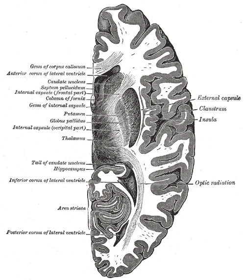
Figure 1Gray742
Long description
Horizontal section of right cerebral hemisphere.

Figure 1: Horizontal section of right cerebral hemisphere.
The structure is divided into three main parts: the anterior limb, the genu, and the posterior limb. The anterior limb lies between the caudate nucleus and the lentiform nucleus. The genu is the knee-like bend where the anterior and posterior limbs meet. The posterior limb extends between the lentiform nucleus and the thalamus. A small retrolenticular part lies behind the posterior limb, and a sublenticular part lies below it. [1]
The fibres within the internal capsule are organised somatotopically. The anterior limb contains frontopontine fibres and thalamocortical fibres. The genu contains corticobulbar fibres, which innervate the cranial nerve nuclei in the brainstem. The posterior limb contains the corticospinal tract, which supplies the body, as well as sensory fibres including the thalamocortical radiations for vision, hearing, and somatosensation. [1]
Damage to the internal capsule, often due to lacunar stroke affecting the lenticulostriate arteries, results in severe capsular syndromes. Because the fibres are tightly packed, even a small lesion can cause profound deficits. A classic presentation is pure motor hemiparesis, affecting the face, arm, and leg on the contralateral side. This occurs because the corticospinal and corticobulbar fibres are compressed or destroyed. Sensory loss may also occur if the thalamocortical fibres in the poster [1]
The blood supply to the internal capsule is critical. The anterior limb and genu are supplied by branches from the anterior cerebral artery and the middle cerebral artery. The posterior limb is supplied by the lenticulostriate branches of the middle cerebral artery and the anterior choroidal artery. Occlusion of these small penetrating arteries leads to lacunar infarcts, which are a common cause of stroke in patients with hypertension. [1]
Clinical pearlCapsular Syndrome
A small lacunar infarct in the posterior limb of the internal capsule causes contralateral pure motor hemiparesis of the face, arm, and leg.
| Part | Location | Main Fibres |
|---|---|---|
| Anterior limb | Between caudate and lentiform nucleus | Frontopontine, thalamocortical |
| Genu | Bend between anterior and posterior limbs | Corticobulbar |
| Posterior limb | Between lentiform nucleus and thalamus | Corticospinal, sensory radiations |
| Retrolenticular part | Behind posterior limb | Optic radiations |
| Sublenticular part | Below posterior limb | Auditory radiations |
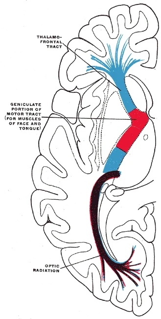
Figure 2Gray746
Long description
Diagram of the tracts in the internal capsule. Motor tract red. The sensory tract (blue) is not direct, but formed of neurons receiving impulses from below in the thalamus and transmitting them to the cortex. The optic radiation (occipitothalamic) is shown in violet.

Figure 2: Diagram of the tracts in the internal capsule (motor tract red).
The internal capsule is a dense band of white matter located between the lentiform nucleus laterally and the caudate nucleus and thalamus medially. It appears as a bright, V-shaped structure in this staining. The anterior limb lies between the caudate nucleus and the lentiform nucleus. The posterior limb lies between the thalamus and the lentiform nucleus. The genu is the bend connecting these two limbs. The retrolenticular part lies posterior to the lentiform nucleus. The sublenticular part lies inferior to the lentiform nucleus. The lentiform nucleus consists of the putamen and globus pallidus. The caudate nucleus has a head, body, and tail. The thalamus is the large oval mass medial to the posterior limb. The internal capsule carries fibres between the cortex and the brainstem or spinal cord. It is a critical pathway for motor and sensory information. Damage to this area causes significant neurological deficits. The anterior limb contains frontopontine fibres and thalamocortical fibres. The genu contains corticobulbar fibres. The posterior limb contains corticospinal fibres and thalamocortical sensory fibres. The retrolenticular part contains optic radiations. The sublenticular part contains auditory radiations. Understanding this anatomy is essential for localising lesions. The lenticulostriate arteries penetrate the anterior limb and genu. The anterior choroidal artery supplies the posterior limb. This vascular supply is important for understanding stroke patterns. The internal capsule is compact and vulnerable to compression. Oedema or haemorrhage in this region can disrupt multiple fibre tracts simultaneously. The lentiform nucleus is lateral to the posterior limb. The caudate nucleus is medial to the anterior limb. The thalamus is medial to the posterior limb. This spatial arrangement is consistent across individuals. The internal capsule is a key landmark in neuroimaging. It is easily identified on CT and MRI scans. The V-shape is characteristic. The angle of the V varies with the level of section. At the level of the centrum semiovale, the limbs are more separated. At the level of the midbrain, the posterior limb is narrower. The internal capsule is surrounded by white matter tracts. These tracts connect different parts of the brain. The internal capsule is the main conduit for these connections. It is essential for voluntary movement and sensation. Lesions here cause contralateral hemiparesis or hemiplegia. The severity depends on the extent of the damage. The internal capsule is a vital structure in neuroanatomy. Its study is fundamental for clinical practice. Each part has specific functions and connections. The anterior limb connects the frontal lobe to the thalamus. The genu connects the cortex to the brainstem. The posterior limb connects the cortex to the spinal cord. The retrolenticular part connects the lateral geniculate nucleus to the visual cortex. The sublenticular part connects the medial geniculate nucleus to the auditory cortex. This organisation is precise and orderly. The internal capsule is a masterpiece of neural design. It efficiently packs millions of fibres into a small space. The density of fibres is high. This makes it susceptible to ischaemic damage. The vascular supply is terminal in many cases. Collateral circulation is limited. This is why strokes here are devastating. The internal capsule is a critical junction. It integrates motor and sensory information. It is essential for coordinated movement. The anterior limb is shorter than the posterior limb. The genu is the narrowest part. The posterior limb is the longest. The retrolenticular part is small but important. The sublenticular part is also small. These proportions are consistent. The internal capsule is a key structure in the brain. Its anatomy is well-defined. The internal capsule is a vital part of the central nervous system. Its study is rewarding and necessary. The internal capsule is a gateway to the body. It controls movement and sensation. Understanding them is key to understanding brain function. The internal capsule is the road through this landscape. It is busy and important. The density of fibres is evident. The internal capsule is a real structure. It is not just a concept. The internal capsule is a critical structure. Its importance cannot be overstated. The internal capsule is a vital part of neuroanatomy. Use it well. [1]
The internal capsule is a dense band of white matter located within the cerebral hemisphere. It serves as the primary conduit for ascending and descending neural pathways. In a coronal cross-section, the internal capsule appears as a V-shaped or angular structure. This shape is defined by its relationship with the basal ganglia nuclei. The anterior limb lies between the caudate nucleus and the putamen. The posterior limb lies between the thalamus and the lentiform nucleus. The genu is the knee-like bend connecting these two limbs. The retrolenticular part lies posterior to the thalamus and lentiform nucleus. The sublenticular part lies inferior to the lentiform nucleus.
The anterior limb of the internal capsule is situated medial to the putamen and lateral to the head of the caudate nucleus. The genu is the anterior angle where the anterior and posterior limbs meet. The posterior limb is the largest part, lying between the thalamus medially and the posterior part of the lentiform nucleus laterally. The retrolenticular part is posterior to the thalamus and lentiform nucleus. The sublenticular part extends inferiorly from the retrolenticular part. These structures are arranged to facilitate the passage of fibres to and from the cerebral cortex.
The anterior limb contains frontopontine fibres and thalamocortical fibres. These fibres connect the frontal lobe to the pons and the thalamus to the frontal cortex. The genu contains corticobulbar fibres. These fibres travel from the motor cortex to the brainstem nuclei. The posterior limb contains the corticospinal tract. This is the major motor pathway. It carries fibres from the primary motor cortex to the spinal cord. The posterior limb also contains thalamocortical fibres. These fibres connect the thalamus to the parietal, temporal, and occipital lobes. The retrolenticular part contains optic radiations. These fibres carry visual information from the lateral geniculate nucleus to the visual cortex. The sublenticular part contains auditory radiations. These fibres carry auditory information from the medial geniculate nucleus to the auditory cortex.
The anterior limb and genu are supplied by the recurrent artery of Heubner and lenticulostriate branches. The posterior limb is supplied by the anterior choroidal artery and lenticulostriate branches. The retrolenticular and sublenticular parts are supplied by the anterior choroidal artery and branches of the posterior cerebral artery. This vascular supply is critical for maintaining the integrity of the white matter tracts.
DefinitionInternal Capsule
A compact band of projection fibres in the cerebrum that connects the cerebral cortex with subcortical structures, including the brainstem and spinal cord.
The arrangement of fibres in the internal capsule is somatotopic. The corticospinal tract fibres are arranged such that those for the upper limb are anterior to those for the lower limb. The corticobulbar fibres are located in the genu. The thalamocortical fibres are distributed throughout the posterior limb. This organization allows for precise localisation of lesions. A lesion in the genu affects facial and tongue movement. A lesion in the posterior limb affects arm and leg movement. A lesion in the retrolenticular part affects vision. A lesion in the sublenticular part affects hearing.
| Part | Fibres | Function |
|---|---|---|
| Anterior limb | Frontopontine, Thalamocortical | Frontal lobe connections |
| Genu | Corticobulbar | Brainstem motor nuclei |
| Posterior limb | Corticospinal, Thalamocortical | Spinal cord motor, Sensory |
| Retrolenticular part | Optic radiations | Vision |
| Sublenticular part | Auditory radiations | Hearing |
The internal capsule is a dense band of white matter that serves as the primary conduit for information travelling between the cerebral cortex and the rest of the central nervous system. It is situated deep within the brain, wedged between the thalamus and the basal ganglia. To understand its connections, one must visualise the brain in a coronal section. This figure shows a coronal view of the basal ganglia. The internal capsule appears as a V-shaped or inverted V-shaped structure of white matter. The anterior limb lies between the caudate nucleus and the lentiform nucleus. The posterior limb lies between the thalamus and the lentiform nucleus. The genu is the bend where the anterior and posterior limbs meet. The retrolenticular part lies posterior to the lentiform nucleus. The sublenticular part lies inferior to the lentiform nucleus. These regions carry specific afferent and efferent fibres.
Afferent connections bring sensory information to the cortex. Fibres from the thalamus travel through the posterior limb. These are thalamocortical fibres. They carry sensory data to the primary sensory cortex. The retrolenticular part carries optic radiations. These fibres connect the lateral geniculate nucleus to the visual cortex. The sublenticular part carries auditory radiations. These fibres connect the medial geniculate nucleus to the auditory cortex. Without these afferent pathways, the cortex would receive no sensory input.
Efferent connections carry motor commands from the cortex. Fibres from the motor cortex travel through the genu and anterior limb. These are corticobulbar and corticospinal fibres. The genu contains corticobulbar fibres. These fibres innervate cranial nerve nuclei in the brainstem. The anterior limb contains frontopontine fibres. These fibres connect the frontal lobe to the pons. The posterior limb contains the corticospinal tract. These fibres descend to the spinal cord. They control voluntary movement of the body. The posterior limb also contains corticorubral fibres. These fibres connect to the red nucleus.
The internal capsule is narrow and vulnerable. A small lesion here can cause significant deficits. This is because many fibres are packed into a small space. The anterior limb carries frontopontine fibres. The genu carries corticobulbar fibres. The posterior limb carries corticospinal fibres. Damage to the posterior limb causes contralateral hemiparesis. This is weakness on the opposite side of the body. Damage to the genu causes facial weakness. This is because the corticobulbar fibres are affected.
The lentiform nucleus lies lateral to the internal capsule. It consists of the putamen and the globus pallidus. The caudate nucleus lies medial to the anterior limb. The thalamus lies medial to the posterior limb. The claustrum lies lateral to the lentiform nucleus. The external capsule separates the claustrum from the lentiform nucleus. The extreme capsule lies lateral to the claustrum. These structures form the basal ganglia circuitry. The internal capsule is the output pathway for this circuitry. It integrates motor planning with execution.
DefinitionInternal Capsule
A dense white matter structure between the thalamus and basal ganglia carrying afferent and efferent fibres.
| Region | Location | Primary Fibres |
|---|---|---|
| Anterior Limb | Between caudate and lentiform | Frontopontine |
| Genu | Bend between limbs | Corticobulbar |
| Posterior Limb | Between thalamus and lentiform | Corticospinal |
| Retrolenticular Part | Posterior to lentiform | Optic radiations |
| Sublenticular Part | Inferior to lentiform | Auditory radiations |
The internal capsule is a dense band of white matter fibres that serves as the primary conduit for motor and sensory pathways between the cerebral cortex and the brainstem. Its blood supply is critical because damage to these vessels results in significant neurological deficits, commonly referred to as capsular strokes. The vascular architecture is segmented, with different arterial branches supplying specific limbs of the capsule. The internal carotid arteries along with the vertebral arteries are the two primary suppliers of blood to the human brain. [1]
The anterior limb of the internal capsule is supplied by branches from the anterior cerebral artery and the anterior communicating artery. These vessels provide oxygenated blood to the front part of the capsule, which contains frontopontine fibres and thalamocortical fibres. The genu, the knee-like bend of the capsule, receives its blood supply from the recurrent artery of Heubner, a branch of the anterior cerebral artery. This artery is crucial for supplying the head of the caudate nucleus and [1]
The posterior limb of the internal capsule, which carries the corticospinal tract, is supplied by the lenticulostriate arteries. These are small, deep penetrating branches that arise from the middle cerebral artery. This type of stroke is known as a capsular stroke. The blood supply to the retrolenticular part of the internal capsule comes from the posterior cerebral artery and t [1]
The corona radiata consists of fibres that fan out from the internal capsule to the cerebral cortex. These fibres are supplied by the same lenticulostriate arteries that supply the internal capsule. The anterior cerebral arteries join together to form an anastomosis called the anterior communicating artery. The blood supply of the capsule is thus a mosaic of small, deep arteries that are susceptible to occlusion or rupture. [1]
Clinical pearlCapsular Stroke
A capsular stroke is often caused by hypertension affecting the lenticulostriate arteries, leading to infarction in the posterior limb of the internal capsule.
The clinical presentation of a capsular stroke depends on the specific part of the capsule affected. Damage to the posterior limb typically results in contralateral hemiparesis or hemiplegia. The blood supply of the capsule is therefore a key area of study for understanding stroke pathophysiology. [1]
| Region | Arterial Supply | Clinical Significance |
|---|---|---|
| Anterior Limb | Anterior cerebral artery, anterior communicating artery | Frontopontine fibre damage |
| Genu | Recurrent artery of Heubner | Facial motor fibre damage |
| Posterior Limb | Lenticulostriate arteries (from MCA) | Corticospinal tract damage |
| Retrolenticular Part | Posterior cerebral artery | Visual and sensory fibre damage |
The lenticulostriate arteries are small and lack collateral circulation, making them highly susceptible to occlusion. This is why capsular strokes are common in patients with chronic hypertension. The blood supply of the internal capsule is thus a critical determinant of neurological outcome in stroke patients.
DefinitionLenticulostriate Arteries
Small penetrating branches of the middle cerebral artery that supply the basal ganglia and internal capsule.
Understanding the blood supply of the internal capsule is essential for localizing lesions in neuroimaging. The specific pattern of deficit can help identify which arterial branch is affected. For example, pure motor hemiparesis suggests a lesion in the posterior limb supplied by the lenticulostriate arteries.
The internal capsule is a critical white matter structure in the brain that serves as the primary conduit for motor and sensory information. It is located between the basal ganglia and the thalamus. Understanding its function requires understanding its anatomy, as the specific location of a lesion determines the clinical deficit. The internal capsule is not a single uniform band but is divided into distinct parts, each carrying different types of neural pathways.
The anterior limb lies between the caudate nucleus medially and the putamen laterally. The genu is the knee-like bend where the anterior and posterior limbs meet. A small region called the retrolenticular part lies posterior to the globus pallidus and putamen, while the sublenticular part lies inferior to the lentiform nucleus.
DefinitionInternal Capsule
A compact bundle of projection fibres that connects the cerebral cortex with the brainstem and spinal cord, passing between the thalamus and the basal ganglia.
The motor functions are primarily carried by the corticospinal and corticobulbar tracts. These fibres travel through the posterior limb and the genu, respectively. Damage to these areas results in contralateral weakness or paralysis. The sensory functions are carried by the thalamocortical radiations, which also pass through the posterior limb. These fibres transmit sensory information from the thalamus to the somatosensory cortex. Lesions here cause contralateral loss of sensation.
| Tract | Location | Primary Function |
|---|---|---|
| Corticospinal tract | Posterior limb | Voluntary motor control of the body |
| Corticobulbar tract | Genu and anterior part of posterior limb | Voluntary motor control of the head and neck |
| Thalamocortical radiations | Posterior limb | Sensory information transmission to the cortex |
| Frontopontine fibres | Anterior limb | Connection between frontal lobe and pons |
| Parietopontine fibres | Retrolenticular part | Connection between parietal lobe and pons |
The genu contains corticobulbar fibres. The posterior limb contains the corticospinal tract and thalamocortical sensory fibres. This organisation means that a single lesion can affect multiple functions depending on its size and exact location. For example, a small lacunar infarct in the posterior limb may cause pure motor hemiparesis, while a larger lesion may also affect sensory pathways.
Clinical pearlClinical Pearl
A lesion in the internal capsule often causes dense contralateral hemiplegia because the fibres are densely packed, unlike in the cerebral cortex where they are more dispersed.
The lenticulostriate arteries, branches of the middle cerebral artery, supply the anterior limb and the genu. This vascular supply is important for understanding the patterns of stroke. This is because the optic radiations pass through the retrolenticular part, which is supplied by this artery.
Exam trapCommon Trap
Do not confuse the internal capsule with the external capsule. The external capsule is lateral to the putamen and contains different fibres, primarily involved in limbic and autonomic functions, not primary motor or sensory pathways.
In summary, the internal capsule is a vital highway for neural communication. Its compact structure means that even small lesions can have significant clinical consequences. The specific deficits depend on which part of the capsule is affected. Understanding this anatomy is essential for localising neurological lesions.
The internal capsule is a critical white matter pathway that contains important ascending and descending tracts. Lesions in this area can result in significant neurological deficits due to the disruption of these neural pathways. The internal capsule is divided into anterior limb, genu, and posterior limb, each containing different fiber tracts that, when damaged, produce distinct clinical syndromes.
The internal capsule lies medial to the lentiform nucleus (putamen and globus pallidus) and lateral to the thalamus and caudate nucleus. The anterior limb separates the head of the caudate from the lentiform nucleus, while the posterior limb separates the thalamus from the globus pallidus and putamen. The genu is the bend between these two limbs where the corticobulbar fibers descend to innervate the cranial nerves.
Lesions in the internal capsule most commonly result from ischemic stroke, hemorrhage, or traumatic injury. The clinical presentation depends on the specific location and extent of the damage. Because the internal capsule contains a high concentration of motor fibers, even small lesions can produce significant weakness. The corticospinal fibers are somatotopically organized, with fibers to the leg located most posteriorly and medially, while fibers to the face are located anteriorly and laterally. This explains why leg weakness may be more prominent in posterior limb lesions affecting the lateral aspect.
The internal capsule is a dense band of white matter situated between the lentiform nucleus and the thalamus/caudate nucleus. It serves as the primary conduit for motor and sensory pathways.
The internal capsule is shaped like a V when viewed from above, with an anterior limb, a genu, and a posterior limb. The genu is the bend where the anterior and posterior limbs meet. The posterior limb lies between the lentiform nucleus and the thalamus. It carries corticospinal, corticobulbar, and sensory fibres.
DefinitionInternal Capsule
A compact bundle of projection fibres connecting the cerebral cortex to the brainstem and spinal cord, located between the basal ganglia and thalamus.
The genu contains corticobulbar fibres. These fibres innervate the cranial nerve nuclei in the brainstem. The posterior limb carries the bulk of motor and sensory information. The corticospinal tract runs through the posterior limb. It descends towards the spinal cord.
| Segment | Location | Primary Contents |
|---|---|---|
| Anterior Limb | Between caudate and lentiform nucleus | Frontopontine fibres, thalamocortical fibres |
| Genu | Bend between anterior and posterior limbs | Corticobulbar fibres |
| Posterior Limb | Between lentiform nucleus and thalamus | Corticospinal fibres, sensory fibres |
Lesions of the internal capsule cause severe contralateral deficits. A small lesion can produce significant weakness because the fibres are densely packed. This is often called a "capsular warning syndrome" if transient.
Exam trapCapsular Hemiparesis
Do not confuse internal capsule lesions with cortical lesions. Internal capsule lesions cause pure motor or pure sensory deficits without cortical signs like aphasia or neglect, as the cortex itself is spared.
The corticospinal tract is particularly vulnerable. Damage here results in contralateral hemiplegia. The face, arm, and leg are affected equally. This is because the fibres are arranged somatotopically but are tightly bundled. The genu carries fibres to the face. The posterior limb carries fibres to the body.
Clinical pearlPure Motor Stroke
A lacunar infarct in the posterior limb of the internal capsule causes pure motor hemiparesis. This is a classic exam presentation for small vessel disease.
Sensory loss may also occur. If the thalamocortical fibres are damaged, contralateral hemianesthesia results. The patient loses sensation on the opposite side of the body. Vision and hearing are typically spared as their pathways do not run through the internal capsule in the same way.
NoteVascular Supply
The lenticulostriate arteries supply the internal capsule. These are small branches of the middle cerebral artery. They are prone to hypertensive damage.
Understanding the anatomy helps predict outcomes. Anterior limb lesions are rare and may cause mild cognitive changes. Genu lesions affect facial movement. Posterior limb lesions are the most common cause of stroke-related disability. Rehabilitation focuses on retraining these dense pathways.
DefinitionLenticulostriate Arteries
Small penetrating arteries arising from the middle cerebral artery that supply the basal ganglia and internal capsule.
The internal capsule is a critical junction. It integrates motor commands and sensory feedback. Damage here disrupts the entire side of the body. Early recognition is vital for management.
Students frequently confuse the internal capsule with the external capsule or the claustrum due to their close anatomical proximity in coronal sections. A critical error is assuming the internal capsule is a single uniform structure; it is actually divided into distinct parts with specific vascular supplies and functional contents. Misidentifying the genu as the posterior limb leads to incorrect localisation of motor deficits. Another common mistake is overlooking the anterior limb's connection to the frontopontine fibres and thalamocortical projections, leading to an incomplete understanding of frontal lobe syndromes. Students often fail to distinguish between the anterior and posterior limbs based on their relationship to the lentiform nucleus, which is essential for interpreting stroke patterns. Confusing the retrolenticular part with the sublenticular part can result in errors when assessing visual or auditory pathway involvement in capsular strokes. Furthermore, assuming that all motor fibres are located in the posterior limb ignores the significant motor contribution of the genu, particularly for the face and upper limb. Finally, neglecting the vascular anatomy, specifically the lenticulostriate arteries, leads to a failure to predict the extent of lacunar infarcts.
Knowledge of the internal capsule is vital at the bedside for localising lesions causing hemiparesis. In the operating theatre, neurosurgeons must navigate around this structure to avoid causing permanent motor or sensory deficits during tumour resection. On imaging, particularly MRI, identifying the characteristic 'V' shape of the internal capsule helps in diagnosing acute ischaemic strokes. In examinations, students are tested on the specific tracts contained within each part of the capsule and the clinical consequences of their disruption. Understanding the blood supply from the lenticulostriate arteries is crucial for predicting the size and location of lacunar infarcts. This knowledge allows clinicians to correlate imaging findings with clinical symptoms, such as pure motor hemiparesis or sensorimotor stroke. It also aids in prognostication, as lesions in the posterior limb often have a different recovery trajectory compared to those in the anterior limb.
DefinitionInternal Capsule
A dense band of white matter in the brain that carries information past the basal ganglia, separating the caudate nucleus and thalamus from the lentiform nucleus.
The internal capsule is a critical white matter structure located deep within the cerebral hemisphere. It serves as the primary conduit for ascending and descending axons connecting the cerebral cortex with the brainstem and spinal cord. The structure is shaped like a bent V or an inverted V, depending on the anatomical plane of view. In coronal sections, it appears as a V-shape with the apex pointing downwards. The internal capsule is divided into four main parts: the anterior limb, the genu, the posterior limb, and the retrolenticular part. Each part contains specific fibre tracts and has a distinct vascular supply.
| Part | Location | Key Fibres | Vascular Supply |
|---|---|---|---|
| Anterior limb | Between caudate nucleus and lentiform nucleus | Frontopontine fibres, thalamocortical fibres | Recurrent artery of Heubner |
| Genu | Bend between anterior and posterior limbs | Corticobulbar fibres | Lenticulostriate arteries |
| Posterior limb | Behind lentiform nucleus | Corticospinal fibres, thalamocortical fibres | Lenticulostriate arteries |
| Retrolenticular part | Behind posterior limb | Optic radiations, auditory radiations | Posterior cerebral artery branches |
The anterior limb lies between the head of the caudate nucleus medially and the lentiform nucleus laterally. It contains frontopontine fibres that connect the frontal lobe to the pons. It also carries thalamocortical fibres projecting from the anterior nucleus of the thalamus to the cingulate gyrus. The genu is the knee-like bend connecting the anterior and posterior limbs. It primarily contains corticobulbar fibres, which innervate the cranial nerve nuclei in the brainstem. These fibres are crucial for controlling muscles of the face, head, and neck. The posterior limb is the largest part, situated behind the lentiform nucleus. It contains the corticospinal tract, which is responsible for voluntary motor control of the body. It also carries thalamocortical fibres from the ventral posterior nucleus of the thalamus to the somatosensory cortex. The retrolenticular part lies behind the posterior limb and contains the optic radiations, which carry visual information from the lateral geniculate nucleus to the visual cortex.
Internal Capsule Lesion Analysis
- Identify DeficitPatient presents with motor or sensory loss
- Question: Is face involved?
Yes
- Check GenuCorticobulbar fibres affected
No
- Check Posterior LimbCorticospinal fibres affected
- Question: Is vision affected?
Yes
- Check Retrolenticular PartOptic radiations affected
No
- Standard Capsular StrokeAnterior limb or main posterior limb
Vascular supply is a key concept for understanding clinical outcomes. The anterior limb and genu are supplied by the recurrent artery of Heubner and the lenticulostriate arteries. The posterior limb and retrolenticular part are supplied by the lenticulostriate arteries and branches of the posterior cerebral artery. Occlusion of the lenticulostriate arteries, often due to hypertension, leads to lacunar infarcts. These small strokes can cause pure motor hemiparesis, pure sensory stroke, or sensorimotor stroke. The size of the infarct is limited by the small calibre of these penetrating arteries. Understanding this vascular anatomy helps in predicting the extent of neurological deficits.
Exam trapVascular Confusion
Do not assume the middle cerebral artery directly supplies the internal capsule; it is the lenticulostriate branches that penetrate the capsule.
Clinical correlation is essential for mastering this topic. A lesion in the posterior limb typically causes contralateral hemiparesis affecting the face, arm, and leg equally. This is because the fibres are densely packed. A lesion in the genu may cause isolated facial weakness. Lesions in the retrolenticular part can cause contralateral homonymous hemianopia. The anterior limb lesions are less common but can cause personality changes or executive dysfunction due to disruption of frontopontine and thalamocortical connections. Recognising these patterns allows for accurate localisation of the lesion.
Clinical pearlClinical Pearl
Pure motor hemiparesis is the most common lacunar syndrome, localising to the posterior limb of the internal capsule.
In summary, the internal capsule is a vital white matter structure. It contains motor, sensory, and association fibres. Its compact arrangement means that small lesions can cause significant deficits. Understanding its anatomy, vascular supply, and clinical correlations is essential for medical students. This knowledge forms the basis for diagnosing and managing cerebrovascular diseases.
The internal capsule is divided into anterior limb, genu, posterior limb, and retrolenticular part. The anterior limb contains frontopontine and thalamocortical fibres. The genu contains corticobulbar fibres for cranial nerve control. The posterior limb contains the corticospinal tract and sensory fibres. The retrolenticular part contains optic and auditory radiations. Lenticulostriate arteries supply the anterior limb, genu, and posterior limb. Occlusion of lenticulostriate arteries causes lacunar infarcts. Lesions cause contralateral motor and sensory deficits.
Answer each one in your head first, then open the answer.
Which part of the internal capsule contains corticospinal fibres?
Show answerHide answer
Posterior limbWhat is the primary vascular supply to the genu of the internal capsule?
Show answerHide answer
Lenticulostriate arteriesWhich fibres are located in the anterior limb of the internal capsule?
Show answerHide answer
Frontopontine fibresWhat clinical deficit is associated with a lesion in the retrolenticular part?
Show answerHide answer
Contralateral homonymous hemianopiaWhat type of stroke is commonly caused by lenticulostriate artery occlusion?
Show answerHide answer
Lacunar infarct
- Betts JG, Young KA, Wise JA, et al. Circulatory Pathways. OpenStax, Anatomy and Physiology 2e (CC BY-NC-SA 4.0). Accessed .
Figure credits
Educational content for medical students and doctors. It is not a substitute for clinical judgement or local guidelines.