Ventricular system
The ventricular system is a network of cavities producing and circulating cerebrospinal fluid.
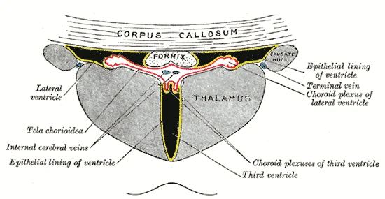
Figure 1Gray723
Long description
Coronal section of lateral and third ventricles.

Figure 1: Coronal section of lateral and third ventricles.
The ventricular system consists of four interconnected cavities within the brain that develop from the original hollow space within the neural tube, the central canal[1]. These cavities produce and circulate cerebrospinal fluid (CSF), which filters blood through specialized membranes called choroid plexuses[1]. The system includes the lateral ventricles (first and second), third ventricle, cerebral aqueduct, and fourth ventricle[1].
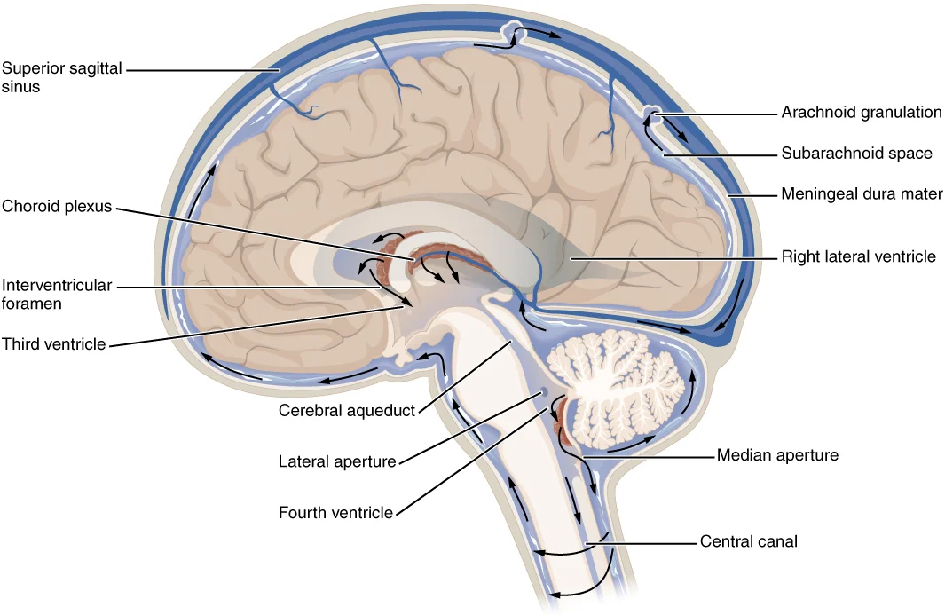
Figure 21317 CFS Circulation
Long description
The choroid plexus in the four ventricles produce CSF, which is circulated through the ventricular system and then enters the subarachnoid space through the median and lateral apertures. The CSF is then reabsorbed into the blood at the arachnoid granulations, where the arachnoid membrane emerges int

Figure 2: The choroid plexus in the four ventricles produce CSF, which is circulated through the ventricular system and then enters the subarachnoid space through the median and lateral apertures.
The lateral ventricles are deep within the cerebrum and take on a C-shaped formation due to the constraints of the skull[1]. They are connected to the third ventricle by two openings called the interventricular foramina (of Monro)[1]. The third ventricle is the space between the left and right sides of the diencephalon, bounded by the medial walls of the hypothalamus and thalamus[1]. The two thalami often touch in the center as the massa intermedia, surrounded by the third ventricle[1].
The third ventricle opens into the cerebral aqueduct (of Sylvius) that passes through the midbrain[1]. The tectum forms the roof and tegmentum forms the floor of the cerebral aqueduct[1]. The aqueduct opens into the fourth ventricle, which is the space between the cerebellum and the pons and upper medulla[1]. The floor of the fourth ventricle is the dorsal surface of the pons and upper medulla[1], and it narrows into the central canal of the spinal cord[1].
The ventricular system connects to the subarachnoid space through three apertures: the single median aperture (of Magendie) and the pair of lateral apertures (of Luschka)[1]. These allow CSF to flow through the ventricles and around the outside of the CNS[1]. CSF is produced within the ventricles by choroid plexuses, which consist of ependymal cells surrounding blood capillaries that filter the blood to make CSF[1]. The fluid is essentially water, small molecules, and electrolytes[1].
From the lateral ventricles, CSF flows into the third ventricle, then through the cerebral aqueduct into the fourth ventricle[1]. From the fourth ventricle, CSF can continue down the central canal of the spinal cord, but most enters the subarachnoid space through the median and lateral apertures[1].
Within the subarachnoid space, CSF flows around all of the CNS, picking up metabolic wastes and acting as a liquid cushion for the brain and spinal cord[1]. The arachnoid granulations are outpocketings of the arachnoid membrane into the dural sinuses that allow CSF to be reabsorbed into the blood, removing metabolic wastes from the CNS[1]. This reabsorption occurs along with the normal circulation of blood through the jugular veins[1].
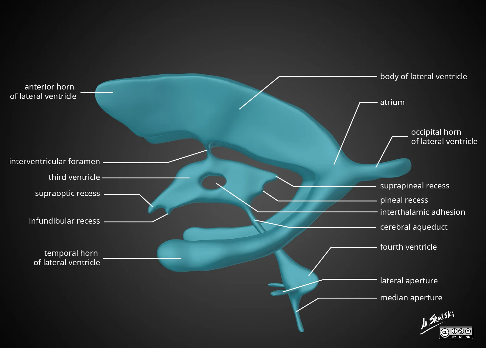
Figure 3Brain-ventricle-anatomy-diagram
Long description
Diagram of the anatomy of the ventricular system of the brain

Figure 3: Diagram of the anatomy of the ventricular system of the brain.
The ventricular system is a set of interconnected, fluid-filled cavities within the brain, derived from the lumen of the embryonic neural tube. It contains cerebrospinal fluid (CSF), which is produced by the choroid plexuses found in all four ventricles. These plexuses appear in dissection as soft, fuzzy, pink structures, and CSF production is tied to the pulse of cardiovascular circulation because the fluid is made from components extracted from the blood. The system comprises two lateral ventricles, the third ventricle, and the fourth ventricle, each connected by narrow channels. [1]
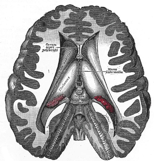
Figure 4Gray716
Long description
Dissection showing the ventricles of the brain.

Figure 4: Dissection showing the ventricles of the brain.
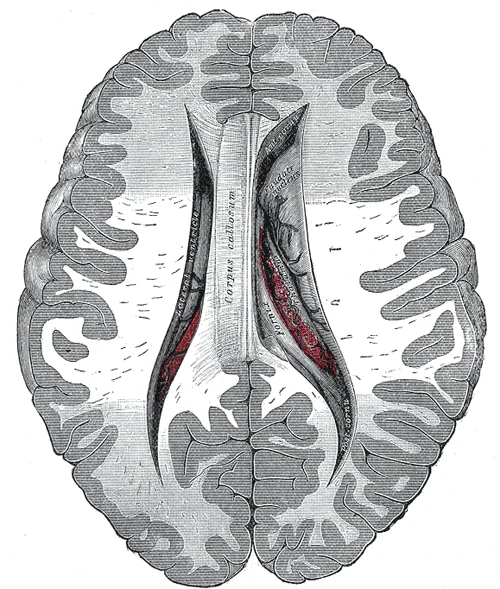
The two lateral ventricles, one in each cerebral hemisphere, are the largest cavities and are C-shaped, curving anteriorly, posteriorly, and inferiorly around the thalamus. Each lateral ventricle has a central body and three horns: the anterior (frontal) horn extends into the frontal lobe, the posterior (occipital) horn into the occipital lobe, and the inferior (temporal) horn into the temporal lobe.
Figure 5Gray737
Long description
Central part and anterior and posterior cornua of lateral ventricles exposed from above.

Figure 5: Central part and anterior and posterior cornua of lateral ventricles exposed from above.
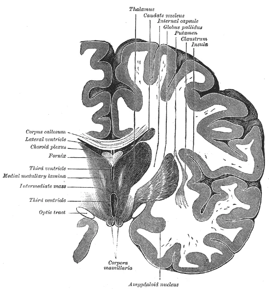
The third ventricle is a narrow, slit-like midline cavity situated within the diencephalon, between the two halves of the thalamus[1]. It is connected posteriorly to the fourth ventricle via the cerebral aqueduct (of Sylvius), a slender canal that runs through the midbrain. The fourth ventricle lies posterior to the pons and the upper half of the medulla oblongata, and anterior to the cerebellum. It narrows inferiorly to become the central canal of the spinal cord, which maintains the tube structure of the neural tube[1]. From the fourth ventricle, CSF also exits through three openings—the two lateral apertures (of Luschka) and the single median aperture (of Magendie)—into the subarachnoid space surrounding the brain and spinal cord.
Figure 6Gray718
Long description
Coronal section of brain through intermediate mass of third ventricle.

Figure 6: Coronal section of brain through intermediate mass of third ventricle.
In the lateral ventricles, the plexus lies along the floor of the central body and the inferior horn. In the third ventricle, it is in the roof; in the fourth, it is in the roof and its lateral recesses. The CSF circulates from the lateral ventricles through the interventricular foramina into the third ventricle, then via the cerebral aqueduct into the fourth ventricle, and out through the apertures into the subarachnoid space, where it cushions the CNS. The entire system is lined by ependymal cells, a type of glial cell that also contributes to CSF production.
Figure 7Gray749
Long description
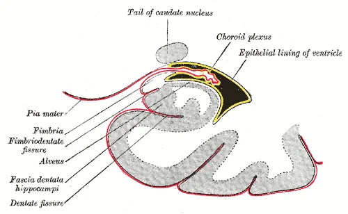
Coronal section of inferior horn of lateral ventricle. (Diagrammatic.)

Figure 7: Coronal section of inferior horn of lateral ventricle (Diagrammatic.).
The ventricular system is a set of interconnected, fluid-filled cavities within the brain, lined by ependyma and filled with cerebrospinal fluid (CSF). In cross-section, each ventricle shows a consistent internal organisation: an inner cavity, a cellular lining, and surrounding neural tissue that differs according to the ventricle’s location)Skip. The walls are formed by the brain parenchyma, and the cavities communicate with each other and with the subarachnoid space.
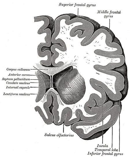
The lateral ventricles, one in each cerebral hemisphere, are the largest cavities. Each has a central body and three horns: anterior (frontal), posterior (occipital), and inferior (temporal). In a coronal cross-section through the body, the cavity appears as a slit-like space, bordered superiorly by the corpus callosum, laterally by the caudate nucleus, and inferiorly by the thalamus. The septum pellucidum forms the medial wall of the anterior horns. [2]
Figure 8Gray743
Long description
Coronal section through anterior cornua of lateral ventricles.

Figure 8: Coronal section through anterior cornua of lateral ventricles.
Figure 9Gray739
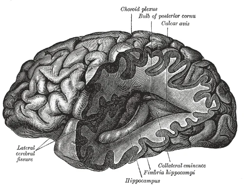
Long description
Posterior and inferior cornua of left lateral ventricle exposed from the side.

Figure 9: Posterior and inferior cornua of left lateral ventricle exposed from the side.
Figure 10Gray738
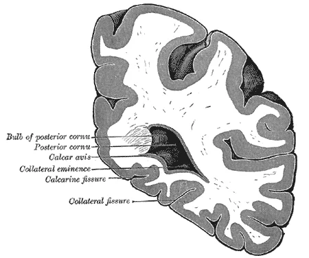
Long description
Coronal section through posterior cornua of lateral ventricle.

Figure 10: Coronal section through posterior cornua of lateral ventricle.
Figure 11Gray736
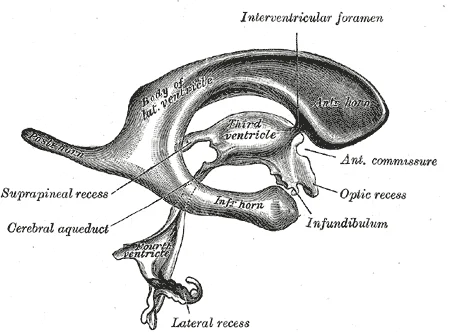
Long description
Drawing of a cast of the ventricular cavities, viewed from the side. (Retzius.)

Figure 11: Drawing of a cast of the ventricular cavities, viewed from the side (Retzius.).
The third ventricle is a narrow, midline cavity between the two thalami. In cross-section it is a vertical slit, with the massa intermedia (when present) crossing its cavity. Its roof is the tela choroidea, its floor is the hypothalamus, and its anterior wall is formed by the lamina terminalis. The interventricular foramina (of Monro) connect each lateral ventricle to the third ventricle at its anterosuperior corner. The third ventricle communicates posteriorly with the fourth ventricle via the cerebral aqueduct (of Sylvius), a narrow canal through the midbrain. [2]
Figure 12Gray750
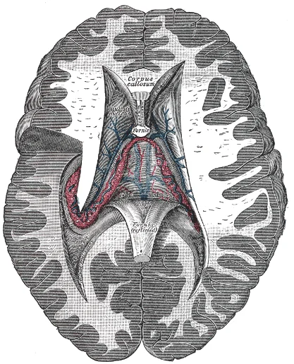
Long description
Tela chorioidea of the third ventricle, and the choroid plexus of the left lateral ventricle, exposed from above.

Figure 12: Tela chorioidea of the third ventricle, and the choroid plexus of the left lateral ventricle, exposed from above.
Figure 13Gray735
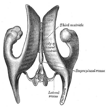
Long description
Drawing of a cast of the ventricular cavities, viewed from above. (Retzius.)

Figure 13: Drawing of a cast of the ventricular cavities, viewed from above (Retzius.).
The fourth ventricle lies in the posterior fossa, between the pons and medulla anteriorly and the cerebellum posteriorly. In cross-section it is diamond-shaped, with a roof formed by the superior and inferior medullary vela, and a floor (the rhomboid fossa) formed by the dorsal surfaces of the pons and medulla. It contains the choroid plexus and has three openings to the subarachnoid space: the median aperture (of Magendie) and the two lateral apertures (of Luschka). These allow CSF to exit the ventricular system. [2]
The ependyma, a single layer of ciliated cuboidal cells, lines the entire system. Beneath it lies a thin subependymal layer of glial cells. The ventricular cavities are not empty spaces but contain CSF under a normal pressure of about 10–15 cm H₂O in the lateral recumbent position (illustrative value, not from passages). Obstruction at any point—for example, at the cerebral aqueduct—causes upstream dilatation (hydrocephalus).
The surrounding gray matter differs by level. Around the lateral ventricles lie the caudate nucleus (medial to the anterior horn), the thalamus (inferior to the body), and the hippocampus (in the floor of the inferior horn). Around the third ventricle are the hypothalamic nuclei in its floor and the medial thalamic nuclei in its walls. Around the fourth ventricle are the cranial nerve nuclei in the floor, arranged in columns: motor nuclei medial, sensory nuclei lateral. The white matter tracts adjacent to the ventricles include the internal capsule (lateral to the thalamus and caudate) and the fornix (running along the medial wall of the lateral ventricle). In the midbrain, the cerebral aqueduct is surrounded by the periaqueductal gray matter, with the oculomotor nucleus (CN III) lying ventrally in the tegmentum.
Figure 14Gray708
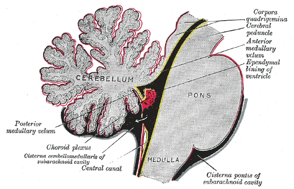
Long description
Scheme of roof of fourth ventricle. The arrow is in the foramen of Magendie.

Figure 14: Scheme of roof of fourth ventricle.
The ventricular system is a network of interconnected cavities within the brain that contains cerebrospinal fluid (CSF). This system serves as a conduit for CSF circulation and provides mechanical protection to the brain's delicate structures.
The lateral ventricles are the largest cavities and are located within each cerebral hemisphere. They communicate with the third ventricle through the interventricular foramina (of Monro). The third ventricle is a midline structure situated between the right and left thalami, connected to the fourth ventricle by the narrow cerebral aqueduct (of Sylvius). The fourth ventricle is diamond-shaped and lies between the pons and medulla anteriorly and the cerebellum posteriorly. It communicates with the subarachnoid space through three apertures: the median aperture (of Magendie) and the two lateral apertures (of Luschka).
CSF flows through this system in a specific direction. It is produced primarily by the choroid plexus in the lateral ventricles, then flows through the interventricular foramina into the third ventricle. From there, it passes through the cerebral aqueduct into the fourth ventricle, and finally exits into the subarachnoid space through the apertures mentioned. This circulation ensures that CSF bathes the entire central nervous system, providing buoyancy and protection.
The fourth ventricle is positioned between the pons, medulla, and cerebellum, with its apertures leading to the subarachnoid space.
DefinitionVentricular System
A network of interconnected cavities within the brain that contains cerebrospinal fluid and provides mechanical protection to neural structures.
The central nervous system (CNS) has a privileged blood supply established by the blood-brain barrier. This specialized arrangement protects the brain and spinal cord from toxins and pathogens in the bloodstream. The arterial blood to the brain comes from two main sources: the internal carotid arteries and the vertebral arteries. [1]
These arteries form the circle of Willis, which provides constant perfusion of the brain even if one vessel is blocked or narrowed. [1]
Branches from the vertebral arteries include the anterior spinal artery, which supplies the anterior aspect of the spinal cord along the anterior median fissure. [1]
After passing through the CNS, blood returns to the circulation through a series of dural sinuses and veins. It drains to the confluence of sinuses, along with the occipital sinuses and straight sinus, which then drains into the transverse sinuses.
The dura mater, a tough outer covering of the CNS, is anchored to the inner surface of the cranial and vertebral cavities. It surrounds the venous spaces known as the dural sinuses, which connect to the jugular veins where blood drains from the head and neck. Blood vessels nourishing the central nervous tissue lie between the pia mater and the nervous tissue itself. [1]
The relationship between these vessels and the ventricular system is clearly demonstrated, showing how the blood supply supports the CSF-producing structures and surrounding neural tissue. [1]
Figure 15Gray734
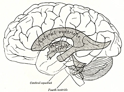
Long description
Scheme showing relations of the ventricles to the surface of the brain.

Figure 15: Scheme showing relations of the ventricles to the surface of the brain.
The ventricular system serves several critical functions in the central nervous system. Primarily, it produces and circulates cerebrospinal fluid (CSF), which acts as a cushion for the brain and spinal cord, providing buoyancy and protection against mechanical injury. CSF also serves as a medium for the transport of nutrients and waste products throughout the nervous system. [2]
The production occurs mainly in the choroid plexuses, which are vascular structures found in each ventricle. [2]
CSF flows through the ventricular system in a specific pathway. It begins in the lateral ventricles, moves through the interventricular foramina into the third ventricle, then passes through the cerebral aqueduct into the fourth ventricle. From the fourth ventricle, CSF exits through the median and lateral apertures to enter the subarachnoid space, where it bathes the entire brain and spinal cord. [2]
The ventricular system also plays a role in maintaining the brain's internal environment by regulating the composition of CSF. This includes maintaining proper ionic balance, pH, and preventing the accumulation of harmful substances. [2]
The ventricular system's design allows for the efficient distribution of CSF throughout the central nervous system, ensuring that all neural tissues receive adequate protection and nourishment. This system is particularly important given the brain's limited ability to store nutrients and its high metabolic demands. [2]
| Lesion Site | Clinical Presentation | Key Features |
|---|---|---|
| Lateral ventricle | Hydrocephalus | Increased intracranial pressure, headache, nausea, vomiting, papilledema [2] |
| Third ventricle | Hydrocephalus | Similar to lateral ventricle lesions, may also cause Parinaud syndrome if involving pineal region |
| Fourth ventricle | Hydrocephalus | Similar presentations, plus cerebellar signs if involving cerebellar peduncles |
| Cerebral aqueduct | Obstructive hydrocephalus | Classic triad: headache, vomiting, papilledema (Papilledema is the swelling of the optic disc due to increased intracranial pressure) |
| Foramina of Luschka and Magendie | Communicating hydrocephalus | Similar presentation to other hydrocephalus types, no obstruction within ventricular system |
| Choroid plexus tumors | Choroid plexus papilloma | Overproduction of CSF causing hydrocephalus, more common in children |
The third ventricle sits between the thalami, and the fourth ventricle is positioned posterior to the pons and medulla, anterior to the cerebellum. The cerebral aqueduct connects the third and fourth ventricles. Lesions at any of these sites can obstruct CSF flow, leading to hydrocephalus with increased intracranial pressure, headache, nausea, vomiting, and papilledema.
In clinical practice, patients with superior colliculus lesions may present with difficulty locating the source of sounds or coordinating head and eye movements toward stimuli.
In children, ventricular system lesions often present differently than in adults due to the flexibility of the developing skull. These tumors often present with macrocephaly in infants and increased intracranial pressure in older children. In contrast, elderly patients with ventricular system lesions may experience more rapid deterioration due to reduced cerebral compliance and pre-existing atrophy.
Lesions affecting the tectal plate, including the colliculi, may also present with Parinaud syndrome when involving the pretectal area, characterized by upward gaze palsy, convergence-retraction nystagmus, and light-near dissociation of the pupillary reflex.
Clinical pearlVentricular Walls
The walls of the third ventricle are formed by the diencephalon. The diencephalon constitutes the walls of the third ventricle and is located deep beneath the cerebrum. [2]
Clinical pearlMidbrain Canal
The cerebral aqueduct is the canal that passes through the center of the midbrain. This narrow channel connects the third and fourth ventricles and is surrounded by the tectum above and tegmentum below. [2]
Clinical pearlMidbrain Boundaries
These regions are named from the Latin words for roof and floor respectively, with the aqueduct passing through their center. [2]
Clinical pearlDiencephalon Components
The diencephalon includes the thalamus, hypothalamus, epithalamus (containing the pineal gland), and subthalamus. The thalamus and hypothalamus are the two major regions, while the epithalamus and subthalamus contain additional important structures. [2]
Exam trapAnatomical Terminology
Dorsal means "back" while ventral means "belly," but in neuroanatomy, they may correspond to posterior and anterior respectively. [2]
Clinical pearlCerebral Division
This deep cleft separates the two distinct halves of the cerebrum and contains the falx cerebri. [2]
Clinical pearlCortical Folding
This extensive folding allows for increased cortical surface area while accommodating the constraints of skull size during birth. [2]
Clinical pearlLobe Boundaries
These sulci serve as important anatomical landmarks for identifying the major cortical regions. [2]
Exam trapOccipital Lobe Borders
Its boundaries are primarily defined functionally rather than by distinct sulcal landmarks. [2]
Clinical pearlBrain Regions
The cerebrum is the largest portion and contains the cerebral cortex and subcortical nuclei. [2]
Clinical pearlCircle of Willis
This arterial ring can maintain perfusion of the brain even if narrowing or blockage limits flow through one part of the system. [1]
Clinical pearlDural Infoldings
There are infoldings of the dura that fit into large crevasses of the brain, including a tent-like structure between the occipital lobes and cerebellum, and another surrounding the pituitary gland. The dura also surrounds and supports the venous sinuses that drain blood from the brain. [1]
A common mistake is confusing the meningeal layers and their functions. The dura mater is a thick fibrous layer that serves as a strong protective sheath over the entire brain and spinal cord, anchored to the inner surface of the cranium and vertebral cavity. Students often mistakenly believe the pia mater is the outermost layer when in fact it is the innermost membrane, a thin fibrous layer that follows the convolutions of the brain and extends into every sulcus. Another error is misunderstanding the order of the meningeal layers, with the pia mater being the innermost layer, directly adjacent to the surface of the CNS. [1]
Students frequently confuse the functions of the cerebrum and cerebellum. The cerebellum, accounting for approximately 10 percent of the brain's mass, is primarily responsible for comparing motor commands from the cerebrum with sensory feedback to coordinate movement, not for initiating voluntary movements as many believe. The pons, often misidentified as simply a bridge structure, actually serves as the main connection between the cerebellum and brain stem while containing gray matter that processes cranial nerve information. [2]
A significant error is misunderstanding the composition of the brain stem. The brain stem consists of the midbrain, pons, and medulla, with the medulla containing significant white matter continuous with the spinal cord. Students often fail to recognize that the reticular formation, a diffuse gray matter region throughout the brain stem, is responsible for regulating sleep and wakefulness. Another common mistake is confusing the primary somatosensory cortex location - it is in the postcentral gyrus (Brodmann's areas 1, 2, and 3) posterior to the central sulcus, not anterior to it. [2]
Exam trapMeninges confusion
The pia mater is the innermost meningeal layer, not the outermost. It extends into all brain sulci and forms the filum terminale.
DefinitionCauda equina
The bundle of spinal nerves resembling a horse's tail that results from nerves growing with the vertebral column while the spinal cord remains relatively short after early childhood.
Knowledge of the ventricular system and surrounding structures is essential for performing a lumbar puncture, a common procedure to withdraw cerebrospinal fluid (CSF). The procedure is performed at the upper lumbar region where the spinal cord does not extend, allowing insertion of a needle through the dura and arachnoid layers without damaging the spinal cord tissue. Understanding the meningeal layers is crucial for this procedure, as the dura mater provides a strong protective barrier that must be penetrated.
In the operating theatre, knowledge of the brain stem components is vital for neurosurgical procedures. The brain stem, composed of the midbrain, pons, and medulla, contains control centers for cardiovascular and respiratory systems, making it a critical area to preserve during surgery. The pons, as the main connection between the cerebellum and brain stem, is particularly important in procedures involving the posterior fossa. The cerebellum's role in coordinating movement means that damage during surgery can result in significant motor deficits. [2]
On imaging studies, understanding the ventricular system helps identify pathological conditions. The cerebellum, which accounts for approximately 10 percent of brain mass, can be assessed for atrophy or lesions. The postcentral gyrus, containing the primary somatosensory cortex, is evaluated in patients with sensory deficits. The medulla, with its significant white matter continuous with the spinal cord, is examined in cases of suspected spinal cord injury or syringomyelia. [2]
Lumbar Puncture Procedure
- Position patientLateral decubitus or sitting position
- Question: Identify landmark?
Yes
- Locate L3-L4 or L4-L5 interspace
- Insert needle midline, cephalad
- Advance through dura and arachnoid
- Collect CSF sample
- Remove needle and apply pressure
No
- Reassess landmarks
- Consider ultrasound guidance
In examinations, students must demonstrate knowledge of the ventricular system and related structures. Common questions test identification of the primary somatosensory cortex location, the composition of the brain stem, and the function of the cerebellum in motor coordination. Understanding the relationship between spinal cord regions and vertebral levels is frequently tested, as is knowledge of the meningeal layers and their functions.
NoteClinical correlation
Damage to the cerebellum results in ataxia and coordination deficits because it compares motor commands with sensory feedback to adjust movement.
Clinical pearlBrain stem functions
The brain stem contains control centers for cardiovascular and respiratory systems and regulates sleep/wakefulness through the reticular formation.
- The pia mater is the innermost meningeal layer that extends into all brain sulci and forms the filum terminale
- The cerebellum accounts for approximately 10 percent of brain mass and coordinates movement by comparing motor commands with sensory feedback
- The brain stem consists of the midbrain, pons, and medulla and contains control centers for cardiovascular and respiratory systems
- The spinal cord does not extend through the lower lumbar region, allowing safe lumbar puncture at the upper lumbar level [1]
- The primary somatosensory cortex is located in the postcentral gyrus (Brodmann's areas 1, 2, and 3)
- The pons serves as the main connection between the cerebellum and brain stem while processing cranial nerve information
- The medulla contains significant white matter continuous with the spinal cord and houses the reticular formation
- The cauda equina forms due to differential growth between the spinal cord and vertebral column
Answer each one in your head first, then open the answer.
Which meningeal layer is the innermost and follows the convolutions of the brain?
Show answerHide answer
Pia materWhat percentage of brain mass does the cerebellum account for?
Show answerHide answer
Approximately 10 percentWhich part of the brain stem contains control centers for cardiovascular and respiratory systems?
Show answerHide answer
Brain stem (midbrain, pons, and medulla)Where is the primary somatosensory cortex located?
Show answerHide answer
Postcentral gyrus (Brodmann's areas 1, 2, and 3)At which vertebral level can a lumbar puncture be performed safely?
Show answerHide answer
Upper lumbar region
- Betts JG, Young KA, Wise JA, et al. Circulation and the Central Nervous System. OpenStax, Anatomy and Physiology 2e (CC BY-NC-SA 4.0). Accessed .
- Betts JG, Young KA, Wise JA, et al. The Central Nervous System. OpenStax, Anatomy and Physiology 2e (CC BY-NC-SA 4.0). Accessed .
Figure credits
- Figure 1: Henry Vandyke Carter, via Wikimedia Commons, Public domain, Public domain.
- Figure 2: OpenStax, via Wikimedia Commons, CC BY 4.0, CC BY 4.0.
- Figure 3: Case courtesy of Dr Matt Skalski, <a href="https://radiopaedia.org/">Radiopaedia.org</a>. From the case <a href="https://radiopaedia.org/cases/37808">rID: 37808</a>, via Wikimedia Commons, CC BY-SA 4.0, CC BY-SA 4.0.
- Figure 4: Henry Vandyke Carter, via Wikimedia Commons, Public domain, Public domain.
- Figure 5: Henry Vandyke Carter, via Wikimedia Commons, Public domain, Public domain.
- Figure 6: Henry Vandyke Carter, via Wikimedia Commons, Public domain, Public domain.
- Figure 7: Henry Vandyke Carter, via Wikimedia Commons, Public domain, Public domain.
- Figure 8: Henry Vandyke Carter, via Wikimedia Commons, Public domain, Public domain.
- Figure 9: Henry Vandyke Carter, via Wikimedia Commons, Public domain, Public domain.
- Figure 10: Henry Vandyke Carter, via Wikimedia Commons, Public domain, Public domain.
- Figure 11: Henry Vandyke Carter, via Wikimedia Commons, Public domain, Public domain.
- Figure 12: Henry Vandyke Carter, via Wikimedia Commons, Public domain, Public domain.
- Figure 13: Henry Vandyke Carter, via Wikimedia Commons, Public domain, Public domain.
- Figure 14: Henry Vandyke Carter, via Wikimedia Commons, Public domain, Public domain.
- Figure 15: Henry Vandyke Carter, via Wikimedia Commons, Public domain, Public domain.
Educational content for medical students and doctors. It is not a substitute for clinical judgement or local guidelines.