Skull osteology
The skull protects the brain and supports facial structures through complex bone articulations.
The skull comprises the cranium and the facial skeleton, forming a protective case for the brain and sensory organs. The brain case contains and protects the brain. The interior space that is almost completely occupied by the brain is called the cranial cavity. This cavity is bounded superiorly by the rounded top of the skull, which is called the calvaria (skullcap), and the lateral and posterior sides of the skull. The bones that form the top and sides of the brain case are usually referred to as the “flat” bones of the skull. [1]
It unites the squamous portion of the temporal bone with the parietal bone. [1]
The occipital bone is the single bone that forms the posterior skull and posterior base of the cranial cavity. [1]
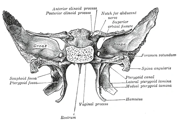
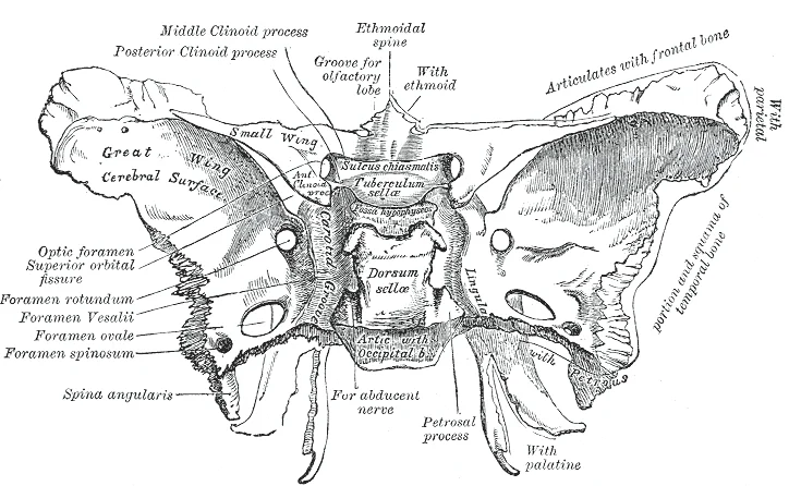
The sphenoid bone is a single, complex bone of the central skull. [1]
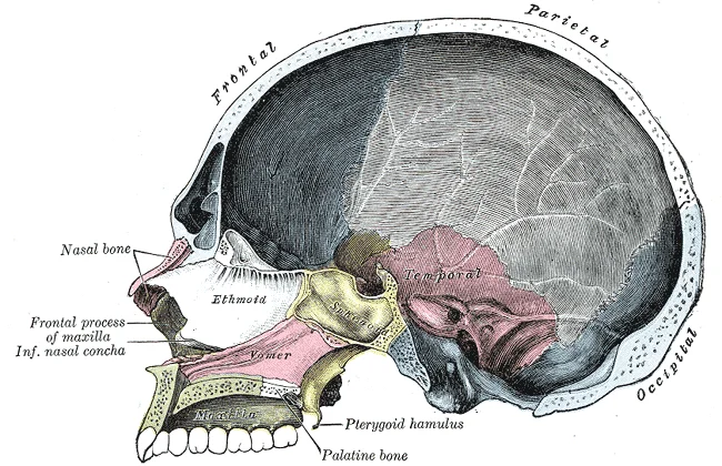
Inside the nasal area of the skull, the nasal cavity is divided into halves by the nasal septum. The upper portion of the nasal septum is formed by the perpendicular plate of the ethmoid bone and the lower portion is the vomer bone. Each side of the nasal cavity is triangular in shape, with a broad inferior space that narrows superiorly. When looking into the nasal cavity from the front of the skull, two bony plates are seen projecting from each lateral wall. The larger of these is the inferior nasal concha, an independent bone of the skull. Located just above the inferior concha is the middle nasal concha, which is part of the ethmoid bone. A third bony plate, also part of the ethmoid bone, is the superior nasal concha. It is much smaller and out of sight, above the middle concha. [1]
Figure 1Gray184
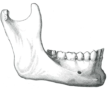
Long description
In the adult. Labelled illustration from Wikimedia Commons, used under its open licence.

Figure 1: In the adult.
Figure 2Gray183
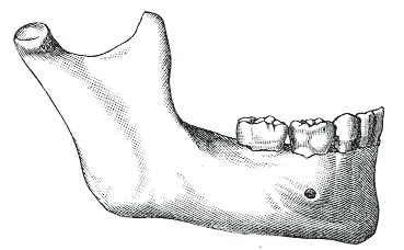
Long description
In childhood. Labelled illustration from Wikimedia Commons, used under its open licence.

Figure 2: In childhood.
Figure 3Gray152
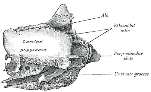
Long description
Ethmoid bone from the right side. Labelled illustration from Wikimedia Commons, used under its open licence.

Figure 3: Ethmoid bone from the right side.
Figure 4Gray151
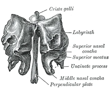
Long description
Ethmoid bone from behind. Labelled illustration from Wikimedia Commons, used under its open licence.

Figure 4: Ethmoid bone from behind.
Figure 5Gray150
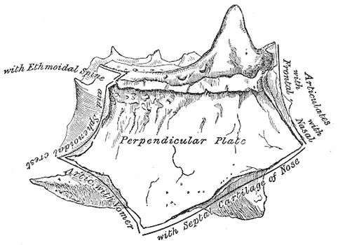
Long description
Perpendicular plate of ethmoid. Shown by removing the right labyrinth.

Figure 5: Perpendicular plate of ethmoid.
On the living patient, several of these bones and their junctions can be felt through the scalp and facial soft tissues, and these palpable landmarks guide clinical examination and imaging interpretation.
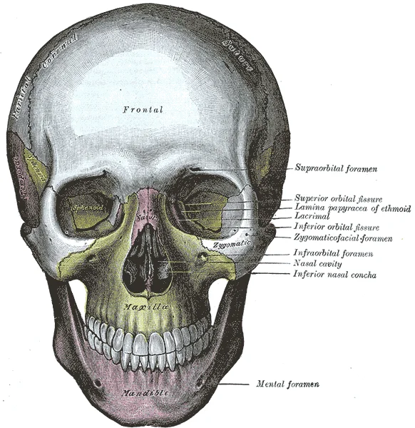
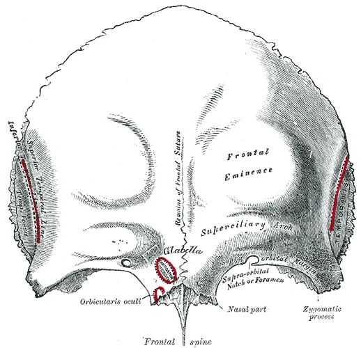
Begin at the forehead: the frontal bone forms the smooth convexity of the brow. Its lower margin, the supraorbital margin, is easily felt just above each eye; at its medial third lies the supraorbital notch or foramen, a groove for the supraorbital nerve and vessels. Running backwards from the brow, the coronal suture is palpable as a slight ridge in many adults; it joins the frontal bone to the right and left parietal bones[1]. The parietal bones form most of the upper lateral skull. The plates from the right and left palatine bones join together at the midline to form the posterior quarter of the hard palate.
Figure 6Gray169
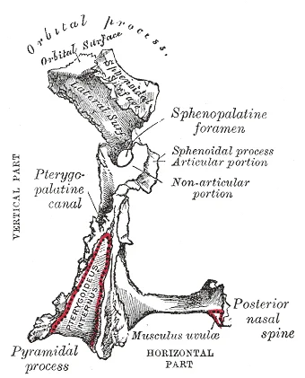
Long description
Left palatine bone. Posterior aspect. Enlarged.

Figure 6: Left palatine bone (posterior aspect).
Figure 7Gray168
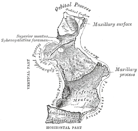
Long description
Left palatine bone. Nasal aspect. Enlarged.

Figure 7: Left palatine bone (nasal aspect).
Figure 8Gray167
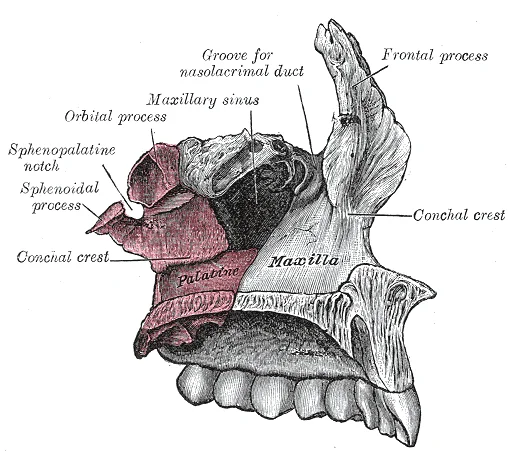
Long description
Articulation of left palatine bone with maxilla.

Figure 8: Articulation of left palatine bone with maxilla.
Figure 9Gray147
Long description
Sphenoid bone. Upper and posterior surfaces.

Figure 9: Sphenoid bone (upper and posterior surfaces).
Figure 10Gray145
Long description
Sphenoid bone. Upper surface. Labelled illustration from Wikimedia Commons, used under its open licence.

Figure 10: Sphenoid bone (upper surface).
Figure 11Gray133
Long description
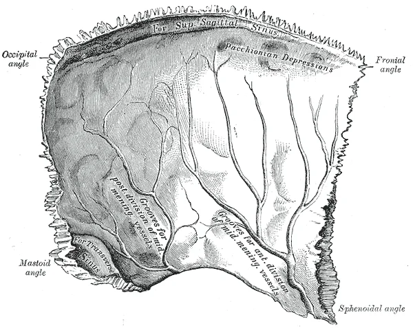
Left parietal bone. Inner surface. Labelled illustration from Wikimedia Commons, used under its open licence.

Figure 11: Left parietal bone (inner surface).
Figure 12Gray132
Long description
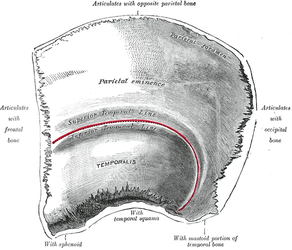
Left parietal bone. Outer surface. Labelled illustration from Wikimedia Commons, used under its open licence.

Figure 12: Left parietal bone (outer surface).
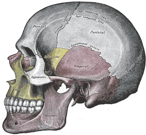
At the back of the head, the lambdoid suture is often palpable as a V-shaped line, named for its resemblance to the Greek letter lambda (Λ); it joins the occipital bone to the parietal and temporal bones[1]. The external occipital protuberance, a midline bump on the occipital bone, is a reliable landmark for the level of the transverse sinuses. Below it, the superior nuchal lines curve laterally and mark the attachment of neck muscles.
Figure 13Gray188
Long description
Side view of the skull. Labelled illustration from Wikimedia Commons, used under its open licence.

Figure 13: Side view of the skull.
On the side of the head, the zygomatic arch (cheekbone) is palpable as a horizontal ridge running from the cheek to just in front of the ear. Above its posterior end lies the pterion, the region where the frontal, parietal, temporal and sphenoid bones meet; this is a thin area of the skull and clinically important because the middle meningeal artery runs deep to it. The mastoid process, a bony prominence behind the ear, is a further surface landmark; its tip marks the insertion of the sternocleidomastoid muscle.
Figure 14Gray191
Long description
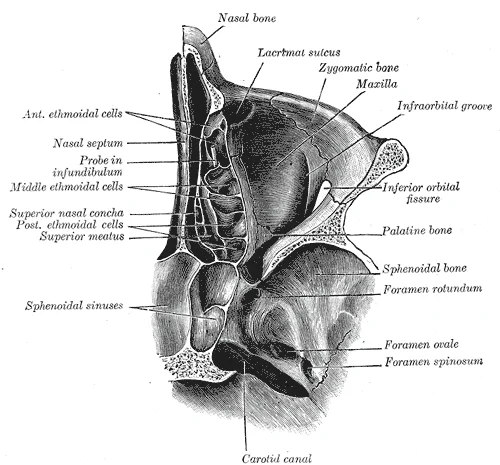
Horizontal section of nasal and orbital cavities.

Figure 14: Horizontal section of nasal and orbital cavities.
Figure 15Gray190
Long description
The skull from the front. Labelled illustration from Wikimedia Commons, used under its open licence.

Figure 15: The skull from the front.
Surface markings of the underlying structures are equally important. Its surface projection is just anterior to the mastoid process, at the anterior edge of the occipital condyle. The pterion, by contrast, overlies the anterior branch of the middle meningeal artery, and a blow here may cause an extradural haematoma.
Figure 16Gray159
Long description
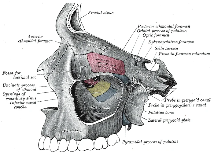
Left maxillary sinus opened from the exterior.

Figure 16: Left maxillary sinus opened from the exterior.
Figure 17Gray194
Long description
Sagittal section of skull. Labelled illustration from Wikimedia Commons, used under its open licence.

Figure 17: Sagittal section of skull.
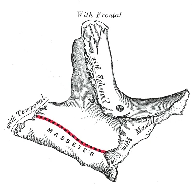
Separating these areas is the bridge of bone called the zygomatic arch. The zygomatic arch is the bony arch on the side of the skull that spans from the area of the cheek to just above the ear canal. It is formed by the junction of two bony processes: a short anterior component, the temporal process of the zygomatic bone (the cheekbone) and a longer posterior portion, the zygomatic process of the temporal bone, extending forward from the temporal bone. Thus the temporal process (anteriorly) and the zygomatic process (posteriorly) join together, like the two ends of a drawbridge, to form the zygomatic arch. One of the major muscles that pulls the mandible upward during biting and chewing arises from the zygomatic arch. [1]
Figure 18Gray186
Long description
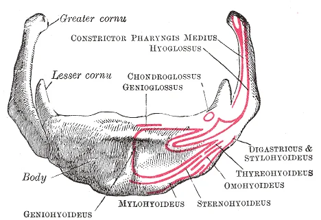
Hyoid bone. Anterior surface. Enlarged. Labelled illustration from Wikimedia Commons, used under its open licence.

Figure 18: Hyoid bone (anterior surface).
Figure 19Gray185
Long description
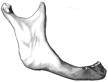
In old age. Side view of the mandible at different periods of life.

Figure 19: In old age.
Figure 20Gray177
Long description
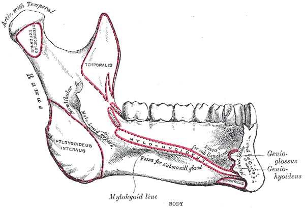
Mandible. Inner surface. Side view. Labelled illustration from Wikimedia Commons, used under its open licence.

Figure 20: Mandible (inner surface).
Figure 21Gray176
Long description
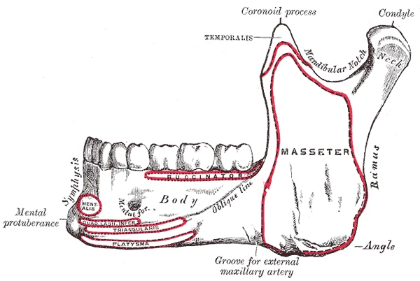
Mandible. Outer surface. Side view. Labelled illustration from Wikimedia Commons, used under its open licence.

Figure 21: Mandible (outer surface).
Figure 22Gray166
Long description
Left zygomatic bone. Temporal surface. Labelled illustration from Wikimedia Commons, used under its open licence.

Figure 22: Left zygomatic bone (temporal surface).
Figure 23Gray165
Long description
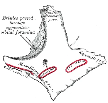
Left zygomatic bone. Malar surface. Labelled illustration from Wikimedia Commons, used under its open licence.

Figure 23: Left zygomatic bone (malar surface).
Figure 24Gray164
Long description
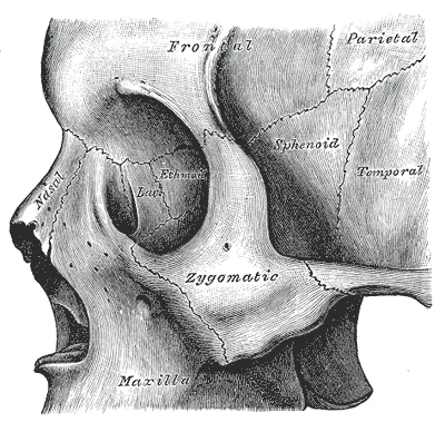
Left zygomatic bone in situ. Labelled illustration from Wikimedia Commons, used under its open licence.

Figure 24: Left zygomatic bone in situ.
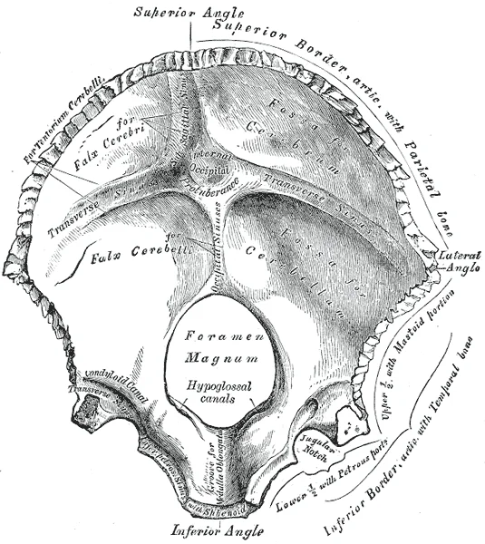
Figure 25Gray130
Long description
Occipita bone. Inner surface. Labelled illustration from Wikimedia Commons, used under its open licence.

Figure 25: Occipita bone (inner surface).
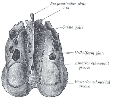
Figure 26Gray149
Long description
Ethmoid bone from above. Labelled illustration from Wikimedia Commons, used under its open licence.

Figure 26: Ethmoid bone from above.
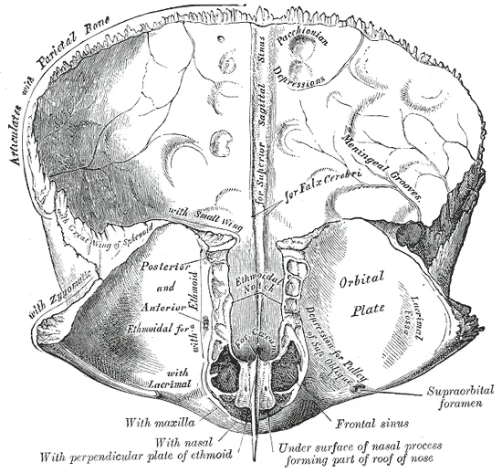
Figure 27Gray135
Long description
Frontal bone. Inner surface. Labelled illustration from Wikimedia Commons, used under its open licence.

Figure 27: Frontal bone (inner surface).
The brain case consists of eight bones. These include the paired parietal and temporal bones, plus the unpaired frontal, occipital, sphenoid, and ethmoid bones. The brain case surrounds and protects the brain, which occupies the cranial cavity inside the skull. It consists of the rounded calvaria and a complex base. The narrow gap between the bones is filled with dense, fibrous connective tissue that unites the bones. The sagittal suture joins the right and left parietal bones; it extends posteriorly from the coronal suture and terminates by joining the lambdoid suture. [1]
The sphenoid also extends laterally to contribute to the sides of the skull. [1]
They also support the cartilages that form the lateral walls of the nose. [1]
The skull is composed of numerous bones that articulate to form a protective case for the brain. These articulations are primarily immovable fibrous joints known as sutures. Sutures interlock the bones tightly, providing stability and resistance to mechanical stress. They are found between the cranial bones, such as the frontal, parietal, temporal, and occipital bones. In contrast, the mandible forms a synovial joint with the temporal bone at the temporomandibular joint, allowing for movement during chewing and speaking. [1]
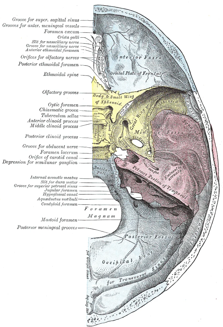
Figure 28Gray193
Long description
Base of the en:skull. Inner or cerebral surface.

Figure 28: Base of the en:skull (inner or cerebral surface).
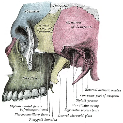
The zygomatic arch creates a boundary for the temporal fossa above and the infratemporal fossa below. The temporomandibular joint is located just anterior to the external acoustic meatus. The condyles of the mandible articulate with the temporal bones to form the temporomandibular joint. The hyoid bone is an independent bone that does not contact any other bone and thus is not part of the skull. [1]
Figure 29Gray189
Long description
Left infratemporal fossa. Labelled illustration from Wikimedia Commons, used under its open licence.

Figure 29: Left infratemporal fossa.
DefinitionSutures
Fibrous joints that connect the bones of the skull, allowing for minimal to no movement.
The base of the skull supports the brain and provides attachment points for muscles and nerves. The floor of the brain case is referred to as the base of the skull. This is a complex area that varies in depth and has numerous openings for the passage of cranial nerves, blood vessels, and the spinal cord. Inside the skull, the base is subdivided into three large spaces, called the anterior cranial fossa, middle cranial fossa, and posterior cranial fossa. From anterior to posterior, the fossae increase in depth. [1]
NoteHyoid Bone
The hyoid bone is unique as it does not articulate with any other bone, suspended by muscles and ligaments.
The paranasal sinuses are hollow, air-filled spaces located within certain bones of the skull. All of the sinuses communicate with the nasal cavity and are lined with nasal mucosa. They serve to reduce bone mass and thus lighten the skull, and they also add resonance to the voice. This second feature is most obvious when you have a cold or sinus congestion. These produce swelling of the mucosa and excess mucus production, which can obstruct the narrow passageways between the sinuses and the nasal cavity. [1]
| Structure | Type | Function |
|---|---|---|
| Sutures | Fibrous | Immovable connection of cranial bones |
| Temporomandibular Joint | Synovial | Allows jaw movement |
| Hyoid Bone | Independent | Supports tongue and larynx |
The skull is not a single rigid shell but a composite of distinct bones united by fibrous joints, creating specific compartments for neural and muscular structures. Understanding the osteology requires identifying how these bones form boundaries for the cranial fossae and facial cavities.
The somewhat larger lateral pterygoid plates serve as attachment sites for chewing muscles that fill the infratemporal space and act on the mandible. Adjacent to these, the ethmoid bone is a single, midline bone that forms the roof and lateral walls of the upper nasal cavity. On the interior of the skull, the ethmoid also forms a portion of the floor of the anterior cranial cavity. The lateral portions of the ethmoid bone are located between the orbit and upper nasal cavity, and thus form the lateral nasal cavity wall and a portion of the medial orbit wall. Located inside this portion of the ethmoid bone are several small, air-filled spaces that are part of the paranasal sinus system of the skull. The temporal bone forms the lower lateral side of the skull. [1]
Figure 30Gray192
Long description
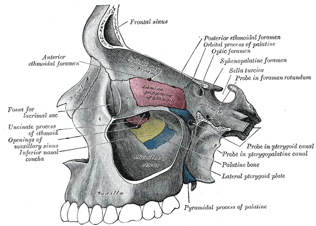
Medial wall of left orbit. Labelled illustration from Wikimedia Commons, used under its open licence.

Figure 30: Medial wall of left orbit.
DefinitionPterygoid Plates
The medial and lateral pterygoid plates are vertical bony projections of the sphenoid bone, serving as attachment sites for masticatory muscles.
The nasal septum consists of both bone and cartilage components. The upper portion of the septum is formed by the perpendicular plate of the ethmoid bone. The lower and posterior parts of the septum are formed by the triangular-shaped vomer bone. In an anterior view of the skull, the perpendicular plate of the ethmoid bone is easily seen inside the nasal opening as the upper nasal septum, but only a small portion of the vomer is seen as the inferior septum. A better view of the vomer bone is seen when looking into the posterior nasal cavity with an inferior view of the skull, where the vomer forms the full height of the nasal septum. The anterior nasal septum is formed by the septal cartilage, a flexible plate that fills in the gap between the perpendicular plate of the ethmoid and vomer bones. [1]
Figure 31Gray134
Long description
Frontal bone. Outer surface. Labelled illustration from Wikimedia Commons, used under its open licence.

Figure 31: Frontal bone (outer surface).
Attached to the lateral wall on each side of the nasal cavity are the superior, middle, and inferior nasal conchae. These are bony plates that curve downward as they project into the space of the nasal cavity. They serve to swirl the incoming air, which helps to warm and moisturize it before the air moves into the delicate air sacs of the lungs. This also allows mucus, secreted by the tissue lining the nasal cavity, to trap incoming dust, pollen, bacteria, and viruses. The largest of the conchae is the inferior nasal concha, which is an independent bone of the skull. The middle concha and the superior conchae, which is the smallest, are both formed by the ethmoid bone. When looking into the anterior nasal opening of the skull, only the inferior and middle conchae can be seen. [1]
Clinical pearlConchae Function
The nasal conchae increase surface area to humidify and filter inhaled air, protecting the lower respiratory tract.
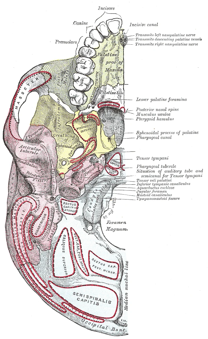
Inside the skull, the floor of the cranial cavity is subdivided into three cranial fossae. Since the brain occupies these areas, the shape of each conforms to the shape of the brain regions that it contains. Each cranial fossa has anterior and posterior boundaries and is divided at the midline into right and left areas by a significant bony structure or opening. Openings here include the large foramen magnum, plus the internal acoustic meatus, jugular foramina, and hypoglossal canals. Additional openings located on the external base of the skull include the stylomastoid foramen and the entrance to the carotid canal. Located on the medial wall of the petrous ridge in the posterior cranial fossa is the internal acoustic meatus. This opening provides for passage of the nerve from the hearing and equilibrium organs of the inner ear, and the nerve that supplies the muscles of the face. Located at the anterior-lateral margin of the foramen magnum is the hypoglossal canal. These emerge on the inferior aspect of the skull at the base of the occipital condyle and provide passage for an important nerve to the tongue. [1]
Figure 32Gray187
Long description
Base of skull. Inferior surface. Labelled illustration from Wikimedia Commons, used under its open licence.

Figure 32: Base of skull (inferior surface).
Figure 33Gray129
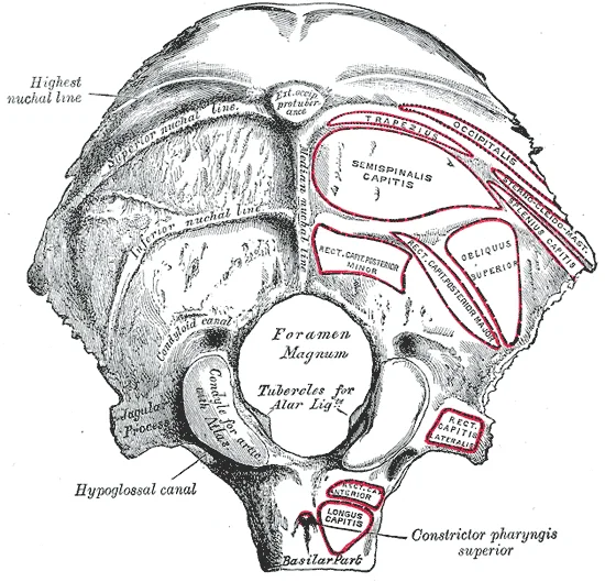
Long description
Occipital bone. Outer surface. Labelled illustration from Wikimedia Commons, used under its open licence.

Figure 33: Occipital bone (outer surface).
Figure 34Gray140
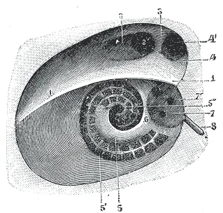
Long description
Diagrammatic view of the fundus of the right internal acoustic meatus. (Testut.) 1. Crista falciformis. 2. Area facialis, with (2’) internal opening of the facial canal. 3. Ridge separating the area facialis from the area cribrosa superior. 4. Area cribrosa superior, with (4’) openings for nerve fil

Figure 34: Diagrammatic view of the fundus of the right internal acoustic meatus (Testut.) 1.
The medial pterygoid muscle is situated deep within the infratemporal fossa, inserting onto the medial surface of the ramus and angle of the mandible. These muscles act on the mandible during chewing. The orbicularis oculi surrounds the orbit, which is the bony socket that houses the eyeball and muscles that move the eyeball or open the upper eyelid. The buccinator lies in the cheek, compressing the cheek against the teeth. The mentalis muscle inserts into the skin of the chin, near the mental foramen. [1]
| Muscle | Origin | Insertion | Action | Nerve Supply |
|---|---|---|---|---|
| Temporalis | Temporal fossa | Coronoid process of mandible | Elevates mandible | Mandibular nerve |
| Masseter | Zygomatic arch | Lateral ramus/angle | Elevates mandible | Mandibular nerve |
| Medial Pterygoid | Sphenoid bone | Medial ramus/angle | Elevates mandible | Mandibular nerve |
| Lateral Pterygoid | Sphenoid bone | Neck of mandible | Protrudes mandible | Mandibular nerve |
| Orbicularis Oculi | Orbital margins | Skin around eye | Closes eyelid | Facial nerve |
| Frontalis | Epicranial aponeurosis | Skin of forehead | Raises eyebrows | Facial nerve |
The supraorbital foramen provides for passage of a sensory nerve to the skin of the forehead. Below the orbit is the infraorbital foramen, which is the point of emergence for a sensory nerve that supplies the anterior face below the orbit. The mandible has two openings, the mandibular foramen on its inner surface and the mental foramen on its external surface near the chin. The nasal conchae are bony projections from the lateral walls of the nasal cavity. The large inferior nasal concha is an independent bone, while the middle and superior conchae are parts of the ethmoid bone. The nasal septum is formed by the perpendicular plate of the ethmoid bone, the vomer bone, and the septal cartilage. [1]
The skull receives its arterial supply from branches of both the external and internal carotid arteries. The external carotid artery supplies the extracranial soft tissues and the bones of the face and calvarium, while the internal carotid artery supplies the intracranial contents and the inner table of the cranial bones. The middle meningeal artery, a branch of the maxillary artery (itself a terminal branch of the external carotid artery), is the principal artery of the dura mater and also supplies the calvarial bones. It enters the skull through the foramen spinosum, a small opening in the greater wing of the sphenoid bone, which lies just anterior to the squamous portion of the temporal bone. Once inside, it runs laterally in the middle cranial fossa, grooving the squamous temporal bone and the greater wing of the sphenoid before dividing into frontal and parietal branches that ascend over the cerebral convexity. [1]
The middle cranial fossa, which is deeper than the anterior fossa and extends from the lesser wings of the sphenoid anteriorly to the petrous ridges posteriorly, is a key conduit for these vessels. The petrous ridges, formed by the petrous portion of the temporal bones, give the fossa its butterfly shape, narrow at the midline and broad laterally. [1]
The paranasal sinuses receive their blood supply from branches of the maxillary artery, particularly the infraorbital and sphenopalatine arteries. The maxillary sinus, the largest of the paranasal sinuses, lies within the maxillary bone just below the orbit. Its arterial supply comes from the posterior superior alveolar artery and the infraorbital artery, both branches of the maxillary artery. The frontal sinus is located just above the eyebrows, within the frontal bone. [1]
Clinical pearlClinical relevance
The middle meningeal artery is the most common source of extradural haemorrhage after head trauma, because it lies in a groove on the inner table of the squamous temporal bone, where it is easily torn by a skull fracture.
The venous drainage of the skull bones accompanies the arterial supply through emissary veins, which connect the intracranial dural venous sinuses with the extracranial veins. The diploic veins, which run within the diploë of the calvarial bones, drain into the dural sinuses and the extracranial veins. This venous network has no valves, allowing blood to flow in either direction, which is clinically important in the spread of infection from the scalp or sinuses to the intracranial cavity.
The venous drainage of the skull is a dual system: an intracranial (dural venous sinuses) and an extracranial (veins of the scalp and face) network, connected by emissary veins that pass through foramina. The dural venous sinuses lie between the two layers of dura mater and receive blood from the brain, the diploë of the skull bones, and the orbit.
The superior sagittal sinus runs in the midline along the attached margin of the falx cerebri, beginning at the crista galli of the ethmoid bone and terminating at the internal occipital protuberance. Here it usually drains into the right transverse sinus. The inferior sagittal sinus runs in the free margin of the falx cerebri and joins the great cerebral vein to form the straight sinus, which continues to the confluence of sinuses. The transverse sinuses pass laterally from the confluence of sinuses.
The cavernous sinuses lie on each side of the sella turcica in the middle cranial fossa, lateral to the hypophyseal fossa. They receive the superior and inferior ophthalmic veins and the superficial middle cerebral veins. The internal carotid artery and abducens nerve pass through the sinus, while the oculomotor, trochlear, ophthalmic and maxillary nerves run in its lateral wall. The cavernous sinus drains via the superior and inferior petrosal sinuses; the superior petrosal sinus runs along the petrous ridge of the temporal bone to the transverse sinus, and the inferior petrosal sinus exits through the jugular foramen. [1]
Emissary veins connect the dural sinuses to extracranial veins. The mastoid emissary vein passes through the mastoid foramen in the mastoid portion of the temporal bone, connecting the sigmoid sinus to the posterior auricular or occipital veins. The parietal emissary vein passes through the parietal foramen. The condylar emissary vein exits via the condylar canal. The venous plexus of the foramen ovale and the foramen spinosum connect the cavernous sinus to the pterygoid plexus. The ophthalmic veins connect the cavernous sinus to the facial vein via the angular vein, providing a route for spread of infection from the face to the cavernous sinus. [1]
The transverse sinuses run laterally from the confluence of sinuses at the internal occipital protuberance, grooving the occipital bone. The superior petrosal sinuses run along the petrous ridge, connecting the cavernous to the transverse sinuses.
The skull transmits twelve pairs of cranial nerves, each passing through specific foramina to reach its target. For the medical student, the clinically relevant nerves are those that supply the muscles of facial expression, mastication, and the sensory skin of the face and scalp. The trigeminal nerve (CN V) is the principal sensory nerve of the face and the motor nerve for mastication. The facial nerve (CN VII) supplies the muscles of facial expression. Both have complex courses through the skull and are vulnerable to injury at specific bony landmarks.
The trigeminal nerve (CN V) emerges from the pons and divides into three divisions: ophthalmic (V1), maxillary (V2), and mandibular (V3). V1 and V2 are purely sensory; V3 carries both sensory fibres and the motor root that supplies the muscles of mastication. The ophthalmic division (V1) passes through the superior orbital fissure to supply the skin of the forehead, upper eyelid, and dorsum of the nose. The maxillary division (V2) exits through the foramen rotundum, crosses the pterygopalatine fossa, and enters the orbit via the inferior orbital fissure, emerging on the face through the infraorbital foramen to supply the skin of the midface, lower eyelid, and upper lip. The mandibular division (V3) leaves the skull through the foramen ovale; its sensory branches supply the skin of the lower face, jaw, and anterior two-thirds of the tongue, while its motor root innervates the four muscles of mastication. [2]
The facial nerve (CN VII) exits the skull through the stylomastoid foramen, then enters the parotid gland and divides into five terminal branches. It supplies all the muscles of facial expression, the platysma, and the posterior belly of the digastric. The olfactory nerve (CN I) fibres pass through the olfactory foramina of the cribriform plate of the ethmoid bone to reach the brain. [1]
In the anterior cranial fossa, the crista galli projects upward at the midline, flanked by the cribriform plates with their sieve-like olfactory foramina. Posteriorly, the clinoid processes of the sphenoid flank the sella turcica; the foramen for CN VI lies dorsolateral to the carotid canal at the base of the clinoid process. The vagus opening is a lateral slit on the medial wall of the foramen magnum, with the small circular CN XII openings near the occipital condyle. [2]
| Nerve | Motor supply | Cutaneous (sensory) supply |
|---|---|---|
| CN V1 (Ophthalmic) | None | Forehead, upper eyelid, dorsum of nose |
| CN V2 (Maxillary) | None | Midface, lower eyelid, upper lip |
| CN V3 (Mandibular) | Muscles of mastication (masseter, temporalis, medial & lateral pterygoid), mylohyoid, anterior belly of digastric | Lower face, jaw, anterior two-thirds of tongue |
| CN VII (Facial) | Muscles of facial expression, platysma, posterior belly of digastric, stylohyoid | Taste to anterior two-thirds of tongue (special sensory) |
| CN VI (Abducens) | Lateral rectus (eye) | None |
| CN XII (Hypoglossal) | Intrinsic and extrinsic muscles of the tongue | None |
The motor root of V3 travels with the sensory fibres through the foramen ovale, then separates to supply the temporalis, masseter, and the medial and lateral pterygoid muscles. The lateral pterygoid attaches to the condylar process of the mandible and the articular disc of the temporomandibular joint, while the temporalis inserts on the coronoid process. A lesion of the mandibular division causes deviation of the jaw toward the paralysed side on opening, due to unopposed action of the opposite lateral pterygoid. The facial nerve, after emerging from the stylomastoid foramen, winds around the styloid process and enters the parotid gland; a lesion here produces ipsilateral paralysis of all facial expression muscles, with loss of the nasolabial fold and inability to close the eye.
The lymphatic drainage of the skull is a complex network that mirrors the vascular supply, ensuring efficient removal of interstitial fluid and immune surveillance for the cranial structures. Understanding this drainage is critical for tracing the spread of infections or metastatic disease from the head and neck region. The lymphatic vessels originate in the superficial and deep tissues, converging towards specific nodal groups that serve as primary filtering stations.
The superficial lymphatics of the scalp and face drain into a series of peripheral nodes. These nodes are strategically located at the margins of the head and neck, acting as the first line of defence.
| Node Group | Location | Drainage Area |
|---|---|---|
| Occipital | Over the occipital bone | Posterior scalp |
| Mastoid (Postauricular) | Between the ear and sternocleidomastoid | Lateral scalp, posterior auricle |
| Parotid | Within or superficial to the parotid gland | Lateral eyelids, temporal region |
| Preauricular (Anterior) | Anterior to the tragus | Lateral eyelids, cheek, temporal region |
| Submandibular | Within the submandibular triangle | Face, nose, upper lip, lateral tongue |
| Submental | Beneath the chin | Tip of tongue, central lower lip, chin |
The occipital nodes lie on the surface of the occipitalis muscle, receiving lymph from the posterior scalp. The mastoid nodes are situated between the mastoid process and the sternocleidomastoid muscle, draining the lateral scalp and the posterior aspect of the auricle. The parotid nodes are embedded within the parotid gland or lie superficial to it, collecting lymph from the lateral aspects of the eyelids and the temporal region. The preauricular nodes are located anterior to the tragus of the ear, draining the lateral eyelids, the cheek, and the temporal region. The submandibular nodes are found within the submandibular triangle, draining the face, nose, upper lip, and the lateral aspect of the tongue. The submental nodes are located beneath the chin, draining the tip of the tongue, the central lower lip, and the chin.
The deep lymphatics of the skull and face converge towards the deep cervical lymph nodes. These nodes form a chain along the internal jugular vein and are crucial for the final filtration of lymph before it enters the venous system.
DefinitionDeep Cervical Nodes
A chain of lymph nodes located along the internal jugular vein, serving as the primary drainage site for the head and neck.
The deep cervical nodes receive lymph from the superficial nodes via efferent vessels. They also receive direct drainage from the deep structures of the head, including the nasopharynx, larynx, and thyroid gland. The efferent vessels from the deep cervical nodes form the jugular lymphatic trunks.
Lymphatic Pathway
- Scalp LymphaticsOriginate in superficial tissues
- Question: Drain to?
Posterior
- Occipital Nodes
Lateral
- Mastoid Nodes
Anterior
- Parotid/Preauricular Nodes
- ConvergenceEfferents join deep cervical chain
- Deep Cervical NodesAlong internal jugular vein
- Jugular TrunkForms from deep cervical efferents
- Venous AngleDrains into junction of internal jugular and subclavian veins
The jugular lymphatic trunks on each side typically drain into the thoracic duct on the left and the right lymphatic duct on the right. The thoracic duct empties into the junction of the left internal jugular and left subclavian veins, while the right lymphatic duct drains into the junction of the right internal jugular and right subclavian veins. This arrangement ensures that lymph from the head and neck returns to the systemic circulation efficiently.
Clinical pearlClinical Relevance
Infection in the scalp can spread to the deep cervical nodes, leading to lymphadenopathy. Metastatic cancer from the scalp or face often spreads to these nodal groups.
Understanding the lymphatic drainage of the skull is essential for clinical practice, particularly in the diagnosis and management of head and neck pathologies. The detailed knowledge of nodal groups and their drainage areas allows for accurate assessment of disease spread and appropriate surgical planning.
The sphenoid sinus is situated within the body of the sphenoid bone, positioned just anterior and inferior to the sella turcica, making it the most posterior of the paranasal sinuses. Adjacent to this, the lateral aspects of the ethmoid bone contain multiple small spaces separated by very thin bony walls. [1]
Figure 35Gray146
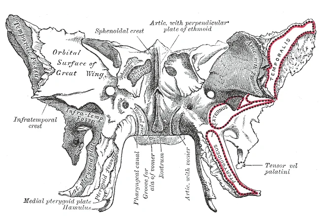
Long description
Sphenoid bone. Anterior and inferior surfaces.

Figure 35: Sphenoid bone (anterior and inferior surfaces).
DefinitionParanasal Sinuses
Air-filled spaces located within the frontal, maxillary, sphenoid, and ethmoid bones.
| Structure | Relation |
|---|---|
| Sphenoid Sinus | Anterior and inferior to sella turcica |
| Ethmoid Air Cells | Between upper nasal cavity and medial orbit |
| Quadrate Concavity | Supports tympanic membrane |
| Vagus Foramen | Dorsal to hypoglossal foramina |
When examining the skull, it is essential to understand how different imaging modalities visualise bony structures. Plain X-ray provides a quick overview of gross anatomy but lacks depth resolution. Computed Tomography (CT) is the gold standard for bone assessment, offering high-resolution cross-sectional images that clearly delineate sutures, sinuses, and the cranial base. Magnetic Resonance Imaging (MRI) is superior for soft tissue, such as the brain and cranial nerves, but it is less effective for detailed osteology unless specific bone marrow sequences are used. Ultrasound has limited utility in adult skull imaging due to the acoustic impedance of bone, though it may be used in infants with open fontanelles.
The sphenoid forms much of the base of the central skull. [1]
DefinitionOsseous Labyrinth
The bony enclosure of the inner ear, containing the semicircular canals and vestibule.
| Structure | Description |
|---|---|
| Anterior Canal | Curved and highly arched in some species. |
| Posterior Canal | Compact arc with compact posterior vestibule. |
| Horizontal Canal | Varies in distance from the vestibule. |
| Ampullae | Anterior, lateral, and posterior ampullae are well developed. |
The skull provides critical surface landmarks for clinical procedures and localises injury patterns. Understanding the osteology is essential for interpreting trauma and nerve palsies.
The temporal bone contains important landmarks for surgical access.
The parietal bone forms the superior vault.
- The bregma marks the junction of the coronal and sagittal sutures and corresponds to the location of the anterior fontanelle in the infant skull.
- The lambda indicates the meeting point of the sagittal and lambdoid sutures, overlying the site of the posterior fontanelle during fetal development.
- The pterion overlies the anterior branch of the middle meningeal artery, making it a critical landmark where trauma can result in an extradural hematoma.
- The sphenoidal angle of the parietal bone features an internal groove that lodges the anterior divisions of the middle meningeal artery.
- The supraorbital margin terminates laterally in the robust zygomatic process, which articulates directly with the zygomatic bone.
- The temporal lines curve upward and backward across the frontal and parietal bones, defining the boundary of the temporal fossa and the origin of the temporalis muscle.
Students frequently confuse the anatomical boundaries of the cranial fossae, particularly when identifying the posterior cranial fossa. A common error is assuming the occipital bone forms the entire floor and anterior boundary. In reality, the posterior cranial fossa is the most posterior and deepest portion of the cranial cavity. It is bounded anteriorly by the petrous ridges, while the occipital bone forms the floor and posterior wall. This distinction is critical for localising lesions on imaging. Another frequent mistake involves the contents of this fossa. Students often overlook that it contains the cerebellum of the brain. The large foramen magnum, or “great aperture”, divides the fossa at the midline. This opening provides for passage of the spinal cord. Confusing the foramen magnum with other occipital foramina can lead to errors in identifying neural structures. [1]
A second pitfall relates to the mandible, particularly in comparative contexts. Students may assume the human mandible is a single bone, whereas in skinks, it comprises four discrete elements: the angular, coronoid, dentary, and splenial. Additionally, there are two partially fused elements: the articular and the surangular. The dentary is curved inward anteriorly and is the longest bone of the lower jaw. Misidentifying the dentary as the entire mandible leads to errors in understanding jaw mechanics. The dentary teeth occupy about one-third of the total jaw length in T. laevis. In T. sulcata, they occupy one-half, and in T. gonwouoi, two-thirds. This variation affects bite force calculations. [2]
Students also struggle with the premaxilla. This bone, normally paired in skinks, contacts the maxilla posteroventrally, nasal posterodorsally, and vomer ventromedially. Trachylepis sulcata and T. gonwouoi possess paired premaxillae, while T. laevis possesses a premaxilla that is fused at the base with a faint suture visible only on the nasal process. Assuming all premaxillae are fused or all are paired is incorrect. The nasal process is triangular in dorsal and ventral view. The base of this process is wide, whereas the distal end tapers to a rounded tip in T. laevis and a pointed tip in T. sulcata and T. gonwouoi.
The jugal bone presents further confusion. The anterior process of the jugal is very short in T. laevis, barely extending beyond the length of the ectopterygoid and only slightly overlapping the posterior process of the maxilla. In contrast, the anterior process in T. sulcata and T. gonwouoi extends well beyond the ectopterygoid, overlaps the posterior process of the maxilla nearly entirely, and narrowly contacts the prefrontal medially and lacrimal laterally. The jugal does not participate in the lacrimal foramen in these two species, but medially it borders a shallow groove that is a continuation of this foramen. A jugal foramen is present at the point of inflection, and this region is swollen in T. gonwouoi. A facet is present anterior to the foramen where the posterior process of the maxilla overlaps laterally, forming a butt-lap joint. This facet is more posterior in T. sulcata and T. gonwouoi than in T. laevis. The jugal is strongly recumbent in T. laevis (144°) but less so in T. sulcata (130°) and T. gonwouoi (125°).
Finally, the squamosal bone is often misdescribed. This is a paired element that contacts the postparietal process of the parietal dorsomedially, supratemporal posteromedially, postfrontal anteromedially, quadrate posteroventrally, and postorbital anteromedially (except for T. laevis, which lacks this element). It overlays the prootic and the otoccipital. The squamosal is a thin, elongate bone that curves posterolaterally, forming a hockey stick shape. This bone plays a role in quadrate suspension in these taxa and interacts with the squamosal notch of the quadrate. The squamosal of T. sulcata and T. gonwouoi is relatively longer than that of T. laevis. The inflection point of the long axis is more posterior in T. laevis and T. gonwouoi as compared to T. sulcata. The curvature of the squamosal is most extreme in T. gonwouoi.
Knowledge of the posterior cranial fossa is essential at the bedside and in the operating theatre. Clinicians must recognise that the posterior cranial fossa is the most posterior and deepest portion of the cranial cavity. This anatomical reality dictates surgical approaches to cerebellar lesions. Surgeons must navigate around the petrous ridges, which bound the fossa anteriorly. The occipital bone forms the floor and posterior wall. Understanding that the fossa contains the cerebellum of the brain helps localise symptoms such as ataxia. The large foramen magnum, or “great aperture”, divides the fossa at the midline. This opening provides for passage of the spinal cord. Compression here can cause life-threatening brainstem signs. [1]
On imaging, radiologists identify the posterior cranial fossa by its boundaries. The petrous ridges are key landmarks. The occipital bone forms the floor and posterior wall. The foramen magnum is the central feature. It provides for passage of the spinal cord. Misidentifying the boundaries can lead to missed diagnoses of posterior fossa masses.
In examinations, students must describe the mandible accurately. The mandible comprises four discrete elements: the angular, coronoid, dentary, and splenial. There are also two partially fused elements: the articular and the surangular. The dentary is curved inward anteriorly and is the longest bone of the lower jaw. Dentary teeth occupy about one-third of the total jaw length in T. laevis. In T. sulcata, they occupy one-half, and in T. gonwouoi, two-thirds.
The premaxilla is another frequent exam topic. This bone, normally paired in skinks, contacts the maxilla posteroventrally, nasal posterodorsally, and vomer ventromedially. Trachylepis sulcata and T. gonwouoi possess paired premaxillae, while T. laevis possesses a premaxilla that is fused at the base with a faint suture visible only on the nasal process. The nasal process is triangular in dorsal and ventral view. The base of this process is wide, whereas the distal end tapers to a rounded tip in T. laevis and a pointed tip in T. sulcata and T. gonwouoi.
The jugal bone’s variations are also examined. The anterior process of the jugal is very short in T. laevis, barely extending beyond the length of the ectopterygoid and only slightly overlapping the posterior process of the maxilla. In T. sulcata and T. gonwouoi, it extends well beyond the ectopterygoid, overlaps the posterior process of the maxilla nearly entirely, and narrowly contacts the prefrontal medially and lacrimal laterally. The jugal does not participate in the lacrimal foramen in these two species, but medially it borders a shallow groove that is a continuation of this foramen. A jugal foramen is present at the point of inflection, and this region is swollen in T. gonwouoi. A facet is present anterior to the foramen where the posterior process of the maxilla overlaps laterally, forming a butt-lap joint. This facet is more posterior in T. sulcata and T. gonwouoi than in T. laevis. The jugal is strongly recumbent in T. laevis (144°) but less so in T. sulcata (130°) and T. gonwouoi (125°).
The squamosal bone’s role in quadrate suspension is clinically relevant in comparative studies. This is a paired element that contacts the postparietal process of the parietal dorsomedially, supratemporal posteromedially, postfrontal anteromedially, quadrate posteroventrally, and postorbital anteromedially (except for T. laevis, which lacks this element). It overlays the prootic and the otoccipital. The squamosal is a thin, elongate bone that curves posterolaterally, forming a hockey stick shape. This bone plays a role in quadrate suspension in these taxa and interacts with the squamosal notch of the quadrate. The squamosal of T. sulcata and T. gonwouoi is relatively longer than that of T. laevis. The inflection point of the long axis is more posterior in T. laevis and T. gonwouoi as compared to T. sulcata. The curvature of the squamosal is most extreme in T. gonwouoi.
Posterior Cranial Fossa Anatomy
- Identify Posterior Cranial FossaMost posterior and deepest portion of cranial cavity
- Question: Bounded Anteriorly By?
Yes
- Petrous Ridges
No
- Re-evaluate Boundaries
- Question: Floor and Posterior Wall?
Yes
- Occipital Bone
No
- Re-evaluate Boundaries
- Question: Midline Division?
Yes
- Foramen Magnum
No
- Check for Variants
- Foramen Magnum FunctionProvides for passage of the spinal cord
The posterior cranial fossa is the most posterior and deepest portion of the cranial cavity. It contains the cerebellum of the brain. The posterior fossa is bounded anteriorly by the petrous ridges. The occipital bone forms the floor and posterior wall. It is divided at the midline by the large foramen magnum. The foramen magnum provides for passage of the spinal cord. The mandible comprises four discrete elements and two partially fused elements. The dentary is the longest bone of the lower jaw.
Answer each one in your head first, then open the answer.
What bone forms the floor and posterior wall of the posterior cranial fossa?
Show answerHide answer
Occipital boneWhat structure divides the posterior cranial fossa at the midline?
Show answerHide answer
Foramen magnumWhat does the foramen magnum provide for?
Show answerHide answer
Passage of the spinal cordWhich bone is the longest in the lower jaw?
Show answerHide answer
DentaryHow many discrete elements comprise the mandible?
Show answerHide answer
Four
- Betts JG, Young KA, Wise JA, et al. The Skull. OpenStax, Anatomy and Physiology 2e (CC BY-NC-SA 4.0). Accessed .
- Paluh DJ, Bauer AM. Comparative skull anatomy of terrestrial and crevice-dwelling Trachylepis skinks (Squamata: Scincidae) with a survey of resources in scincid cranial osteology. PloS one 2017;12(9):e0184414. doi:10.1371/journal.pone.0184414. Accessed .
Figure credits
- Figure 1: Henry Vandyke Carter, via Wikimedia Commons, Public domain, Public domain.
- Figure 2: Henry Vandyke Carter, via Wikimedia Commons, Public domain, Public domain.
- Figure 3: Henry Vandyke Carter, via Wikimedia Commons, Public domain, Public domain.
- Figure 4: Henry Vandyke Carter, via Wikimedia Commons, Public domain, Public domain.
- Figure 5: Henry Vandyke Carter, via Wikimedia Commons, Public domain, Public domain.
- Figure 6: Henry Vandyke Carter, via Wikimedia Commons, Public domain, Public domain.
- Figure 7: Henry Vandyke Carter, via Wikimedia Commons, Public domain, Public domain.
- Figure 8: Henry Vandyke Carter, via Wikimedia Commons, Public domain, Public domain.
- Figure 9: Henry Vandyke Carter, via Wikimedia Commons, Public domain, Public domain.
- Figure 10: Henry Vandyke Carter, via Wikimedia Commons, Public domain, Public domain.
- Figure 11: Henry Vandyke Carter, via Wikimedia Commons, Public domain, Public domain.
- Figure 12: Henry Vandyke Carter, via Wikimedia Commons, Public domain, Public domain.
- Figure 13: Henry Vandyke Carter, via Wikimedia Commons, Public domain, Public domain.
- Figure 14: Henry Vandyke Carter, via Wikimedia Commons, Public domain, Public domain.
- Figure 15: Henry Vandyke Carter, via Wikimedia Commons, Public domain, Public domain.
- Figure 16: Henry Vandyke Carter, via Wikimedia Commons, Public domain, Public domain.
- Figure 17: Henry Vandyke Carter, via Wikimedia Commons, Public domain, Public domain.
- Figure 18: Henry Vandyke Carter, via Wikimedia Commons, Public domain, Public domain.
- Figure 19: Henry Vandyke Carter, via Wikimedia Commons, Public domain, Public domain.
- Figure 20: Henry Vandyke Carter, via Wikimedia Commons, Public domain, Public domain.
- Figure 21: Henry Vandyke Carter, via Wikimedia Commons, Public domain, Public domain.
- Figure 22: Henry Vandyke Carter, via Wikimedia Commons, Public domain, Public domain.
- Figure 23: Henry Vandyke Carter, via Wikimedia Commons, Public domain, Public domain.
- Figure 24: Henry Vandyke Carter, via Wikimedia Commons, Public domain, Public domain.
- Figure 25: Henry Vandyke Carter, via Wikimedia Commons, Public domain, Public domain.
- Figure 26: Henry Vandyke Carter, via Wikimedia Commons, Public domain, Public domain.
- Figure 27: Henry Vandyke Carter, via Wikimedia Commons, Public domain, Public domain.
- Figure 28: Henry Vandyke Carter, via Wikimedia Commons, Public domain, Public domain.
- Figure 29: Henry Vandyke Carter, via Wikimedia Commons, Public domain, Public domain.
- Figure 30: Henry Vandyke Carter, via Wikimedia Commons, Public domain, Public domain.
- Figure 31: Henry Vandyke Carter, via Wikimedia Commons, Public domain, Public domain.
- Figure 32: Henry Vandyke Carter, via Wikimedia Commons, Public domain, Public domain.
- Figure 33: Henry Vandyke Carter, via Wikimedia Commons, Public domain, Public domain.
- Figure 34: Henry Vandyke Carter, via Wikimedia Commons, Public domain, Public domain.
- Figure 35: Henry Vandyke Carter, via Wikimedia Commons, Public domain, Public domain.
Educational content for medical students and doctors. It is not a substitute for clinical judgement or local guidelines.