Cranial fossae
The cranial fossae form the floor of the cranial cavity, divided into anterior, middle, and posterior sections.
The cranial fossae constitute the floor of the cranial cavity, located within the intracranial part of the skull. This region is formed through coordinated cranial base development and neurocranial accommodation, creating complex endocranial surfaces that differ fundamentally from external cranial morphology. The fossae are resilient to taphonomic changes and possess unique internal morphology, making them suitable for examination via routine CT imaging in disaster victim identification contexts. They are generally divided into three distinct sections: the anterior, middle, and posterior cranial fossae.
The anterior cranial fossa is the shallowest of the three regions. Its key anatomical landmarks include the crista galli, the ethmoid plate, and the optic canals. The crista galli is a midline ridge that separates the two olfactory foramina. The length of the crista galli is the most sexually dimorphic structure in this region, with sex estimation accuracy reaching up to 83.7%. However, the overall accuracy for the anterior fossa region is lower, at 67.5%, compared to other fossae. [2]
The middle cranial fossa has a characteristic butterfly shape. Its central depression is known as the sella turcica, which houses the pituitary gland. This region contains several important foramina, including the foramen rotundum, foramen ovale, and foramen spinosum. The distance of the foramen ovale is a key metric for sex estimation in this area, with an accuracy of up to 63.9%. The middle fossa region as a whole offers a sex classification accuracy of 77.1%. [2]
The posterior cranial fossa contains the foramen magnum, which is the largest foramen of the skull. The foramen magnum is located anterior to the clivus. The clivus is a sloping surface that forms the anterior wall of the posterior fossa. The length of the clivus is a significant dimorphic trait, providing sex estimation accuracy of up to 69%. The posterior region generally yields the highest classification accuracy among the three fossae, at 79.9%. [2]
| Fossa | Key Landmarks | Sex Estimation Accuracy |
|---|---|---|
| Anterior | Crista galli, ethmoid plate | 67.5% (region), 83.7% (crista galli) |
| Middle | Sella turcica, foramen ovale | 77.1% (region), 63.9% (foramen ovale) |
| Posterior | Foramen magnum, clivus | 79.9% (region), 69% (clivus) |
NoteForensic relevance
Traits with sex estimation accuracy exceeding 80% are regarded as high quality, while those with lower accuracy should not be used as a single indicator. [2]
The cranial fossae form the floor of the cranial cavity and are arranged in a stepped fashion from anterior to posterior. They house the brain and provide attachment points for meninges and cranial nerves. Understanding their surface anatomy requires correlating internal bony landmarks with external palpable features and radiological views. [2]
The anterior cranial fossa is the most superior and shallowest depression. It is bounded anteriorly by the frontal bone and posteriorly by the lesser wings of the sphenoid and the sulcus chiasmatis. The middle cranial fossa lies inferior to the anterior fossa and is deeper. It is bounded laterally by the temporal bones and contains the sella turcica centrally. The posterior cranial fossa is the largest and deepest depression, located at the base of the skull. It is bounded anteriorly by the dorsum sellae and clivus and posteriorly by the occipital bone. The foramen magnum is centrally located within this fossa.
Palpable landmarks on the external surface correspond to these internal structures. The glabella, located between the eyebrows, overlies the frontal sinus and the anterior cranial fossa. The external occipital protuberance is a prominent midline bump on the back of the head, corresponding to the internal occipital protuberance within the posterior cranial fossa. The mastoid process, palpable behind the ear, relates to the temporal bone and the middle cranial fossa. The zygomatic arch, formed by the zygomatic process of the temporal bone, marks the lateral boundary of the middle cranial fossa.
The cranial fossae are not merely passive containers; they guide the passage of cranial nerves and vessels. The anterior cranial fossa supports the frontal lobes. The middle cranial fossa houses the temporal lobes. The posterior cranial fossa contains the cerebellum and brainstem. The boundaries between these fossae are defined by specific bony ridges and foramina. The lesser wings of the sphenoid separate the anterior and middle fossae. The petrous part of the temporal bone separates the middle and posterior fossae. The foramen magnum in the posterior fossa allows the passage of the spinal cord and vertebral arteries.
DefinitionCranial Fossae
Three stepped depressions in the base of the skull that house the brain and transmit cranial nerves and vessels.
The surface markings are critical for surgical approaches and radiological interpretation. The pterion, where the frontal, parietal, temporal, and sphenoid bones meet, overlies the middle meningeal artery and the middle cranial fossa. The inion, corresponding to the external occipital protuberance, marks the attachment of the nuchal ligament and overlies the posterior cranial fossa. The supraorbital margin, palpable above the eyes, relates to the anterior cranial fossa. These landmarks help clinicians localize intracranial pathology based on external examination or imaging.
The resilience of the cranial fossae allows them to provide crucial information even in fragmented remains, as they are preferentially preserved compared with other skeletal parts[1]. This preservation is due to their protected intracranial location and relatively thick structure[1]. In forensic contexts, the cranial fossae are often intact even when other bones are commingled or thermally altered[1]. This makes them valuable for biological profiling, including sex estimation, which can narrow down identification by up to 50%[1].
| Landmark | Location | Underlying Fossa |
|---|---|---|
| Glabella | Midline, between eyebrows | Anterior |
| Pterion | Lateral skull, junction of four bones | Middle |
| External Occipital Protuberance | Posterior midline, base of skull | Posterior |
| Mastoid Process | Behind the ear | Middle (lateral boundary) |
Figure 1Gray195
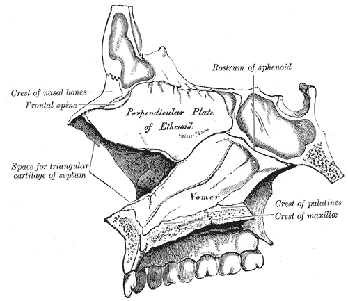
Long description
Medial wall of left nasal fossa. Labelled illustration from Wikimedia Commons, used under its open licence.

Figure 1: Medial wall of left nasal fossa.
The cranial cavity is divided into three distinct compartments known as the anterior, middle, and posterior cranial fossae. These fossae are formed by specific bones of the skull base and house the corresponding lobes of the brain. In this inferior view, the anterior cranial fossa is the most superior and anterior compartment, formed primarily by the frontal bone, the ethmoid bone, and the lesser wings of the sphenoid bone. It houses the frontal lobes of the cerebrum. The middle cranial fossa lies inferior and posterior to the anterior fossa, forming a deeper depression. It is bounded anteriorly by the lesser wings and anterior clinoid processes of the sphenoid, and posteriorly by the petrous part of the temporal bone and the dorsum sellae. This fossa contains the temporal lobes. The posterior cranial fossa is the deepest and most posterior compartment, formed by the occipital bone, the temporal bones, and the basilar part of the occipital bone. It houses the cerebellum, pons, and medulla oblongata. [1]
Figure 2Gray199
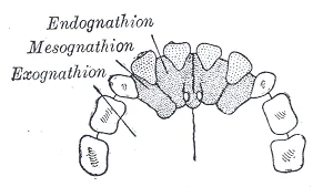
Long description
The premaxilla and its sutures. (After Albrecht.)

Figure 2: The premaxilla and its sutures (After Albrecht.).
DefinitionCranial Fossae
The anterior, middle, and posterior cranial fossae are sequential depressions in the base of the skull that support the frontal, temporal, and hindbrain structures respectively.
Key landmarks within these fossae include the sella turcica, a saddle-shaped depression in the sphenoid bone that houses the pituitary gland. The foramen magnum, located in the posterior fossa, allows the passage of the medulla oblongata and associated meninges. The optic canals are situated in the anterior fossa near the midline, transmitting the optic nerves. The middle cranial fossa contains the foramen ovale, foramen spinosum, and foramen lacerum, each transmitting specific vessels and nerves. The posterior fossa contains the jugular foramina and hypoglossal canals. Understanding these bony boundaries is essential for localising neurological deficits and interpreting imaging studies.
The cranial fossae are not joints in the traditional synovial sense but are bony depressions formed by the articulation of specific skull bones. These fossae house the brain and are separated by rigid sutures. The anterior cranial fossa is formed by the frontal, ethmoid, and sphenoid bones. The middle cranial fossa is formed by the sphenoid and temporal bones. The posterior cranial fossa is formed by the occipital and temporal bones. These bones are connected by fibrous joints known as sutures. Sutures are immovable joints that provide stability and protection for the brain. They do not allow for movement. The only 'movement' is the slight flexibility during birth or pathological conditions. [1]
The boundary is marked by the lesser wing of the sphenoid bone.
The joints of the cranial fossae are primarily synostoses in adults. In children, they are synchondroses or sutures. The sutures include the coronal, sagittal, and lambdoid sutures. These sutures fuse over time. The spheno-occipital synchondrosis is a cartilaginous joint that fuses in early adulthood. This fusion is critical for skull stability. The temporomandibular joint is a synovial joint but is not part of the cranial fossae. It is located inferior to the temporal bone. The cranial fossae themselves are static structures. They do not move relative to each other. The brain sits within these fossae. The dura mater lines the inner surface of the skull. It forms folds that separate the brain compartments. These folds include the falx cerebri and tentorium cerebelli. The tentorium cerebelli separates the occipital lobes from the cerebellum. It attaches to the petrous part of the temporal bone. This attachment is firm and stable.
DefinitionSuture
A fibrous joint between the bones of the skull that allows no movement in adults.
The stability of these joints is essential for protecting the brain. Any disruption, such as a fracture, can lead to serious complications. Fractures of the cranial fossae can damage the brain or cranial nerves. The anterior fossa is thin and prone to fracture. The middle fossa is deeper and houses the pituitary gland. The posterior fossa is the deepest and contains the brainstem and cerebellum. The joints between these bones must remain intact to maintain intracranial pressure and protect neural structures.
The cranial cavity is partitioned into three distinct fossae: anterior, middle, and posterior. These compartments are defined by the bony contours of the skull base and reinforced by specific dural folds. The falx cerebri separates the cerebral hemispheres, while the tentorium cerebelli divides the cerebrum from the cerebellum. The falx cerebri attaches anteriorly to the crista galli of the ethmoid bone and posteriorly blends with the tentorium cerebelli. The tentorium cerebelli forms a horizontal shelf that supports the occipital lobes and covers the cerebellum. [1]
The middle cranial fossa lies centrally, containing the temporal lobes. The posterior cranial fossa is located inferiorly, accommodating the cerebellum and brainstem. The petrous part of the temporal bone forms the lateral boundary between the middle and posterior fossae.
The dura mater is thick and fibrous, adhering tightly to the inner table of the skull except at the venous sinuses. The periosteal layer of the dura forms the outer covering, while the meningeal layer forms the inner lining. At the foramen magnum, the dura continues as the spinal dura mater. The falx cerebri and tentorium cerebelli are formed by the meningeal layer folding inward. The superior sagittal sinus runs along the attached margin of the falx cerebri, draining blood from the cerebral hemispheres. The straight sinus is formed at the junction of the falx cerebri and tentorium cerebelli, draining the deep cerebral veins. [1]
In clinical practice, understanding these compartments is crucial for localising lesions. A fracture of the middle cranial fossa may damage the middle meningeal artery, leading to an epidural haematoma. The anterior cranial fossa is thin and susceptible to fractures, which can result in cerebrospinal fluid rhinorrhoea if the cribriform plate is breached. The posterior cranial fossa is the deepest and most protected, but fractures here can compress the brainstem, causing rapid neurological deterioration. [1]
Clinical pearlClinical Relevance
The tentorial incisure allows the midbrain to pass through; swelling above the tentorium can cause uncal herniation, compressing the oculomotor nerve.
The compartments also influence the spread of infection. Meningitis can spread along the dural reflections, particularly in the subdural space, which is a potential space between the periosteal and meningeal layers. Understanding the precise anatomy of these fascial compartments aids in surgical planning and interpretation of neuroimaging studies. [1]
The sternocleidomastoid originates from the manubrium and clavicle, inserting onto the mastoid process. When both contract, the head flexes forward; individually, they cause rotation to the opposite side. The trapezius acts as an antagonist, causing extension and hyperextension of the neck. These muscles are innervated by the accessory nerve, also known as the spinal accessory nerve. The accessory nerve innervates the sternocleidomastoid and trapezius muscles. Movement of the head and neck using these muscles is controlled by the accessory nerve. To test these muscles, the patient is asked to flex and extend the neck or shrug the shoulders against resistance, testing the strength of the muscles. Lateral flexion of the neck toward the shoulder tests both at the same time. Any difference on one side versus the other would suggest damage on the weaker side.
| Muscle | Origin | Insertion | Action | Nerve Supply |
|---|---|---|---|---|
| Sternocleidomastoid | Manubrium, Clavicle | Mastoid Process | Head flexion, contralateral rotation | Accessory Nerve |
| Trapezius | Occipital Bone, Spine | Clavicle, Scapula | Neck extension, shoulder shrug | Accessory Nerve |
| Superior Rectus | Common Tendon | Superior Eye | Elevates eye | Oculomotor Nerve |
| Inferior Rectus | Common Tendon | Inferior Eye | Depresses eye | Oculomotor Nerve |
The oculomotor nerve controls the extraocular muscles. To elevate the eyes, the oculomotor nerve on either side stimulates the contraction of both superior rectus muscles. To depress the eyes, the oculomotor nerve on either side stimulates the contraction of both inferior rectus muscles. The oculomotor, trochlear, and abducens nerves control the extraocular muscles and are connected by the medial longitudinal fasciculus to coordinate gaze. Testing conjugate gaze is as simple as having the patient follow a visual target, like a pen tip, through the visual field ending with an approach toward the face to test convergence and accommodation. Coordination of the skeletal muscles for convergence and coordination of the smooth muscles of the ciliary body for accommodation are referred to as the accommodation–convergence reflex. [1]
Clinical pearlClinical Pearl
Damage to the accessory nerve affects head orientation and shoulder shrug strength, often tested alongside vestibulo-ocular reflex assessments.
Base of skull fractures can compromise these nerves. The trigeminal system conveys somatosensation of the face to the brain stem at the level of the pons. It is the sensory component of the jaw-jerk reflex, a stretch reflex of the masseter muscle. These are both relayed to the thalamus and, ultimately, to the postcentral gyrus of the parietal lobe. [1]
Exam trapCommon Trap
Do not confuse the spinal accessory nerve with cervical spinal nerves; while they contribute to scapular elevation, the primary motor input for sternocleidomastoid and trapezius is cranial.
The motor response, through contraction of the muscles of the pharynx, is mediated through the vagus nerve. Normally, the vagus nerve is considered autonomic in nature. The vagus nerve directly stimulates the contraction of skeletal muscles in the pharynx and larynx to contribute to the swallowing and speech functions. Further testing of vagus motor function has the patient repeating consonant sounds that require movement of the muscles around the fauces. The patient is asked to say “lah-kah-pah” or a similar set of alternating sounds while the examiner observes the movements of the soft palate and arches between the palate and tongue. [1]
The arterial supply to the cranial fossae is derived primarily from the internal carotid and vertebral arteries, which form the Circle of Willis at the base of the brain. This arrangement ensures collateral circulation, although the fossae themselves receive blood via branches that penetrate the dura mater or accompany cranial nerves.
The anterior cranial fossa is supplied mainly by branches of the internal carotid artery. The anterior cerebral arteries ascend from the terminal bifurcation of the internal carotid arteries. They run anteriorly along the medial aspects of the frontal lobes, within the interhemispheric fissure. Branches such as the orbitofrontal and frontopolar arteries supply the underlying frontal bone and dura. The ethmoidal arteries, branches of the ophthalmic artery (from the internal carotid), supply the cribriform plate and adjacent frontal sinus mucosa.
The middle cranial fossa receives its blood supply from the middle meningeal artery, a branch of the maxillary artery, which itself arises from the external carotid artery. The middle meningeal artery enters the skull through the foramen spinosum. It ascends between the dura mater and the inner surface of the skull. The anterior branch runs laterally across the pterion, a region of thin bone where the frontal, parietal, temporal, and sphenoid bones meet. This vessel is clinically significant because trauma to the pterion can rupture it, leading to an epidural haematoma. Additional supply comes from small branches of the internal carotid and vertebral arteries that penetrate the dura near the cavernous sinus and petrous apex.
The posterior cranial fossa is supplied by branches of the vertebral and basilar arteries. The posterior inferior cerebellar arteries, branches of the vertebral arteries, supply the inferior surface of the cerebellum and the adjacent dura. The anterior inferior cerebellar arteries, arising from the basilar artery, supply the anterior inferior cerebellar surface. The superior cerebellar arteries, also from the basilar artery, supply the superior surface. The occipital artery, a branch of the external carotid artery, contributes to the supply of the posterior scalp and may anastomose with meningeal branches. The vertebral arteries themselves give off spinal branches and contribute to the basilar artery, which then divides into the posterior cerebral arteries.
Clinical pearlClinical Pearl
The pterion is the thinnest part of the lateral skull and overlies the anterior branch of the middle meningeal artery. Trauma here can cause rapid epidural haemorrhage.
The venous drainage of the cranial fossae is a complex network that ultimately empties into the dural venous sinuses. These sinuses are located between the layers of the dura mater and serve as the primary conduits for deoxygenated blood from the brain. Understanding this drainage is critical, as infections or thromboses in these areas can have severe neurological consequences. The drainage pattern differs slightly between the anterior, middle, and posterior cranial fossae due to the varying anatomical structures present in each region.
In the anterior cranial fossa, the superior sagittal sinus runs along the midline, attached to the falx cerebri. It receives blood from the superior cerebral veins, which drain the superior surfaces of the frontal and parietal lobes. It joins the great cerebral vein to form the straight sinus.
In the middle cranial fossa, the cavernous sinuses are prominent. These paired structures lie on either side of the sella turcica. They receive blood from the ophthalmic veins, central retinal vein, and superficial middle cerebral vein. The cavernous sinuses are connected to each other by anterior and posterior communicating veins. They drain posteriorly into the superior and inferior petrosal sinuses. The superior petrosal sinus runs along the upper border of the petrous part of the temporal bone, draining into the transverse sinus.
It runs along the junction of the falx cerebri and tentorium cerebelli. The transverse sinuses run laterally from the confluence of sinuses along the attached margin of the tentorium cerebelli. They curve inferiorly to become the sigmoid sinuses. The sigmoid sinuses follow the S-shaped groove in the petrous part of the temporal bone and exit the skull through the jugular foramen to become the internal jugular veins.
DefinitionDural Venous Sinuses
Special venous channels located between the periosteal and meningeal layers of the dura mater, responsible for draining blood from the brain and cerebrospinal fluid into the internal jugular veins.
| Sinus | Location | Drains Into |
|---|---|---|
| Superior Sagittal | Midline, attached to falx cerebri | Confluence of sinuses |
| Inferior Sagittal | Free edge of falx cerebri | Great cerebral vein |
| Cavernous | Either side of sella turcica | Superior/Inferior petrosal sinuses |
| Straight | Junction of falx and tentorium | Confluence of sinuses |
| Transverse | Attached margin of tentorium | Sigmoid sinus |
| Sigmoid | Sigmoid sinus groove in temporal bone | Internal jugular vein |
The pituitary gland sits in the sella turcica of the sphenoid bone, directly inferior to the optic chiasm. The oculomotor, trochlear, and abducens nerves control eye movements, including conjugate gaze and convergence, with the medial recti adducting eyes for near vision.
DefinitionOptic Pathway
The optic nerves enter the cranium through the optic canals and meet at the optic chiasm, where fibers sort to process opposite visual field halves.
The optic nerve (CN II) transmits visual information. Deficits in visual field perception suggest damage along the pathway between the orbit and diencephalon. Loss of peripheral vision may result from a pituitary tumor pressing on the optic chiasm. [1]
DefinitionVestibulocochlear Nerve
The vestibulocochlear nerve carries equilibrium and auditory sensations from the inner ear to the medulla.
The vestibulocochlear nerve (CN VIII) carries both equilibrium and auditory sensations from the inner ear to the medulla. Problems with balance, such as vertigo, and deficits in hearing may both point to problems with the inner ear. Within the petrous region of the temporal bone is the bony labyrinth of the inner ear. The vestibule is the portion for equilibrium, composed of the utricle, saccule, and the three semicircular canals. The cochlea is responsible for transducing sound waves into a neural signal. The sensory nerves from these two structures travel side-by-side as the vestibulocochlear nerve, though they are really separate divisions. [1]
The wood of the applicator can be snapped so that a pointed end is opposite the soft cotton-tipped end. While the patient’s eyes are closed, the examiner touches the two ends of the applicator to the patient’s face, alternating randomly between them. The patient must identify whether the stimulus is sharp or dull. These stimuli are processed by the trigeminal system separately. [1]
| Nerve | Motor Supply | Cutaneous/Sensory Supply |
|---|---|---|
| Optic (II) | None | Visual field perception |
| Vestibulocochlear (VIII) | None | Equilibrium and auditory sensations |
| Trigeminal (V) | Muscles of mastication | Face via sensory discrimination |
| Oculomotor (III) | Medial recti, lens shape | None |
| Hypoglossal (XII) | Tongue muscles | None |
This may appear similar to testing visual field deficits related to the optic nerve, but the difference is that the patient is asked to not move the eyes while the examiner moves a stimulus into the peripheral visual field. Here, the extent of movement is the point of the test. The examiner is watching for conjugate movements representing proper function of the related nuclei and the MLF. Failure of one eye to abduct while the other adducts in a horizontal movement is referred to as internuclear ophthalmoplegia. When this occurs, the patient will experience diplopia, or double vision, as the two eyes are temporarily pointed at different stimuli. [1]
DefinitionConvergence and Accommodation
When the two eyes move to look at something closer to the face, they both adduct, which is referred to as convergence. [1]
As visual stimuli move closer to the face, the two medial recti muscles cause the eyes to move in the one nonconjugate movement that is part of gaze control. When the two eyes move to look at something closer to the face, they both adduct, which is referred to as convergence. To keep the stimulus in focus, the eye also needs to change the shape of the lens, which is controlled through the parasympathetic fibers of the oculomotor nerve. The change in focal power of the eye is referred to as accommodation. Accommodation ability changes with age; focusing on nearer objects, such as the written text of a book or on a computer screen, may require corrective lenses later in life. [1]
DefinitionHypoglossal Nerve
Loss of function of the tongue can interfere with speech and swallowing. [1]
Loss of function of the tongue can interfere with speech and swallowing. Additionally, because the location of the hypoglossal nerve and nucleus is near the cardiovascular center, inspiratory and expiratory areas for respiration, and the vagus nuclei that regulate digestive functions, a tongue that protrudes incorrectly can suggest damage in adjacent structures that have nothing to do with controlling the tongue. [1]
The lymphatic drainage of the cranial fossae is not a direct process, as the brain parenchyma itself lacks conventional lymphatic vessels. Instead, drainage occurs via the meninges and the perivascular spaces. This distinction is critical for understanding the spread of infection or malignancy. The superficial and deep cervical lymph nodes serve as the primary recipients for this drainage.
The anterior cranial fossa drains primarily through the ethmoidal and frontal regions. Lymph from the dura mater here follows the venous sinuses. Specifically, the superior sagittal sinus area drains towards the occipital nodes, while the frontal region drains anteriorly. This route bypasses the superficial nodes.
The middle cranial fossa is the largest of the three. It houses the temporal lobes. Lymphatic drainage here is complex. The temporalis muscle and the overlying scalp contribute significantly. The dura mater of the middle fossa drains primarily to the deep cervical nodes. However, there is a notable connection to the parotid nodes. This is clinically relevant in cases of temporal lobe abscesses. The sphenoid sinus area drains to the retropharyngeal nodes. This pathway is important for understanding the spread of sinusitis.
The posterior cranial fossa contains the cerebellum and brainstem. Its drainage is predominantly posterior. The occipital region drains to the occipital nodes. The cerebellar dura mater drains to the deep cervical nodes. The foramen magnum area has a unique drainage pattern. It connects with the spinal lymphatics. This connection allows for the spread of infection from the spinal meninges to the cranial cavity. This is a common site for otogenic spread.
Clinical pearlClinical Pearl
Infections from the anterior fossa can reach the brain via the cribriform plate, bypassing the blood-brain barrier.
| Fossa | Primary Drainage Site | Key Structures Involved |
|---|---|---|
| Anterior | Deep Cervical Nodes | Olfactory bulbs, ethmoidal sinuses |
| Middle | Deep Cervical & Parotid | Temporal dura, sphenoid sinus |
| Posterior | Occipital & Deep Cervical | Cerebellar dura, mastoid air cells |
Understanding these pathways is essential for localising the source of metastatic disease or infectious processes. The lack of direct drainage from the brain tissue means that pathology must involve the meninges or adjacent structures to utilise these lymphatic routes.
The anterior cranial fossa forms the floor of the anterior cranial compartment. It rests above the orbits and nasal cavity. The frontal sinus lies anterior to the frontal bone of this fossa. The olfactory nerves pass through the cribriform foramina. These nerves transmit smell from the nasal mucosa to the brain.
Figure 3Gray196
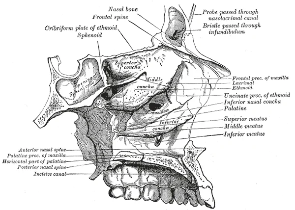
Long description
Roof, floor, and lateral wall of left nasal cavity.

Figure 3: Roof, floor, and lateral wall of left nasal cavity.
The middle cranial fossa is deeper and wider. It houses the temporal lobes. The sella turcica sits in the centre. The pituitary gland rests in the hypophyseal fossa of the sella. The cavernous sinuses lie lateral to the sella. The internal carotid arteries run through these sinuses. Several foramina pierce the sphenoid bone here. The optic canal transmits the optic nerve and ophthalmic artery. The foramen spinosum transmits the middle meningeal artery and vein. [1] [2]
The posterior cranial fossa is the deepest and smallest. It houses the cerebellum and brainstem. The foramen magnum is the largest opening. It transmits the medulla oblongata, vertebral arteries and spinal accessory nerve. The jugular foramen lies lateral to the foramen magnum. It transmits the internal jugular vein and cranial nerves nine, ten and eleven. The hypoglossal canal transmits the hypoglossal nerve. These nerves relate closely to the petrous part of the temporal bone. [2] [1]
| Foramen | Fossa | Key Contents |
|---|---|---|
| Cribriform plate | Anterior | Olfactory nerves |
| Optic canal | Middle | Optic nerve, ophthalmic artery |
| Superior orbital fissure | Middle | CN III, IV, V1, VI |
| Foramen rotundum | Middle | CN V2 |
| Foramen ovale | Middle | CN V3, accessory meningeal artery |
| Foramen spinosum | Middle | Middle meningeal artery and vein |
| Foramen magnum | Posterior | Medulla, vertebral arteries, CN XI |
| Jugular foramen | Posterior | Internal jugular vein, CN IX, X, XI |
| Hypoglossal canal | Posterior | CN XII |
Fractures of the base of the skull often involve these foramina. Anterior fossa fractures may cause CSF rhinorrhoea. Middle fossa fractures may cause CSF otorrhoea or facial nerve palsy. Posterior fossa fractures are rare but serious. They may compress the brainstem. The relations to the sinuses are critical. The sphenoid sinus lies below the sella turcica. Fractures here can damage the pituitary gland. The ethmoid sinuses lie below the anterior fossa. Fractures here can damage the olfactory nerves. Understanding these relations helps in diagnosis and management.
Understanding the cranial fossae requires correlating surface landmarks with deep bony architecture visible on imaging. The anterior cranial fossa forms the floor of the anterior cranial compartment. On axial CT, it appears as a shallow depression anterior to the planum sphenoidale. The frontal lobes rest directly upon the orbital plates of the frontal bone. These plates are thin and susceptible to fracture. The cribriform plate of the ethmoid bone lies centrally. It transmits olfactory nerve fibres from the nasal cavity to the brain. The crista galli projects superiorly from this plate. It serves as an attachment point for the falx cerebri. On MRI, the cerebrospinal fluid spaces are hyperintense on T2-weighted images. This contrast helps delineate the subarachnoid space around the frontal poles.
The middle cranial fossa is deeper and more complex. It is bounded anteriorly by the lesser wings of the sphenoid and posteriorly by the petrous temporal bones. The sella turcica sits in the centre. It houses the pituitary gland. The optic canals transmit the optic nerves. These canals are located just anterior to the sella. The superior orbital fissures lie lateral to the sella. They transmit cranial nerves III, IV, V1, and VI. On CT, the petrous ridges appear as dense, curved structures. They separate the middle from the posterior fossa. The temporal lobes occupy the majority of this fossa. They rest against the squamous and petrous parts of the temporal bone. MRI is superior for visualising the cavernous sinus. This venous channel lies lateral to the sella. It contains the internal carotid artery and cranial nerves.
The posterior cranial fossa is the largest and deepest. It accommodates the cerebellum and brainstem. The tentorium cerebelli separates it from the occipital lobes above. The foramen magnum is the largest opening. It transmits the medulla obliquely into the spinal canal. The clivus slopes downwards from the dorsum sellae to the foramen magnum. The petrous temporal bones form the lateral walls. The internal acoustic meatus lies on the posterior surface of the petrous bone. It transmits the facial and vestibulocochlear nerves. On MRI, the fourth ventricle is clearly visible anterior to the cerebellum. It communicates with the central canal of the medulla. The transverse sinuses run along the attached margin of the tentorium. They drain blood from the brain.
The middle fossa is central, showing the temporal lobes and sella turcica. The posterior fossa is at the bottom, containing the cerebellum and brainstem. The superior orbital fissures are lateral to the sella.
The cranial fossae form the floor of the cranial cavity, providing structural support for the brain and serving as critical landmarks for neurosurgical procedures. Understanding their boundaries is essential for interpreting imaging and managing trauma. The anterior cranial fossa supports the frontal lobes. Its floor is formed by the orbital plates of the frontal bone, the cribriform plate of the ethmoid bone, and the body of the sphenoid. The cribriform plate transmits the olfactory nerves. Injury here can cause cerebrospinal fluid rhinorrhoea. The middle cranial fossa supports the temporal lobes. It is deeper and wider than the anterior fossa. The central part is the sella turcica, which houses the pituitary gland. The sphenoid bone forms the bulk of this fossa. The greater wing of the sphenoid forms the lateral wall. Key foramina transmit important nerves and vessels. The foramen rotundum transmits the maxillary nerve. The foramen ovale transmits the mandibular nerve. The foramen spinosum transmits the middle meningeal artery. Fractures through these foramina can cause specific nerve palsies. The posterior cranial fossa is the deepest and largest. It houses the cerebellum and brainstem. The occipital bone forms the posterior wall. The sphenoid and temporal bones form the anterior and lateral walls. The foramen magnum transmits the medulla oblongata and vertebral arteries. The jugular foramen transmits the glossopharyngeal, vagus, and accessory nerves. The hypoglossal canal transmits the hypoglossal nerve.
Surface landmarks are vital for clinical examination. The internal occipital protuberance marks the confluence of sinuses. Fractures here can cause epidural haematoma.
The anterior fossa is at the top, the middle in the centre, and the posterior at the bottom. The sella turcica is central in the middle fossa. The foramen magnum is central in the posterior fossa.
Measurable sexual dimorphism traits are found in the CFS, particularly the crista galli, and can be used as indicators in forensic sex estimation. This anatomical variation is relevant in forensic identification but has limited clinical relevance for acute management. [2]
| Foramen | Structure Transmitted | Clinical Significance |
|---|---|---|
| Rotundum | Maxillary nerve (V2) | Loss of sensation in mid-face |
| Ovale | Mandibular nerve (V3) | Weakness of muscles of mastication |
| Spinosum | Middle meningeal artery | Epidural haematoma if fractured |
The anterior cranial fossa is a shallow depression formed primarily by the frontal, ethmoid, and sphenoid bones. It houses the frontal lobes of the cerebrum. A key landmark is the crista galli, a vertical ridge of the ethmoid bone. This structure serves as the attachment point for the falx cerebri. Measurements of the crista galli show sexual dimorphism in some populations. Female lengths in these latter populations range from 11.06 to 14.57 mm. One study by Komut and Golpinar found accuracy rates for linear measurements between 81.2% and 83.7%. [2]
The middle cranial fossa is deeper and houses the temporal lobes. It is bounded laterally by the greater wings of the sphenoid and the temporal bones. The sella turcica sits centrally within this fossa. It houses the pituitary gland. Dimensions of the sella turcica vary by sex. In the Colombian population, mean height is 47.75 mm in males and 45.12 mm in females. Mean length is 38.73 mm in males and 37.15 mm in females. These differences are statistically significant with a p-value of 0.000. [2]
The posterior cranial fossa is the deepest and largest fossa. It houses the cerebellum, pons, and medulla oblongata. The foramen magnum is its central opening. Shape analysis of the foramen magnum reveals population-specific trends. In the Indian population, 29.09% of males have a round foramen magnum. Another 25.45% of males have an oval shape. In females, the most common shape is hexagonal, occurring in 27.27% of cases. A round shape is found in 25.45% of females. Geometric morphometric analysis in Bosnia and Herzegovina showed classification accuracy based on form was 62.25% in males. Accuracy based on shape was 62.41% in males and 58.9% in females. [2]
Clinical relevance extends to nearby neural structures. The vestibular and cochlear nuclei are close neighbors. Damage to structures near these nuclei can cause deficits in both hearing and balance. Eye movement control involves the lateral and medial rectus muscles. The medial rectus is controlled by the oculomotor nucleus in the midbrain. These nuclei are not in the fossae but are closely related to the brainstem structures resting in the posterior fossa. [1]
Clinical pearlCrista Galli Measurement
Remember that crista galli dimensions vary significantly by population and sex, with Colombian males averaging longer lengths than Taiwanese males.
Exam trapSella Turcica Dimensions
Do not assume uniform sella turcica size; Colombian males have significantly greater mean height and length than females.
DefinitionPosterior Fossa Contents
The posterior cranial fossa contains the cerebellum, pons, and medulla, bounded by the occipital bone and temporal bones.
NoteForamen Magnum Shape
In Indian populations, hexagonal is the most common foramen magnum shape in females, while round is most common in males.
Clinical pearlVestibular and Cochlear Proximity
Damage near the vestibular and cochlear nuclei can affect both hearing and balance due to their anatomical proximity.
DefinitionVisual Acuity Standard
Normal visual acuity is defined by a letter subtending 5 minutes of an arc at 20 feet. [1]
Note20/60 Vision Meaning
20/60 vision means a patient sees at 20 feet what a normal eye sees at 60 feet. [1]
Clinical pearlForamen Magnum Classification
Classification accuracy for foramen magnum shape in Bosnia and Herzegovina was approximately 62% for both sexes.
Exam trapAnterior Fossa Boundaries
The anterior cranial fossa is formed by the frontal, ethmoid, and sphenoid bones, not the temporal bones.
DefinitionMiddle Fossa Landmark
The sella turcica is the central landmark of the middle cranial fossa, housing the pituitary gland.
NoteVisual Field Testing
Visual field boundaries can be assessed clinically by asking patients to report when fingers disappear from peripheral vision. [1]
DefinitionCommon pitfalls
Students often confuse the anatomical boundaries of the cranial fossae with adjacent structures, misinterpret measurements, or overlook population‑specific variations that affect sex estimation.
-
Assuming the cranial fossae are identical in all populations
The width of the foramen magnum varies markedly between groups: 30.1–32.66 mm in males and 27.75–31.0 mm in females in several Asian populations, whereas the Colombian mean is 22.66 mm in males and 20.97 mm in females.
Correct: Always reference population‑specific reference ranges when using these measurements for sex estimation. -
Treating the foramen ovale distance as a single metric
The right‑left distance differs by sex and ethnicity: 57.3–62.5 mm in males and 55.6–60.1 mm in females in Chinese, Taiwanese, and Iranian groups, while in Colombians the mean is 8.64 mm in females versus 8.13 mm in males.
Correct: Use both length and breadth, and compare with the appropriate population data. -
Ignoring the clivus dimensions
The maximum width of the clivus is 63.7 mm in Chinese males and 48.11 mm in Colombian males, with a similar pattern for females (61.7 mm vs 43.48 mm). [2] Correct: Include clivus width and length in a multivariate sex‑estimation model rather than relying on a single parameter. -
Overlooking the influence of sex hormones on bone growth
Androgens promote radial bone expansion, while estrogens restrict periosteal expansion, leading to larger male dimensions in the cranial fossae. [2] Correct: Explain the biological basis for dimensional differences rather than attributing them solely to mechanical load. -
Assuming linear measurements alone are sufficient
The accuracy of sex classification using foramen magnum width alone is 67.3 % and 63.2 % for length. [2] Correct: Combine multiple measurements (e.g., foramen magnum, clivus, foramen ovale) to improve classification accuracy. -
Neglecting the role of skeletal plasticity
Past gender‑based activities influenced bone morphology, contributing to observed size differences.
Correct: Consider historical and cultural context when interpreting cranial fossa dimensions. -
Failing to use population‑specific reference data
The accuracy of sex determination is greatly enhanced with data from the same ancestral group.
Correct: Always match the sample to the appropriate reference population. -
Misidentifying the boundaries of the cranial fossae
The anterior cranial fossa is bounded anteriorly by the frontal bone, laterally by the sphenoid, and posteriorly by the ethmoid; the middle fossa is bordered by the temporal bone and the sphenoid; the posterior fossa is limited by the occipital bone and the foramen magnum.
Correct: Use clear anatomical landmarks when describing each fossa.
DefinitionClinical relevance
Knowledge of cranial fossa anatomy guides neuro‑imaging interpretation, surgical planning, and forensic sex estimation.
- Neurological examination: The trigeminal nerve exits the skull through the foramen ovale and the foramen spinosum. Discrimination of light touch versus pain at these foramina helps localise lesions in the brain stem, as pain is mediated by the spinal trigeminal nucleus in the medulla. [1]
- Cranial nerve testing: Assessing olfactory function (anosmia) informs about potential damage to the olfactory bulb within the anterior cranial fossa, which can affect flavour perception.
- Surgical approach to the middle cranial fossa: The temporal bone and sphenoid form the lateral boundary; accurate knowledge of the foramen ovale and spinosum is essential to avoid nerve injury during procedures such as trigeminal neuralgia blocks.
- Posterior fossa surgery: The foramen magnum is the gateway to the spinal cord; its width and length influence the choice of craniotomy size and the risk of brainstem compression.
- Computed tomography (CT): Measurements of the foramen magnum width (30.1–32.66 mm in Asian males) and length (34.5–37.6 mm in Asian males) are routinely taken to assess for cranial base anomalies.
- Magnetic resonance imaging (MRI): The clivus and its dimensions (maximum width 63.7 mm in Chinese males) are visualised to evaluate midline skull base lesions.
- Forensic imaging: Radiographic measurements of the foramen ovale distance (57.3–62.5 mm in Asian males) aid in sex estimation when skeletal remains are incomplete.
- Multiple‑choice questions: Students are asked to match cranial fossa boundaries, recognise the significance of foramen dimensions, and calculate sex‑estimation accuracy percentages.
- Practical exams: Identification of foramina on dry skulls and measurement of the foramen magnum width and length are common tasks.
- Sex estimation: Combining foramen magnum width (accuracy 67.3 %) and clivus length (accuracy 69 % in UAE males) improves classification rates.
- Population identification: Differences in clivus width and foramen ovale distance between Chinese and Colombian populations highlight the need for population‑specific reference data.
Clinical decision tree for cranial fossa assessment
- Patient presents with cranial neuropathyAssess sensory discrimination
- Question: Painful stimulus recognised?
Yes
- Spinal trigeminal nucleus likely intact
No
- Consider medullary lesion
- The foramen magnum width ranges 30.1–32.66 mm in Asian males and 27.75–31.0 mm in Asian females. [2]
- Foramen ovale distance differs by sex and ethnicity; e.g., 57.3–62.5 mm in Asian males. [2]
- Clivus maximum width is 63.7 mm in Chinese males and 48.11 mm in Colombian males.
- Sex estimation accuracy improves when multiple cranial fossa measurements are combined. [2]
- Population‑specific reference data are essential for accurate forensic classification. [2]
- Trigeminal nerve testing distinguishes light touch from pain via the spinal trigeminal nucleus.
- Olfactory dysfunction indicates possible anterior cranial fossa pathology.
- Surgical planning in the middle and posterior fossae relies on precise anatomical boundaries.
Answer each one in your head first, then open the answer.
What is the typical width of the foramen magnum in Asian males?
Show answerHide answer
30.1–32.66 mmWhich nucleus mediates painful stimuli from the foramen ovale?
Show answerHide answer
Spinal trigeminal nucleusName one factor that contributes to sex differences in cranial fossa dimensions.
Show answerHide answer
Sex hormones (androgens/estrogens)What is the accuracy of sex classification using foramen magnum width alone?
Show answerHide answer
67.3 %Why is population‑specific data important in forensic anthropology?
Show answerHide answer
It improves classification accuracy by accounting for intra‑population variation
- Betts JG, Young KA, Wise JA, et al. The Cranial Nerve Exam. OpenStax, Anatomy and Physiology 2e (CC BY-NC-SA 4.0). Accessed .
- Haslan H, Zulkiflee NDI, Alias A, et al. Sexual dimorphism of cranial fossae in forensic anthropology: A systematic review. Journal of Taibah University Medical Sciences 2026;21(3):393-410. doi:10.1016/j.jtumed.2026.03.014. Accessed .
Figure credits
Educational content for medical students and doctors. It is not a substitute for clinical judgement or local guidelines.