Facial muscles
The facial region contains muscles of expression, mastication and tongue control, innervated mainly by the trigeminal and facial nerves.
The facial region is the anterior part of the head bounded superiorly by the skull base, inferiorly by the mandible, laterally by the zygomatic arches, and medially by the nasal bones. It is encompassed by the facial muscles, which are divided into the muscles of mastication and the muscles of facial expression. The muscles of mastication originate from the temporal bone, the sphenoid bone, and the mandible, and insert onto the mandible or the maxilla, allowing jaw movements. The muscles of facial expression originate from the skull and insert into the skin of the face, enabling expressions such as smiling, frowning and blinking.
The trigeminal nerve (cranial nerve V) supplies the muscles of mastication and provides cutaneous sensation to the face. The hypoglossal nerve (cranial nerve XII) controls the extrinsic and intrinsic muscles of the tongue, except for the palatoglossus which is supplied by the vagus nerve. [2] [3]
This reflex demonstrates the integration of sensory input and motor output within the cranial nerve pathways. [3]
The boundaries of the facial region are clinically relevant when assessing cranial nerve function. For example, a deficit in the hypoglossal nerve produces a tongue that deviates toward the side of the lesion when protruded, indicating unilateral weakness. Similarly, loss of facial nerve function results in asymmetry of facial expressions, such as inability to raise the corner of the mouth on the affected side. [3]
| Muscle | Origin | Insertion | Action | Nerve |
|---|---|---|---|---|
| Masseter | Zygomatic arch | Ramus of mandible | Elevates mandible | Trigeminal (V3) |
| Temporalis | Temporal fossa | Coronoid process | Elevates, retracts mandible | Trigeminal (V3) |
| Frontalis | Galea aponeurotica | Skin of forehead | Raises eyebrows | Facial (VII) |
| Orbicularis oculi | Orbital margin | Skin around eye | Closes eyelids | Facial (VII) |
| Zygomaticus major | Zygomatic bone | Skin of cheek | Raises mouth corner | Facial (VII) |
| Buccinator | Alveolar processes | Cheek | Compresses cheek | Facial (VII) |
| Depressor anguli oris | Mandible | Skin of mouth | Lowers mouth corner | Facial (VII) |
| Levator labii superioris | Maxilla | Skin of upper lip | Elevates upper lip | Facial (VII) |
| Genioglossus | Mandible | Tongue | Protrudes tongue | Hypoglossal (XII) |
| Hyoglossus | Hyoid bone | Tongue | Retracts, depresses tongue | Hypoglossal (XII) |
The facial region’s anatomy, innervation and functional boundaries form the foundation for clinical assessment of cranial nerve integrity and for understanding the mechanics of facial expression and mastication.
Lateral to the orbit, the orbicularis oculi forms a circular sphincter around the eye, with its palpebral part lying just beneath the skin of the eyelids. The zygomaticus major and minor muscles originate from the zygomatic bone and insert into the angle of the mouth, forming prominent bands that elevate the lip. The buccinator muscle lies deep to the zygomaticus but its fibers can be inferred along the cheek, compressing the cheek against the teeth. The orbicularis oris surrounds the mouth, forming a complex sphincter that closes and protrudes the lips. The surface markings correspond to the underlying muscle bellies and tendons, which can be palpated during clinical examination. Understanding this layout is essential for identifying nerve supply and potential sites of injury. [3]
Figure 1Gray381
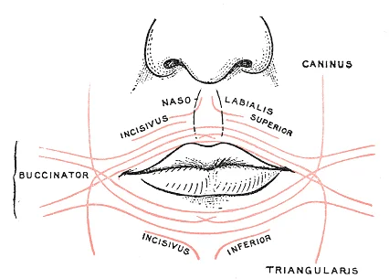
Long description
Scheme showing arrangement of fibers of Orbicularis oris.

Figure 1: Scheme showing arrangement of fibers of Orbicularis oris.
DefinitionFacial Expression
Muscles derived from the second pharyngeal arch, innervated by the facial nerve, responsible for moving the skin of the face.
The facial nerve (CN VII) is responsible for the muscles involved in facial expressions.
| Muscle | Origin | Insertion | Action | Nerve Supply |
|---|---|---|---|---|
| Frontalis | Epicranial aponeurosis | Skin of eyebrows | Elevates eyebrows | Facial nerve |
| Orbicularis oculi | Frontal process of maxilla | Skin around orbit | Closes eyelids | Facial nerve |
| Zygomaticus major | Zygomatic bone | Angle of mouth | Elevates angle of mouth | Facial nerve |
| Orbicularis oris | Maxilla and mandible | Skin of lips | Closes and protrudes lips | Facial nerve |
Clinical assessment involves asking the patient to perform specific movements, such as smiling, closing eyes tightly, and puffing out cheeks. Asymmetry may indicate facial nerve palsy. The surface anatomy provides the roadmap for identifying these muscles during physical examination.
The reader should observe the frontal bone superiorly, which serves as the origin for the frontalis muscle. This muscle lies immediately beneath the galea aponeurotica and inserts into the skin of the eyebrows. Its contraction elevates the eyebrows and wrinkles the forehead, a key component of expressive facial movement.
The mandible forms the lower boundary of the facial skeleton. The platysma, a broad, thin sheet of muscle, arises from the fascia over the pectoralis major and deltoid, ascending along the neck to insert into the inferior border of the mandible and the skin of the lower face. It tenses the skin of the neck and depresses the mandible. The masseter, a powerful muscle of mastication, originates from the zygomatic arch and inserts into the ramus and angle of the mandible. It elevates the mandible, closing the jaw. The temporalis muscle, originating from the temporal fossa, inserts into the coronoid process of the mandible, also contributing to jaw closure and retraction.
The nasal bones form the bridge of the nose. The nasalis muscle originates from the maxilla and inserts into the aponeurosis across the dorsum of the nose. It compresses the nasal cartilage, narrowing the nostrils. The orbicularis oculi surrounds the orbit, originating from the frontal process of the maxilla and inserting into the skin around the eye. It closes the eyelids, protecting the cornea and contributing to blinking. The orbicularis oris encircles the mouth, originating from the maxilla and mandible and inserting into the modiolus at the angle of the mouth. It compresses the lips, essential for speech and sucking.
The buccinator muscle originates from the alveolar processes of the maxilla and mandible and the pterygomandibular raphe, inserting into the modiolus. It compresses the cheeks against the teeth, aiding in chewing and blowing. The mentalis muscle originates from the mandible and inserts into the skin of the chin, elevating and protruding the lower lip. These muscles act on the facial bones to produce expression and facilitate oral functions. The precise bony landmarks ensure efficient force transmission for both aesthetic and functional movements.
The SMAS lies deep to the subcutaneous fat and superficial to the parotidomasseteric fascia. It covers the parotid gland and the masseter muscle. The facial nerve branches run within this plane. The zygomatic arch provides a bony landmark.
The muscles of facial expression are unique because they originate from bone or fascia and insert directly into the skin, allowing for subtle movements that facilitate non-verbal communication. These muscles are innervated by the facial nerve (cranial nerve VII). Understanding their anatomy requires grouping them by functional compartments and recognising their relationship with sensory innervation and vascular supply. [2]
Figure 2Expression of the Emotions Figure 1
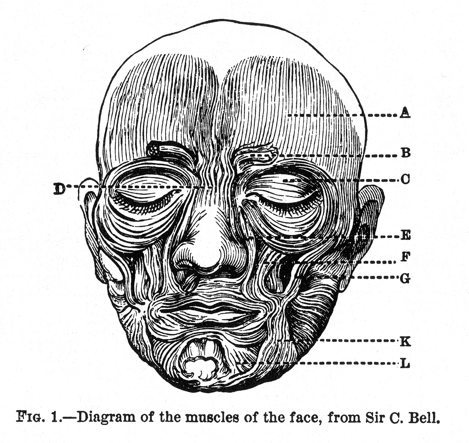
Long description
Figure 1 from Charles Darwin's The Expression of the Emotions in Man and Animals, showing human facial muscles (see also figure 2 and 3 (both by Henle), the latter of which has the key). Caption reads "FIG. 1.—Diagram of

Figure 2: Figure 1 from Charles Darwin's The Expression of the Emotions in Man and Animals, showing human facial muscles (see also figure 2 and 3 (both by Henle), the latter of which has the key) (caption reads "fig).
Figure 3Gray380
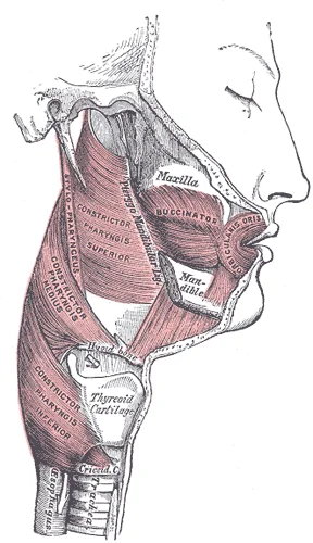
Long description
Muscles of the pharynx and cheek. Labelled illustration from Wikimedia Commons, used under its open licence.

Figure 3: Muscles of the pharynx and cheek.
| Muscle | Origin | Insertion | Action | Nerve Supply |
|---|---|---|---|---|
| Frontalis | Galea aponeurotica | Skin of eyebrows | Raises eyebrows, wrinkles forehead | Temporal branch of facial nerve |
| Orbicularis oculi | Frontal process of maxilla | Skin around orbit | Closes eyelids | Zygomatic and buccal branches of facial nerve |
| Orbicularis oris | Maxilla and mandible | Skin of lips | Closes and purses lips | Buccal and marginal mandibular branches of facial nerve |
| Zygomaticus major | Zygomatic bone | Angle of mouth | Draws angle of mouth up and out | Zygomatic and buccal branches of facial nerve |
| Buccinator | Alveolar processes of maxilla and mandible | Orbicularis oris | Compresses cheek against teeth | Buccal branch of facial nerve |
| Platysma | Fascia of pectoralis major and deltoid | Skin of lower face and mandible | Tenses skin of neck, depresses mandible | Cervical branch of facial nerve |
The facial nerve branches are critical landmarks. The temporal branch supplies the frontalis and orbicularis oculi. The zygomatic branch supplies the orbicularis oculi and zygomaticus muscles. The buccal branch supplies the buccinator and orbicularis oris. The marginal mandibular branch supplies the depressor anguli oris and mentalis. The cervical branch supplies the platysma.
Figure 4Gray378
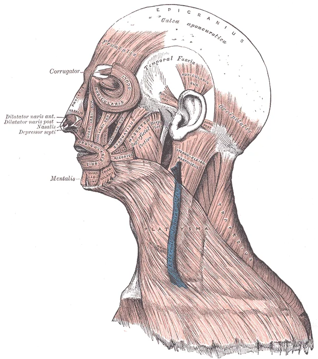
Long description
Muscles of the head, face, and neck. Labelled illustration from Wikimedia Commons, used under its open licence.

Figure 4: Muscles of the head, face, and neck.
This sensory map overlaps with the motor supply of the facial nerve.
The blood supply to the face is rich, coming from the external carotid artery. The facial artery is a major branch. It gives off the inferior labial, superior labial, and lateral nasal branches. The angular artery is the terminal branch. The danger triangle of the face is bounded by the corners of the mouth and the bridge of the nose. Infections here can spread to the cavernous sinus via the angular vein, which connects to the ophthalmic vein. This is a clinical trap.
Exam trapDanger Triangle
Infections in the danger triangle can spread to the cavernous sinus via the angular vein, leading to serious complications.
The muscles of expression are thin and variable. They allow for the complex movements of the face. The facial nerve branches must be identified carefully during surgery to avoid paralysis. The trigeminal territories help localise sensory deficits. The blood supply ensures rapid healing but also poses a risk of haematoma. Understanding these relationships is essential for clinical practice.
The external carotid artery ascends in the neck and gives off several branches. The first major branch relevant to the face is the facial artery. It arises from the external carotid artery in the neck. It courses anteriorly towards the face. It passes deep to the submandibular gland. It then winds over the inferior border of the mandible. This point is a key landmark. The artery is palpable here. It then enters the face. It courses superiorly and anteriorly. It runs alongside the nose. It gives off branches to the lower lip and cheek. These branches anastomose with branches from the ophthalmic artery. The angular artery supplies the medial canthus of the eye. It anastomoses with the dorsal nasal branch of the ophthalmic artery. This anastomosis is clinically significant. It provides collateral circulation. [1]
Another important branch is the superficial temporal artery. It arises from the external carotid artery. It ascends anterior to the ear. It passes superficial to the zygomatic arch. It divides into frontal and parietal branches. These branches supply the scalp and the temporalis muscle. The maxillary artery is the other terminal branch of the external carotid artery. It arises deep in the parotid gland. It courses anteriorly. It gives off the infraorbital artery. This artery exits the infraorbital foramen. It supplies the lower eyelid and the side of the nose. It anastomoses with the angular artery.
The arterial supply ensures that the muscles of facial expression receive adequate blood flow. These muscles are thin and lie in the subcutaneous tissue. They are involved in expression. The vessels run deep to the muscles. They are protected by the muscle layer. The anastomoses between the external and internal carotid systems provide redundancy. This is important for tissue survival.
The labels identify each vessel. The reader should note the course of the facial artery. It is a key landmark. The angular artery is also important. It connects to the ophthalmic system. The superficial temporal artery supplies the scalp. The maxillary artery supplies the deep structures. The reader can trace the flow of blood. This aids in understanding the supply. The labels are clear. The structures are well-defined. The relationships are clear.
The facial muscles receive their arterial supply primarily from branches of the external carotid artery, notably the facial artery and the superficial temporal artery. Understanding the venous drainage is critical because of the unique connections between the facial veins and the intracranial cavity.
The venous system of the face is characterised by a lack of valves in many of its tributaries. This anatomical feature permits bidirectional blood flow. Consequently, septic emboli from an infected lesion in the danger triangle can travel posteriorly into the cranial cavity. The cavernous sinus is a paired dural venous sinus located on either side of the sella turcica. It receives blood from the superior and inferior ophthalmic veins, which drain the orbit and the medial aspect of the face. It is formed by the union of the supratrochlear and supraorbital veins. It lies superficial to the buccinator and masseter muscles. The deep facial vein provides a crucial link between the superficial facial vein and the deep pterygoid plexus. The pterygoid plexus surrounds the lateral pterygoid muscle and the maxillary artery. Emissary veins connect this plexus to the cavernous sinus and the sphenoparietal sinus. These connections bypass the dural sinuses' usual unidirectional flow. [1]
The clinical significance of this anatomy cannot be overstated. A boil or abscess in the upper lip or nose is particularly dangerous. The infection can spread via the facial vein to the angular vein. From there, it enters the superior ophthalmic vein. The superior ophthalmic vein drains directly into the cavernous sinus. Once in the cavernous sinus, the infection can spread to the contralateral side via intercavernous sinuses. It can also extend into the internal jugular vein. This can lead to severe complications such as meningitis, brain abscess, or septic shock. Therefore, manipulation of lesions in the danger triangle is strictly contraindicated.
Clinical pearlDanger Triangle
The area from the corners of the mouth to the bridge of the nose is the danger triangle. Infections here can spread to the cavernous sinus via valveless veins.
| Structure | Origin/Location | Termination/Connection | Clinical Note |
|---|---|---|---|
| Angular Vein | Medial angle of eye | Facial vein | Start of facial drainage |
| Facial Vein | Angular vein | Internal jugular vein | Valveless, risk of retrograde spread |
| Deep Facial Vein | Facial vein | Pterygoid plexus | Connects superficial to deep system |
| Pterygoid Plexus | Infratemporal fossa | Cavernous sinus (via emissary veins) | Route for intracranial spread |
| Superior Ophthalmic Vein | Orbit | Cavernous sinus | Direct link from face to sinus |
The cranial nerves are the twelve nerves that arise from the brain or brain‑stem and supply the head and neck. Five of these nerves are purely motor, three are purely sensory and four are mixed. The motor nerves innervate the extra‑ocular muscles, the muscles of mastication, the tongue, the pharynx and the neck. The sensory nerves carry general sensation from the face, scalp and oral cavity, and special senses such as smell, sight and hearing. Mixed nerves contain both sensory and motor fibres and are responsible for facial expression, taste and autonomic control of salivary glands and the upper aerodigestive tract. [2] [3] The spinal nerves are all mixed nerves. They emerge from the spinal cord through intervertebral foramina, reorganise in plexuses and give rise to systemic nerves that supply the trunk and limbs. The thoracic spinal nerves do not form a plexus but give rise directly to intercostal nerves.
The facial nerve (CN VII) is responsible for the muscles involved in facial expressions. [2]
DefinitionFacial Nerve (CN VII)
The facial nerve is a mixed cranial nerve that provides motor innervation to the muscles of facial expression and sensory innervation to the anterior two‑thirds of the tongue and parasympathetic fibres to the lacrimal, submandibular and sublingual glands.
| Branch | Muscles supplied | Skin territory (sensory) |
|---|---|---|
| Temporal | Frontalis, corrugator supercilii | Upper eyelid, forehead |
| Zygomatic | Orbicularis oculi, orbicularis oris | Upper cheek, upper lip |
| Buccal | Buccinator, orbicularis oris | Lower cheek, lower lip |
| Marginal mandibular | Depressor anguli oris, depressor labii inferioris | Lower lip, chin |
| Cervical | Platysma | Neck skin |
Table 1 – Motor and cutaneous distribution of the facial nerve branches.
The scalp is supplied by the supratrochlear, supraorbital, zygomaticotemporal, auriculotemporal, posterior auricular, occipital and greater occipital nerves. These are branches of the ophthalmic (CN V₁), maxillary (CN V₂) and mandibular (CN V₃) divisions of the trigeminal nerve, and the cervical plexus (C₁–C₃). [2]
Figure 7 – Nerves of facial muscles and layers of the scalp. Lateral view, axial plane. The figure shows the skin, subcutaneous tissue, galea, pericranium and skull. The facial nerve branches (temporal, zygomatic, buccal, marginal mandibular, cervical) are labelled. Scalp sensory nerves (supratrochlear, supraorbital, zygomaticotemporal, auriculotemporal, posterior auricular, occipital, greater occipital) are labelled on the surface. The danger area is marked over the temporal region where the superficial temporal artery lies just beneath the galea.
Figure 5Gray789
Long description
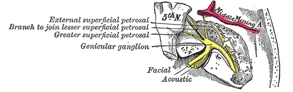
The course and connections of the facial nerve in the temporal bone.

Figure 5: The course and connections of the facial nerve in the temporal bone.
The lymphatic system provides the primary route for immune surveillance and fluid return from the facial muscles. Understanding this drainage is critical for predicting the spread of infection or malignancy in the head and neck region. The flow of lymph follows a predictable pattern, moving from superficial tissues to deep nodal groups before entering the venous circulation.
Posteriorly, the postauricular nodes lie behind the ear, receiving drainage from the scalp and external ear. The deep cervical nodes ultimately drain into the jugular trunk, which empties into the venous angle. This visualisation demonstrates how the facial muscles' overlying skin and subcutaneous tissue drain into these specific nodal basins.
The submental nodes receive lymph from the central lower lip, the floor of the mouth, and the tip of the tongue. These nodes drain primarily into the submandibular nodes. The submandibular nodes are larger and receive drainage from the lateral lower lip, the cheek, the nose, and the upper lip. They also receive input from the submental nodes. From the submandibular nodes, lymph flows into the deep cervical nodes.
The preauricular and parotid nodes drain the lateral aspects of the eyelids, the temporal region, and the external ear. These nodes also drain into the deep cervical chain. The postauricular nodes drain the posterior scalp and the external auditory meatus, also connecting to the deep cervical nodes.
The deep cervical lymph nodes form a vertical chain along the internal jugular vein. They are the final common pathway for most head and neck lymphatic drainage. The jugular trunk, formed by the confluence of deep cervical drainage, typically empties into the junction of the internal jugular and subclavian veins. On the right side, this may enter the right lymphatic duct, while on the left, it enters the thoracic duct. [1]
Clinical pearlClinical Relevance
Malignancies of the face often metastasize to the regional lymph nodes before spreading distantly. Palpation of these nodes is a key part of the physical examination.
The facial muscles are situated within the superficial fascia, directly deep to the skin and superficial to the deep facial structures. They are arranged in groups that encircle the natural orifices of the face, allowing for the expression of emotion and the manipulation of food. Understanding their relations requires visualising the face as a series of concentric layers. The most superficial layer is the skin, followed by the subcutaneous tissue containing the facial muscles, the deep fascia, and finally the underlying bone or cartilage.
Deep to the skin lies the orbicularis oculi muscle, which encircles the orbit. Medial to the orbicularis oculi is the nasalis muscle, which lies over the nasal cartilage. Below the eye, the zygomaticus major muscle originates from the zygomatic bone and inserts into the corner of the mouth.
| Muscle | Origin | Insertion | Action | Nerve Supply |
|---|---|---|---|---|
| Orbicularis oculi | Frontal process of maxilla | Skin around orbit | Closes eyelids | Facial nerve |
| Zygomaticus major | Zygomatic bone | Angle of mouth | Pulls angle up and out | Facial nerve |
| Buccinator | Alveolar processes | Orbicularis oris | Compresses cheek | Facial nerve |
| Frontalis | Epicranial aponeurosis | Skin of forehead | Raises eyebrows | Facial nerve |
The spaces surrounding these muscles are critical for the spread of infection or fluid. The superficial facial spaces contain the muscles and the neurovascular bundles. The facial artery and vein pass anterior to the masseter and then curve upwards towards the medial canthus of the eye.
DefinitionFacial Muscles
Striated muscles located in the subcutaneous tissue of the face, responsible for facial expression and controlled by the facial nerve.
The relations of the facial muscles to the underlying bones determine their mechanical advantage. The maxilla and mandible provide attachment points for the muscles of mastication and expression. The nasal bones and cartilages provide support for the nasalis and levator labii superioris alaeque nasi. The mandible provides the insertion for the depressor anguli oris and depressor labii inferioris. The frontal bone provides the superior attachment for the frontalis. The temporalis muscle passes deep to the zygomatic arch, which acts as a pulley.
Exam trapNerve Supply
The masseter is supplied by the trigeminal nerve, not the facial nerve. All other facial expression muscles are supplied by the facial nerve.
The venous drainage of the face is significant due to the lack of valves in the facial veins. Blood from the facial vein can flow backwards into the cavernous sinus. This is a potential route for infection to spread from the face to the brain. These arteries supply the lips and nose.
NoteClinical Note
Infection in the 'danger triangle' of the face can spread to the cavernous sinus via the facial vein.
Facial muscles are soft-tissue structures that are poorly visualised on plain X-ray due to the lack of inherent contrast with surrounding fat and connective tissue. They appear as indistinct soft-tissue densities superimposed over the facial bones. Consequently, plain radiography is rarely useful for assessing the integrity or pathology of these muscles directly. [2]
Computed tomography (CT) provides superior anatomical detail by exploiting differences in tissue density. On CT, facial muscles appear as intermediate-density structures, darker than bone but lighter than subcutaneous fat. They are best visualised in axial and coronal planes. The masseter, temporalis, and buccinator muscles can be clearly delineated. CT is particularly valuable for identifying mass lesions, such as abscesses or tumours, which may distort or displace normal muscle architecture. The relationship of these muscles to the mandible and zygomatic arch is clearly defined, aiding in surgical planning. [4]
Magnetic resonance imaging (MRI) is the gold standard for soft-tissue evaluation of the face. It offers excellent contrast resolution, allowing for the differentiation of individual muscle bundles and their fascial planes. On T1-weighted images, muscle tissue appears with intermediate signal intensity. On T2-weighted images, normal muscle shows low signal intensity, while pathological changes such as oedema or inflammation result in high signal intensity. MRI is essential for assessing neuromuscular junction disorders, myopathies, and infiltrative diseases. It can also visualise the facial nerve (CN VII) and its branches within the parotid gland and facial clefts, providing critical information for diagnosing nerve compression or trauma.
Ultrasound is a dynamic, real-time imaging modality that is increasingly used for superficial structures. It allows for the assessment of muscle thickness, contractility, and blood flow using Doppler techniques. Ultrasound is particularly useful for guiding injections into specific facial muscles, such as the masseter for bruxism or the orbicularis oculi for blepharospasm. It can differentiate between muscle, fat, and fluid collections. The high-frequency transducer provides detailed images of the superficial facial muscles, including the frontalis, corrugator, and orbicularis oculi. However, its utility is limited by the presence of air in the paranasal sinuses and the overlying bone, which obstructs the ultrasound beam.
The caption states: Coronal MRI showing the spatial relationships of major facial muscles.
DefinitionRadiological Anatomy
The visualisation of facial muscles relies on CT for bony relationships, MRI for soft-tissue detail, and ultrasound for dynamic assessment.
| Modality | Best For | Limitations |
|---|---|---|
| X-ray | Bony landmarks | Poor soft-tissue contrast |
| CT | Bony relationships, masses | Radiation exposure |
| MRI | Soft-tissue detail, nerves | Cost, availability |
| Ultrasound | Dynamic assessment, guidance | Operator dependent, air/bone interference |
They originate from bone or fascia and insert directly into the skin, allowing them to move the skin rather than joints. This arrangement enables the expression of emotion and the control of facial orifices. Clinically, understanding their anatomy is vital for managing facial trauma and nerve injuries.
The temporal branch runs superiorly towards the forehead. The zygomatic branch runs laterally towards the eye. The buccal branch runs horizontally towards the mouth. The marginal mandibular branch runs along the lower jaw. The cervical branch runs inferiorly into the neck. These structures are arranged in a fan-like distribution from the parotid gland.
Injuries to the face often involve lacerations that may sever these muscles or the overlying skin. Because the muscles insert into the skin, wounds can gape widely if the muscle fibres are cut. Surgical repair requires precise alignment of the muscle fibres to prevent scarring and restore function. Nerve palsies, particularly of the facial nerve (cranial nerve VII), result in paralysis of these muscles. A complete lesion causes inability to close the eye, drooping of the mouth, and loss of forehead wrinkling. Partial lesions may affect only specific branches, such as the marginal mandibular branch, causing asymmetry when smiling.
Surface landmarks are critical for locating the facial nerve branches during surgery. The tragus of the ear serves as a key landmark. The zygomatic branch emerges 0.5 cm below the zygomatic arch. The marginal mandibular branch emerges 1 cm below the angle of the mandible. These landmarks help surgeons avoid iatrogenic injury during procedures such as facelifts or parotidectomy.
Lacerations here can bleed profusely due to the rich vascular supply and the inability of the vessel walls to constrict because they are held open by the galea. Facial palsy can also result from trauma to the temporal bone, where the facial nerve runs through the facial canal. Fractures of the temporal bone can compress or sever the nerve, leading to immediate or delayed onset of facial weakness.
| Muscle | Origin | Insertion | Action | Nerve Supply |
|---|---|---|---|---|
| Orbicularis oculi | Frontal process of maxilla, medial palpebral ligament | Skin and muscle around orbit | Closes eyelids | Facial nerve |
| Orbicularis oris | Maxilla and mandible | Skin and muscle of lips | Closes and purses lips | Facial nerve |
| Zygomaticus major | Zygomatic bone | Angle of mouth | Elevates angle of mouth | Facial nerve |
| Buccinator | Alveolar processes of maxilla and mandible | Orbicularis oris | Compresses cheek | Facial nerve |
| Platysma | Fascia of pectoralis major and deltoid | Inferior border of mandible, skin of lower face | Depresses mandible, tenses skin of neck | Facial nerve |
They are innervated by the facial nerve (cranial nerve VII). Understanding their anatomy requires distinguishing between the muscles of facial expression and those of mastication, which are innervated by the trigeminal nerve.
These muscles are unique because they originate from bone or fascia and insert directly into the skin. This arrangement allows them to move the skin rather than joints. They are derived from the second pharyngeal arch.
| Muscle | Origin | Insertion | Action | Nerve Supply |
|---|---|---|---|---|
| Orbicularis oculi | Medial palpebral ligament | Skin of lateral eyelid | Closes eyelids | Facial nerve (CN VII) |
| Orbicularis oris | Maxilla and mandible | Skin of lips | Closes and protrudes lips | Facial nerve (CN VII) |
| Frontalis | Epicranial aponeurosis | Skin of forehead | Raises eyebrows | Facial nerve (CN VII) |
| Buccinator | Alveolar processes | Modiolus at corner of mouth | Compresses cheek | Facial nerve (CN VII) |
Lesions of the facial nerve result in distinct patterns of weakness. A peripheral lesion affects both the upper and lower face on the ipsilateral side. This includes inability to wrinkle the forehead or close the eye. A central lesion, such as from a stroke, typically spares the forehead. This is because the upper face receives bilateral cortical innervation.
Exam trapForehead sparing
A central facial palsy spares the upper face due to bilateral cortical input to the facial nucleus. A peripheral palsy affects the entire half of the face.
| Branch | Area of Distribution |
|---|---|
| Temporal | Forehead and upper eyelid |
| Zygomatic | Lower eyelid and side of nose |
| Buccal | Upper lip and nose |
| Marginal mandibular | Lower lip and chin |
| Cervical | Neck muscles (platysma) |
This pulse point is clinically significant for assessing circulation.
Clinical pearlDanger triangle
Infections in the nose and upper lip can spread to the cavernous sinus via valveless facial veins. This can cause life-threatening cavernous sinus thrombosis.
Students frequently confuse the embryological origins and nerve supplies of the muscles of facial expression with those of the muscles of mastication. A critical distinction is that the muscles of facial expression are derived from the second pharyngeal arch, whereas the muscles of mastication arise from the first pharyngeal arch. Consequently, they are innervated by different cranial nerves. The muscles of facial expression are innervated by the facial nerve (cranial nerve VII), while the muscles of mastication are innervated by the mandibular division of the trigeminal nerve (cranial nerve V3). A common error is to attribute the action of closing the jaw to the facial nerve; this is incorrect. Jaw closure is performed by the masseter, temporalis, and medial pterygoid muscles, which are all supplied by V3. [3]
Another frequent mistake involves the peripheral versus central lesions of the facial nerve. In a peripheral lesion, such as Bell’s palsy, the entire half of the face is affected, including the forehead. This is because the upper face receives bilateral cortical innervation, but the lower face receives only contralateral innervation. However, in a central lesion, such as a stroke affecting the motor cortex, the upper face is spared because the bilateral input to the forehead muscles remains intact from the unaffected hemisphere. Students often forget this distinction, leading to misdiagnosis. The ability to wrinkle the forehead is a key clinical sign in localizing the lesion.
A third pitfall is the misunderstanding of the branching pattern of the facial nerve. The nerve exits the skull via the stylomastoid foramen and then divides into terminal branches. These branches do not follow a simple linear path but form a plexus within the parotid gland. It is crucial to remember that the marginal mandibular branch runs inferior to the mandible and innervates the muscles of the lower lip and chin. Injury to this branch results in an asymmetrical smile, where the affected side of the lower lip fails to dep [2]
Furthermore, students often overlook the sensory component of the facial nerve. While primarily motor, the facial nerve carries taste sensation from the anterior two-thirds of the tongue via the chorda tympani. A lesion proximal to the takeoff of the chorda tympani will result in loss of taste on the ipsilateral anterior tongue. This is a subtle but important clinical finding that can help localize the level of the facial nerve lesion. Additionally, the nerve provides parasympathetic innervation to the lacrimal, submandibular, and sublingual glands. Dry eye and reduced salivation are therefore signs of facial nerve dysfunction.
Finally, a common error is to confuse the actions of the orbicularis oculi and orbicularis oris. The orbicularis oculi is a sphincter muscle that closes the eyelids. It is responsible for blinking and squinting. The orbicularis oris surrounds the mouth and is responsible for closing the lips and protruding them for kissing or whistling. These muscles are essential for facial expression and oral competence. Weakness in the orbicularis oris can lead to drooling and difficulty with speech articulation. Understanding these specific actions is vital for assessing facial nerve function in clinical examinations.
Knowledge of facial muscle anatomy is indispensable at the bedside for the assessment of cranial nerve function. The facial nerve (CN VII) is routinely tested by asking the patient to perform a series of facial movements. These include raising the eyebrows, closing the eyes tightly, showing the teeth, and puffing out the cheeks. Each movement tests the integrity of specific branches of the facial nerve. For example, the ability to close the eyes tightly tests the zygomatic and buccal branches, while raising the eyebrows tests the temporal branch. Any asymmetry or weakness in these movements indicates a potential lesion of the facial nerve or its central pathways.
In the operating theatre, understanding the anatomy of the facial muscles is critical for procedures involving the parotid gland, such as parotidectomy. The facial nerve runs through the parotid gland, and its branches are at risk of injury during surgery. Surgeons must carefully identify and preserve these branches to prevent postoperative facial paralysis. The temporal branch is particularly vulnerable as it crosses the zygomatic arch. Injury to this branch can result in the inability to raise the eyebrow on the affected side. Similarly, the marginal mandibular branch is at risk during submandibular gland excision, and injury can lead to asymmetry of the lower lip.
On imaging, particularly MRI and CT scans, the facial muscles are visualized to assess for trauma, tumors, or inflammatory conditions. The masseter and temporalis muscles are often evaluated in cases of temporomandibular joint disorders. The orbicularis oculi and orbicularis oris muscles are assessed in cases of facial trauma to determine the extent of soft tissue injury. Imaging can also reveal atrophy of the facial muscles in cases of chronic denervation. The symmetry of the muscles is a key indicator of normal function. Asymmetry may suggest a underlying neurological or muscular pathology.
In examinations, students are often tested on the origin, insertion, action, and nerve supply of the muscles of facial expression. Tables summarizing these details are commonly used for revision. For instance, the frontalis muscle originates from the epicranial aponeurosis and inserts into the skin of the eyebrows. Its action is to raise the eyebrows and wrinkle the forehead, and it is supplied by the temporal branch of the facial nerve. Similarly, the buccinator muscle originates from the alveolar processes of the maxilla and mandible and the pterygomandibular raphe, and inserts into the modiolus. Its action is to compress the cheek against the teeth, and it is supplied by the buccal branch of the facial nerve.
Facial Nerve Assessment
- Start: Patient ObservationObserve resting facial symmetry
- Question: Forehead wrinkling?
Yes
- Upper face intact
No
- Peripheral lesion likely
| Muscle | Origin | Insertion | Action | Nerve Supply |
|---|---|---|---|---|
| Frontalis | Epicranial aponeurosis | Skin of eyebrows | Raise eyebrows, wrinkle forehead | Temporal branch of CN VII |
| Orbicularis oculi | Orbital margin | Skin around eye | Close eyelids | Zygomatic and buccal branches of CN VII |
| Orbicularis oris | Interlacing fibers | Skin of lips | Close and protrude lips | Buccal and marginal mandibular branches of CN VII |
| Buccinator | Alveolar processes | Modiolus | Compress cheek against teeth | Buccal branch of CN VII |
Facial muscles are derived from the second pharyngeal arch and innervated by CN VII. Central lesions spare the forehead due to bilateral cortical innervation. Peripheral lesions affect the entire half of the face, including the forehead. The facial nerve has five terminal branches: temporal, zygomatic, buccal, marginal mandibular, and cervical. The marginal mandibular branch is at risk during submandibular gland surgery. The chorda tympani carries taste from the anterior two-thirds of the tongue. Clinical testing involves assessing eyebrow elevation, eye closure, and smile symmetry. Imaging is used to evaluate muscle atrophy and soft tissue injuries.
Answer each one in your head first, then open the answer.
Which cranial nerve innervates the muscles of facial expression?
Show answerHide answer
Facial nerve (CN VII)Which branch of the facial nerve is at risk during submandibular gland excision?
Show answerHide answer
Marginal mandibular branchWhat is the action of the orbicularis oris muscle?
Show answerHide answer
Close and protrude lipsWhich part of the face is spared in a central facial lesion?
Show answerHide answer
The foreheadWhat sensory function does the chorda tympani provide?
Show answerHide answer
Taste from the anterior two-thirds of the tongue
- Betts JG, Young KA, Wise JA, et al. Circulatory Pathways. OpenStax, Anatomy and Physiology 2e (CC BY-NC-SA 4.0). Accessed .
- Betts JG, Young KA, Wise JA, et al. The Peripheral Nervous System. OpenStax, Anatomy and Physiology 2e (CC BY-NC-SA 4.0). Accessed .
- Betts JG, Young KA, Wise JA, et al. The Cranial Nerve Exam. OpenStax, Anatomy and Physiology 2e (CC BY-NC-SA 4.0). Accessed .
- Betts JG, Young KA, Wise JA, et al. Sensory Perception. OpenStax, Anatomy and Physiology 2e (CC BY-NC-SA 4.0). Accessed .
Figure credits
- Figure 1: Henry Vandyke Carter, via Wikimedia Commons, Public domain, Public domain.
- Figure 2: This is Plate II from Charles Bell's Anatomy and Philosophy of Expression (1844, third edition. See footnote on p.2 of Expression's introduction)., via Wikimedia Commons, Public domain, Public domain.
- Figure 3: Henry Vandyke Carter, via Wikimedia Commons, Public domain, Public domain.
- Figure 4: Henry Vandyke Carter, via Wikimedia Commons, Public domain, Public domain.
- Figure 5: Henry Vandyke Carter, via Wikimedia Commons, Public domain, Public domain.
Educational content for medical students and doctors. It is not a substitute for clinical judgement or local guidelines.