Scalp
The scalp is a five-layered region bounded by the face, neck, and skull.
The scalp is a distinct anatomical region comprising five specific layers that cover the cranial vault. It is bounded anteriorly by the face, and laterally and posteriorly by the neck. Understanding these boundaries is critical for neurosurgeons aiming to maximize patient recovery and minimize post-procedural complications during interventions. The region serves as the protective envelope for the brain, with its structural integrity maintained by the specific arrangement of its constituent tissues. [1]
Knowledge of the nerves supplying the scalp is vital for procedures such as nerve blocks. For instance, effective nerve block can be achieved at a point 2–5 cm laterally and 2–3 cm below or above the inion. This anatomical knowledge ensures that interventions are both safe and effective, reducing the risk of complications associated with improper needle placement. [1]
The scalp is a distinct anatomical region covering the cranium, comprising five layers[1].
The external occipital protuberance serves as a key midline landmark at the posterior aspect of the skull. From here, the superior nuchal lines extend laterally. The mastoid process is palpable posterior to the ear, while the zygomatic arch and temporal lines define the lateral boundaries.
These locations where the GON pierces the various muscles can potentially cause nerve compression[1].
This emergence point is approximately 6–7 cm lateral from the midline and 4–6 cm caudal to a line connecting the lowest points of the external auditory canals[1]. During lateral suboccipital craniotomy, the LON typically runs along the caudal part of the surgical field, approximately 2–3 cm from the caudal end of the skin incision[1]. [2]
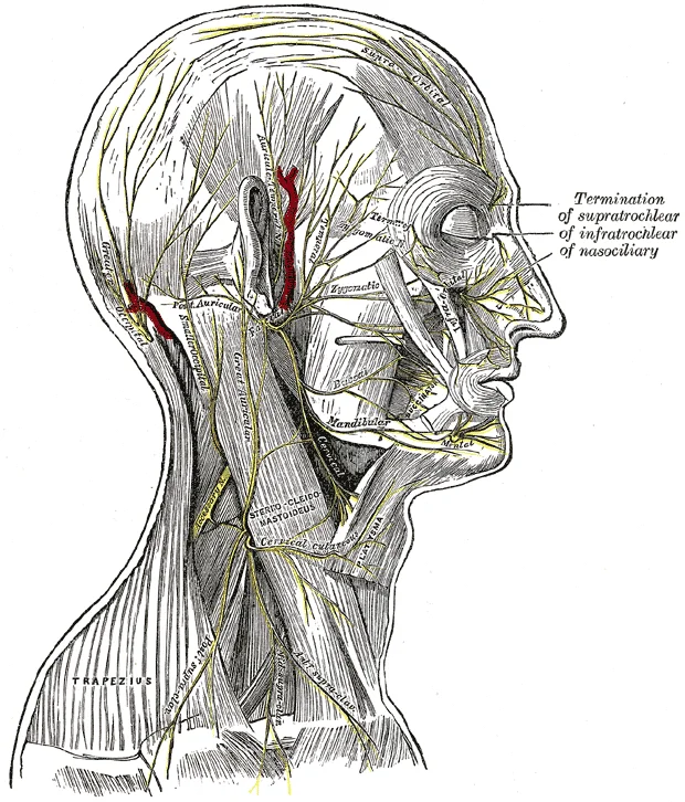
The lesser occipital nerve is traced along the posterior border of the sternocleidomastoid. This visual guide helps correlate surface landmarks with the underlying neural pathways that must be preserved or blocked during neurosurgical procedures.
The inion serves as the prominent external occipital protuberance on the occipital bone, acting as a key surface landmark. The superior nuchal lines extend laterally from the inion, providing attachment for the trapezius and sternocleidomastoid muscles.
This structural organisation is critical for understanding both the spread of pathology and the mechanics of movement.
Figure 1Gray1196
Long description
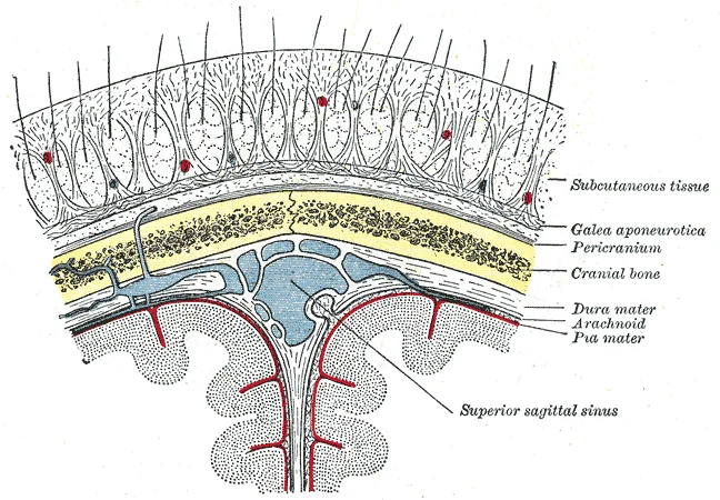
Layers of the scalp, skull and meninges, including the superior sagittal sinus

Figure 1: Layers of the scalp, skull and meninges, including the superior sagittal sinus.
At the most superficial level lies the skin, which is thick and densely packed with hair follicles. Beneath this is the dense connective tissue layer, also known as the subcutaneous tissue. This layer is tightly bound to the skin and contains the neurovascular bundles that supply the scalp. It is within this layer that blood vessels are held open by fibrous septa, preventing significant retraction during lacerations and leading to profuse bleeding.
Deep to the connective tissue is the galea aponeurotica, or epicranial aponeurosis. This is a tough, fibrous sheet that connects the frontalis muscle anteriorly to the occipitalis muscle posteriorly. The galea allows the frontalis and occipitalis muscles to move the scalp independently of the skull. [1]
The deepest layer is the pericranium, which is the periosteum of the skull bones. It is firmly attached to the cranial sutures but can be easily stripped from the diaphyses of the bones. Laterally, this layer ends with the origin of the temporalis fascia along the superior temporal line. [1]
Several nerves traverse these layers. Pain in the region supplied by the LON often manifests as a cervicogenic headache resulting from physical exertion. This nerve is also 2.5 cm lateral to the occipital artery over the occiput and 7 cm lateral to the external occipital protuberance. According to Tubbs et al., the LON branches into medial and lateral parts at approximately the midpoint between the inion and the intermastoid line. [1]
Figure 2Gray790
Long description
The nerves of the scalp, face, and side of neck.

Figure 2: The nerves of the scalp, face, and side of neck.
This arrangement allows for the expression of emotion and protection of the underlying cranium. The structures are grouped into anterior and posterior compartments, linked by the galea aponeurotica.
It provides structural continuity, allowing the contraction of the anterior and posterior bellies to move the entire scalp. The lateral boundaries are formed by the temporal fascia. This layer is crucial for the transmission of force during facial expression.
The scalp receives its blood supply from five major arteries, which form anastomotic networks to ensure robust perfusion. These vessels arise from the external carotid artery and its branches, as well as the internal carotid system via the ophthalmic artery. Understanding their origins and courses is critical for surgical planning and understanding haemorrhage patterns.
It courses posteriorly and superiorly, deep to the sternocleidomastoid muscle. It pierces the posterior belly of the digastric muscle and the occipital fascia. It ascends on the surface of the semispinalis capitis muscle. It supplies the occipital scalp and anastomoses with the posterior auricular and superficial temporal arteries.
It courses posteriorly and superiorly, deep to the parotid gland. It supplies the posterior auricular region and the mastoid area. It anastomoses with the occipital and superficial temporal arteries.
The anastomoses between these vessels are clearly marked, demonstrating the rich collateral circulation.
The rich anastomotic network of the scalp arteries means that ligation of one vessel rarely causes ischaemia. However, haemorrhage from scalp wounds can be significant due to the high pressure of the external carotid system. This layer is also a potential space for haematoma formation.
| Artery | Origin | Course | Branches |
|---|---|---|---|
| Superficial Temporal | External Carotid | Over zygomatic arch | Frontal, Parietal |
| Occipital | External Carotid | Deep to SCM, then superficial | Ascending, Transverse |
| Posterior Auricular | External Carotid | Deep to parotid | Auricular, Mastoid |
| Supraorbital | Ophthalmic | Through supraorbital foramen | Frontal |
| Supratrochlear | Ophthalmic | Medial to supraorbital | Medial Forehead |
The venous drainage of the scalp is a critical anatomical pathway that connects superficial structures to deeper intracranial systems. Understanding this network is essential for appreciating how infections or thromboses can spread from the face and scalp into the cranial cavity.
Posteriorly, the occipital veins descend towards the nape of the neck.
The major tributaries include the supratrochlear veins, which drain the medial forehead and join to form the angular vein. The superficial temporal veins drain the lateral scalp and temple region, eventually forming the retromandibular vein. The occipital veins drain the posterior scalp, draining into the internal jugular vein or the suboccipital venous plexus. These veins are accompanied by their arterial counterparts but lack valves, facilitating retrograde flow.
Figure 3Gray564
Long description
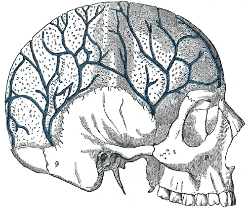
Veins of the diploë as displayed by the removal of the outer table of the skull.

Figure 3: Veins of the diploë as displayed by the removal of the outer table of the skull.
The clinical significance of this anatomy lies in the potential for serious complications. Because the veins are valveless, an infection in the 'danger triangle' of the face or the scalp can spread retrograde into the cavernous sinus. This can lead to cavernous sinus thrombosis, a life-threatening condition characterized by fever, headache, and ophthalmoplegia.
Clinical pearlValveless Veins
The scalp veins are valveless, allowing bidirectional flow and potential spread of infection to intracranial sinuses.
| Vein | Drainage Area | Termination |
|---|---|---|
| Supratrochlear | Medial forehead | Angular vein |
| Supraorbital | Upper eyelid, forehead | Supratrochlear vein |
| Superficial temporal | Lateral scalp, temple | Retromandibular vein |
| Occipital | Posterior scalp | Internal jugular vein |
Figure 4Gray805
Long description
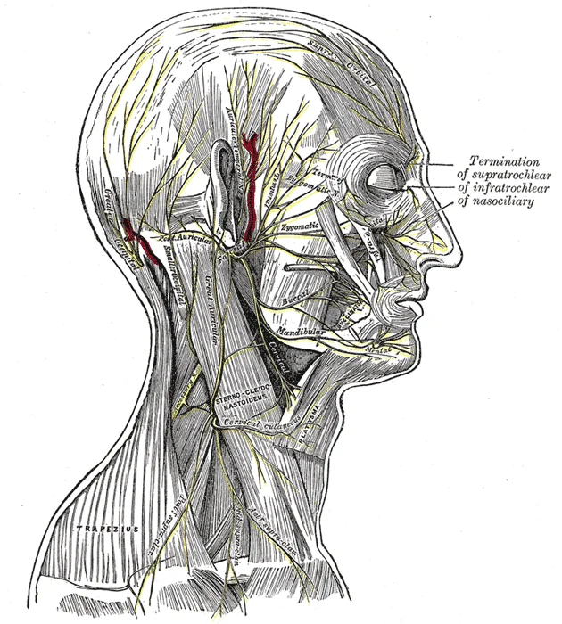
The nerves of the scalp, face, and side of neck.

Figure 4: The nerves of the scalp, face, and side of neck.
The scalp receives its motor and cutaneous supply from a complex network of cervical and cranial nerves. Understanding this innervation requires distinguishing between the muscles of the scalp and the skin that covers them. These muscles are responsible for facial expression and tension on the scalp, respectively. The cutaneous supply, however, is more intricate, involving branches from both the trigeminal nerve and the upper cervical spinal nerves. [1]
Occipital neuralgia is typified by intense pain localized to the occipital skin area typically innervated by the GON and LON[1].
Patients with ON suffer from a shooting or stabbing pain in the neck that radiates over the cranium. On physical examination, tenderness along the course of the GON and LON can be observed. Sometimes hypoesthesia or dysesthesia can occur. The pain is located in the occipital area and may spread toward the vertex. Though usually unilateral, it may be bilateral. [2]
Paroxysmal stabbing pain, with or without persistent aching between paroxysms, in the distribution of the greater, lesser, and/or third occipital nerve. [2]
Cervicogenic headache, which involves pain referral from cervical structures, appears to be produced by convergent excitation that is evoked by stimulation of these nerves and that results in excitation of second-order neurons in the trigeminocervical complex. [2]
Surgical treatment of ON can be considered when a patient does not respond adequately to medical therapies, such as repeated injections, or minimally invasive procedures, such as PRF treatment. However, the results were variable. Recently, there have been a few positive reports on peripheral nerve stimulation, a less invasive surgery, of the GON or LON. Of these approaches, both occipital neurolysis and occipital nerve stimulation (ONS) have been used commonly in the clinical field, recently. [2]
DefinitionDermatome
A dermatome represents an area of skin supplied by a single nerve. [1]
| Structure | Innervation/Feature | Clinical Relevance |
|---|---|---|
| Greater Occipital Nerve (GON) | C2 dorsal ramus | Occipital neuralgia, radiating pain |
| Lesser Occipital Nerve (LON) | C2, C3 ventral rami | Lateral scalp sensation |
| Third Occipital Nerve | C3 dorsal ramus | Suboccipital skin, C2-C3 joint |
| Frontal Nerve | V1 (Trigeminal) | Anterior scalp sensation |
| Zygomaticotemporal Nerve | V2 (Trigeminal) | Lateral scalp sensation |
The lymphatic drainage of the scalp follows a predictable pattern that correlates with the vascular supply and regional nodal basins. Understanding this pathway is essential for identifying the primary site of metastasis in cutaneous malignancies or the source of regional lymphadenopathy. The drainage is generally divided into anterior and posterior groups, which empty into distinct nodal stations.
The anterior portion of the scalp, including the forehead and the anterior scalp above the orbital ridges, drains primarily into the preauricular and parotid lymph nodes. These nodes are situated in front of the ear and within the parotid gland. From here, the efferent vessels pass to the upper deep cervical lymph nodes. This pathway is clinically significant because lesions on the forehead or upper eyelid may present with swelling in the preauricular region before involving the neck.
The efferent vessels from the occipital nodes descend to join the upper deep cervical lymph nodes. This route is critical when evaluating masses at the back of the head, as the occipital nodes are the first echelon of drainage.
The lateral aspects of the scalp, particularly the temporal regions, also contribute to the preauricular and parotid nodal groups. Additionally, some drainage from the temporal area may pass directly to the upper deep cervical nodes. This convergence means that pathology in the temporal region can manifest in multiple nodal stations, complicating clinical examination.
All the aforementioned pathways ultimately converge on the upper deep cervical lymph nodes. These nodes form a chain along the internal jugular vein and the vagus nerve. They serve as the final common pathway for lymphatic drainage from the entire scalp. Enlargement of these nodes, known as cervical lymphadenopathy, is a common clinical sign that warrants investigation into potential scalp, ear, or nasal pathology. The deep cervical nodes then drain into the jugular trunk, which empties into the venous system at the junction of the internal jugular and subclavian veins.
Clinical pearlClinical Pearl
Always examine the preauricular and occipital nodes when assessing scalp lesions, as they are the first line of defence.
These layers are continuous with the facial structures anteriorly and the cervical structures posteriorly. Understanding the spatial relationships within these layers is critical for surgical interventions and understanding the spread of pathology. [1]
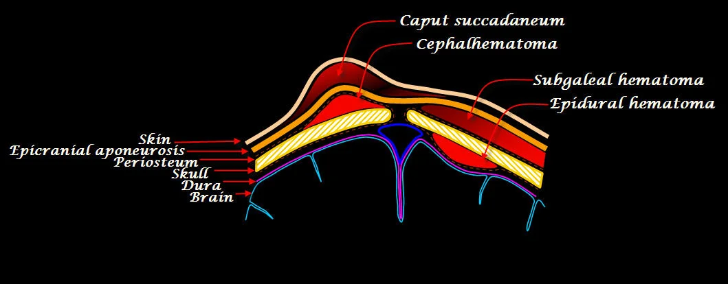
Figure 5Scalp hematomas
Long description
Diagram of the infant scalp showing the locations of the common hematomata of the scalp in relation the layers of the scalp. Newborn Scalp Hematomata

Figure 5: Diagram of the infant scalp showing the locations of the common hematomata of the scalp in relation the layers of the scalp (newborn scalp hematomata).
The most superficial layer is the skin, which is thick and densely hair-bearing. Beneath this lies the dense connective tissue, also known as the subcutaneous layer. This layer contains the neurovascular bundles that supply the scalp. The vessels and nerves run within this dense connective tissue, which is why lacerations in this layer bleed profusely; the tissue holds the vessel ends open. The third layer is the galea aponeurotica, a tough fibrous sheet connecting the frontalis and occipitalis muscles. This layer allows for the movement of the scalp over the skull.
Deep to the aponeurosis is the loose areolar tissue, often called the 'danger space'. This potential space allows the scalp to slide freely over the cranial bones. Crucially, it contains emissary veins that connect the superficial veins of the scalp with the dural venous sinuses within the cranium. Infection can spread through this space to the intracranial cavity. The deepest layer is the pericranium, the periosteum of the skull bones, which is firmly attached at the sutures.
It is supplied by the temporal branch of the facial nerve. The occipitalis originates from the superior nuchal line and inserts into the epicranial aponeurosis. Together, they form the occipitofrontalis muscle.
| Muscle | Origin | Insertion | Action | Nerve Supply |
|---|---|---|---|---|
| Frontalis | Epicranial aponeurosis | Skin of eyebrows | Raises eyebrows | Temporal branch of facial nerve |
| Occipitalis | Superior nuchal line | Epicranial aponeurosis | Pulls scalp posteriorly | Posterior auricular branch of facial nerve |
The sensory innervation of the scalp is derived from both cranial and spinal nerves. The anterior scalp is supplied by the supraorbital and supratrochlear nerves, branches of the ophthalmic division of the trigeminal nerve. The posterior scalp is supplied by the greater occipital nerve (C2 dorsal root ganglion) and the lesser occipital nerve (C2-C3).
This anatomical pathway explains how extracranial triggers can cause intracranial pain.
This highlights the clinical relevance of the greater and lesser occipital nerves in posterior scalp procedures.
The radiological assessment of the scalp and its underlying structures relies on a combination of modalities, each offering distinct advantages for visualising soft tissue, bone, and neural elements. While the scalp itself is primarily composed of soft tissue layers, imaging often extends to the craniocervical junction to evaluate associated pathologies that may present with scalp or head pain.
A simple X-ray serves as an initial screening tool, primarily useful to rule out underlying bony pathologies such as arthritis and craniocervical instability. It provides a rapid overview of the skeletal framework but offers limited detail regarding the soft tissue layers of the scalp. The orientation places the anterior aspect of the head to the left, allowing the viewer to trace the relationship between the occipital bone and the upper cervical vertebrae. This view is essential for identifying gross structural abnormalities that might mimic or accompany scalp disorders. [2]
Computed tomography (CT) of the craniocervical junction is indicated when neoplastic or degenerative osseous pathology is suspected. CT provides superior resolution of bone compared to X-ray, enabling the detection of subtle erosions or masses. The labelled structures include the occipital condyles and the atlas, which are critical for assessing stability. The scan plane is typically axial or sagittal, with the superior aspect of the image corresponding to the top of the head. This modality helps differentiate between soft tissue swelling and bony destruction, a distinction that is vital for accurate diagnosis. [2]
Magnetic resonance imaging (MRI) is considered the most important tool in the diagnosis of disorders affecting the surrounding cervical and occipital soft tissues. It enables detailed visualization of the nerves, muscles, and ligaments that are not well seen on CT or X-ray. The view is often sagittal, with the anterior direction to the left, allowing the observer to see the course of the occipital nerves as they emerge from the skull. The relationship between the semispinalis capitis muscle and the underlying nerve is clearly depicted, highlighting potential sites of compression. [2]
Ultrasound has a limited but specific role in this context. While segments of the greater occipital nerve may be visualized with ultrasound, it is not possible to visualize the nerve in its entirety to allow determination of all sites of compression. The orientation is typically transverse, with the superficial aspect at the top of the image. This modality is useful for dynamic assessment but lacks the comprehensive view provided by MRI. [3]
The scalp presents a critical interface for neurovascular supply and surgical access, where anatomical relationships dictate both injury patterns and therapeutic interventions. Understanding the surface landmarks is essential for identifying nerve entrapment sites and planning regional blocks.
DefinitionOccipital Neuralgia
Paroxysmal shooting or stabbing pain in the dermatomes of the greater or lesser occipital nerve. [2]
It lies in close proximity to the occipital artery, a relationship that can contribute to neuralgia. [1]
Occipital Nerve Block
- Assess PainParoxysmal shooting or stabbing pain in the dermatomes of the greater or lesser occipital nerve
- Question: Response to Medical Therapy?
Yes
- Continue Management
No
- Consider InjectionInjections are typically administered in regions innervated by the GON and LON
Trauma to the posterior head, such as whiplash injuries, can result in neuralgia. The clinical presentation includes allodynia and tenderness to palpation of the nerve or the soft tissue of the suboccipital space. Compression of the occipital nerve can mimic primary occipital neuralgia, creating areas of overlap in clinical presentation. Nerve palsies may arise from direct trauma or entrapment within the muscular layers. [3]
Management follows a step-wise approach, beginning with medication and progressing to invasive procedures for refractory cases. Minimally invasive methods include occipital nerve blocks, chemical or radiofrequency ablation, or transcutaneous electrical nerve stimulation. Local injection with corticosteroid can improve symptoms, though generally only temporarily. Injections are typically administered in regions innervated by the GON and LON. [2] [1]
| Procedure | Anatomical Target | Clinical Note |
|---|---|---|
| Dorsal Rhizotomy | C2 Root | Destructive; risk of painful neuroma |
| Microvascular Decompression | Occipital Artery | Addresses proximity of artery to GON |
| Occipital Neurectomy | GON/LON | Rarely performed due to risks |
| C2 Gangliotomy | C2 Ganglion | Carries grave risks of causalgia |
Clinicians should always remember that destructive procedures carry grave risks: once an anatomic structure is destroyed, it cannot be easily recovered, if at all, and with any destructive procedure there is always the risk of the development of painful neuroma or causalgia, conditions that may be even harder to control than the original complaint. [2]
The scalp consists of five layers, often remembered by the mnemonic SCALP: Skin, Connective tissue, Aponeurosis, Loose areolar tissue, and Pericranium. The loose areolar tissue layer allows for significant mobility of the upper scalp layers and serves as a potential space where fluid or blood can accumulate, such as in a subgaleal haematoma. This layer is also the sit
Injury to these branches can result in an inability to wrinkle the forehead or raise the eyebrows.
DefinitionEmissary Veins
Valveless veins connecting extracranial scalp veins with intracranial dural sinuses, allowing potential spread of infection.
NoteVascular Supply
The scalp's rich arterial anastomosis leads to profuse bleeding from wounds but promotes rapid healing.
Clinical pearlSupraorbital Nerve
A branch of CN V1 that exits the supraorbital foramen to supply sensation to the central forehead and scalp.
DefinitionGalea Aponeurotica
The tendinous sheet connecting the frontalis and occipitalis muscles, forming the middle layer of the scalp.
NoteConnective Tissue
The dense connective tissue layer contains the neurovascular structures and is responsible for the profuse bleeding of scalp lacerations.
Clinical pearlPericranium
The periosteum of the skull, attached at sutures, serving as the outer layer of the cranial bones.
Exam trapInfection Spread
Infections in the loose areolar tissue can spread to the intracranial cavity via emissary veins, bypassing normal barriers.
DefinitionSupratrochlear Nerve
A branch of CN V1 supplying the medial forehead and scalp, often involved in frontal headaches.
NoteAnastomosis
Arteries from both external and internal carotid systems anastomose in the scalp, ensuring robust blood supply.
Students frequently confuse the clinical presentation of Unremitting Head and/or Neck Pain (UHNP) with acute episodic conditions, leading to diagnostic oversight. A primary error is focusing exclusively on the severe, paroxysmal headache while ignoring the background neck discomfort. Patients often lack awareness of the constant mild discomfort in the neck, particularly if they have longstanding symptoms or attentional issues. Consequently, clinicians must actively question the frequency and duration of pain in the neck is less often as carefully assessed as head pain [3]
Another significant pitfall is misdiagnosing UHNP as Occipital Neuralgia without verifying specific criteria. Occipital Neuralgia requires pain that is recurring in paroxysmal attacks lasting seconds to minutes, severe in intensity, and shooting, stabbing, or sharp in quality. Furthermore, the pain must be temporarily eased by an anesthetic block of the greater occipital nerve (GON). In contrast, UHNP is characterised by unremitting discomfort rather than paroxysmal attacks. Students should also note that allodynia in occipital nerve compression is generally occipital, not in the trigeminal distributions, so standard checklists weighted toward trigeminal symptoms may fail to detect it. [3]
A third error involves the management of electrode implantation for pain control. Poor outcomes are reported following up to 30% of implantations. Therefore, a test stimulation before permanent implantation is recommended in many studies. Permanent implantation is usually recommended when 50% pain reduction occurs on test stimulation. Skipping this step risks permanent hardware placement in patients who will not benefit. [2]
Finally, students may overlook the inflammatory component in the transformation of migraine. The gradual development of inflammation and progressively worsening compression of the occipital nerves is consistent with the concept of ‘transformation’ of episodic migraine to chronic migraine. The only class of pharmacological agents found to have a protective effect in this transformation was nonsteroidal anti-inflammatory drugs. Recognising this inflammatory process helps explain why certain prophylactic strategies are effective. [3]
In clinical practice, recognising UHNP is essential for managing patients with chronic head and neck pain. The diagnostic criterion of reduced cervical range of motion and worsening of pain with provocative maneuvers is commonly encountered in patients with UHNP. However, assigning a time of onset to a lesion that cannot be diagnosed on imaging is difficult, making history taking paramount. Clinicians must distinguish these symptoms from Occipital Neuralgia to ensure appropriate treatment pathways, such as nerve blocks versus decompression surgery. [3]
In the operating theatre, knowledge of occipital nerve anatomy and compression is vital for surgical planning. Indirect results of nerve decompression may result in additional improvement that extends beyond the elimination of UHNP, such as improved sleep from the reduction in occipital allodynia. Surgeons must understand that the goal is not just pain relief but also the restoration of function and sleep quality. The hypothesis that inflammation leads to compression aligns with evidence-based research, supporting surgical interventions that address mechanical compression and inflammation. [3]
On imaging, while lesions causing UHNP may not always be visible, understanding the anatomical course of the occipital nerves helps in interpreting subtle signs of compression. The higher likelihood of allodynia in these patients is consistent with the concept of occipital nerve compression. Although imaging may not show the compression directly, the clinical correlation with reduced cervical range of motion supports the diagnosis.
In examinations, students are tested on the specific criteria for Occipital Neuralgia versus UHNP. They must recall that Occipital Neuralgia pain is eased by an anesthetic block of the GON. They must also know that for electrode implantation, a 50% pain reduction on test stimulation is the threshold for permanent implantation. Additionally, understanding the role of nonsteroidal anti-inflammatory drugs in preventing the transformation from episodic to chronic migraine is a key teaching point.
Clinical pearlDiagnostic Clue
UHNP presents with constant mild discomfort described as ‘tightness’ or ‘pressure’ in the posterior neck and sub-occipital space, often overlooked by patients. [3]
| Feature | Unremitting Head/Neck Pain (UHNP) | Occipital Neuralgia |
|---|---|---|
| Pain Quality | Constant mild discomfort, tightness, pressure | Paroxysmal, shooting, stabbing, sharp |
| Duration | Unremitting, longstanding | Seconds to minutes per attack |
| Diagnostic Criterion | Reduced cervical range of motion, worsening with provocative maneuvers | Eased by anesthetic block of GON |
| Allodynia Location | Generally occipital | May be detected by standard checklists |
Evaluation of Chronic Occipital Pain
- Patient presents with chronic occipital painAssess pain quality and duration
- Question: Is pain paroxysmal (seconds-minutes) and sharp?
Yes
- Suspect Occipital NeuralgiaCheck for GON block response
No
- Suspect UHNPCheck for constant pressure/tightness
- Consider electrode implantationOnly if 50% pain reduction on test stimulation
UHNP presents with constant mild discomfort described as ‘tightness’ or ‘pressure’ in the posterior neck and sub-occipital space. Patients often lack awareness of mild, yet constant discomfort in the neck, especially with longstanding symptoms. Occipital Neuralgia requires pain that is recurring in paroxysmal attacks lasting from seconds to minutes. The pain of Occipital Neuralgia must be temporarily eased by an anesthetic block of the GON. Permanent electrode implantation is usually recommended when 50% pain reduction occurs on test stimulation. Poor outcomes after electrode implantation are a common problem that has been reported following up to 30% of implantations. [2] The gradual development of inflammation and progressively worsening compression of the occipital nerves is consistent with the concept of ‘transformation’ of episodic migraine to chronic migraine. Nonsteroidal anti-inflammatory drugs were the only class found to have a protective effect in the ‘transformation’ from EM to CM.
Answer each one in your head first, then open the answer.
What is the recommended threshold for permanent electrode implantation based on test stimulation?
Show answerHide answer
50% pain reductionWhat percentage of electrode implantations are reported to have poor outcomes?
Show answerHide answer
Up to 30%What are the two key characteristics of pain quality for Occipital Neuralgia?
Show answerHide answer
Recurring in paroxysmal attacks lasting from seconds to minutes; severe in intensity; shooting, stabbing or sharp in qualityWhat diagnostic maneuver is commonly encountered in patients with UHNP?
Show answerHide answer
Reduced cervical range of motion and worsening of pain with provocative maneuversWhich class of drugs was found to have a protective effect in the transformation from episodic to chronic migraine?
Show answerHide answer
Nonsteroidal anti-inflammatory drugs
- Kemp WJ, Tubbs RS, Cohen-Gadol AA. The innervation of the scalp: A comprehensive review including anatomy, pathology, and neurosurgical correlates. Surgical neurology international 2011;2:178. doi:10.4103/2152-7806.90699. Accessed .
- Choi I, Jeon SR. Neuralgias of the Head: Occipital Neuralgia. Journal of Korean medical science 2016. Accessed .
- Blake P, Burstein R. Emerging evidence of occipital nerve compression in unremitting head and neck pain. The journal of headache and pain 2019. Accessed .
Figure credits
- Figure 1: Henry Vandyke Carter, via Wikimedia Commons, Public domain, Public domain.
- Figure 2: Henry Vandyke Carter, via Wikimedia Commons, Public domain, Public domain.
- Figure 3: Henry Vandyke Carter, via Wikimedia Commons, Public domain, Public domain.
- Figure 4: Henry Vandyke Carter, via Wikimedia Commons, Public domain, Public domain.
- Figure 5: AMH Sheikh, via Wikimedia Commons, CC BY-SA 3.0, CC BY-SA 3.0.
Educational content for medical students and doctors. It is not a substitute for clinical judgement or local guidelines.