Parotid gland
The parotid gland is the largest salivary gland, situated anterior to the ear and draining via Stensen’s duct.
The parotid gland lies superficial to the masseter muscle, extending from the zygomatic arch to the angle of the mandible. It is divided into superficial and deep lobes by the facial nerve, which traverses the gland in a fan‑shaped distribution. The superficial lobe occupies the majority of the gland’s volume and lies anterior to the sternocleidomastoid, while the deep lobe lies posterior to the masseter and inferior to the zygomatic arch. The parotid duct (Stensen’s duct) originates from the gland’s anterior surface, courses over the masseter, and opens into the oral cavity opposite the second maxillary molar. The gland is palpable as a soft, oval mass over the mandibular angle; a surface landmark is the mandibular angle and the posterior border of the ear.
The gland contains numerous intraglandular lymph nodes, particularly in the deep lobe, and is richly vascularised by branches of the external carotid artery. The facial nerve’s intimate relationship with the gland accounts for the technical difficulty of parotid surgery and the risk of postoperative facial paralysis.
Benign tumours constitute the majority of parotid neoplasms, with pleomorphic adenoma being the most common histological type and Warthin tumour the second most frequent. Parotid tumours occur at a rate of 1:100 000 and represent 2–3 % of head and neck tumours, accounting for 80 % of salivary gland tumours. [2]
Surgical management of benign lesions typically involves a superficial parotidectomy with facial nerve preservation, which is considered the best treatment for pleomorphic adenoma. In malignant cases, a subtotal or total parotidectomy may be required, sometimes with deliberate sacrifice of facial nerve branches if tumour invasion mandates it; adjuvant radiotherapy may follow, while chemotherapy is rarely indicated. The most feared postoperative complication is facial paralysis, occurring in 15% of cases with only 1.9% permanent paralysis. [2]
They are more common in males over 50 who smoke, with a male:female ratio of about 1.5:1, and the average age at presentation is around 60 years. Approximately 15 % are multifocal within the same gland, and 10 % are bilateral; 10–27 % of patients develop multiple tumours, with 5–30 % bilateral. [3]
Surgical excision remains the primary treatment for Warthin tumours, with indications including uncertain diagnosis, cosmetic concern, pain or infection, or patient preference for removal.
The parotid gland is situated in the preauricular and retromandibular regions. It is encapsulated by the parotid sheath, a thickened layer of the deep cervical fascia. The gland is divided into two distinct lobes by the plane of the facial nerve. The deep lobe is smaller and lies medial to the nerve. Consequently, superficial parotidectomy with facial nerve preservation is the most often indicated surgical procedure. [2] [1]
Figure 1Gray1023
Long description
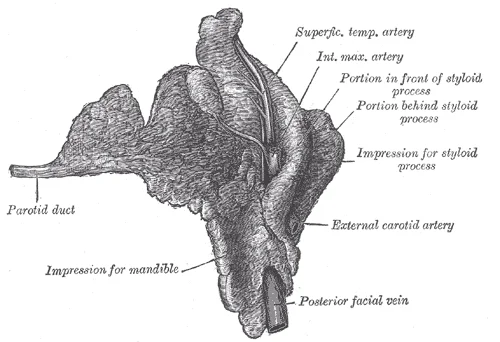
Right parotid gland. Deep and anterior aspects.

Figure 1: Right parotid gland (deep and anterior aspects).
The superficial lobe is lateral to these nerve branches, while the deep lobe extends medially. The sternocleidomastoid muscle lies posteriorly, and the mandible is inferior. The parotid duct emerges from the anterior border of the superficial lobe.
The gland secretes a watery solution containing salivary amylase. This serous composition distinguishes it from the submandibular and sublingual glands, which produce mucus-rich saliva. The unique cellular makeup of the parotid gland dictates this secretion type. [1]
DefinitionParotid Lobes
The parotid gland has a superficial lobe, lateral to the facial nerve, that comprises 4/5 of the glandular parenchyma, and a smaller deep lobe. [2]
The facial nerve exits the skull via the stylomastoid foramen and enters the posterior aspect of the gland. It divides the gland into superficial and deep components. The nerve then branches into five terminal divisions: temporal, zygomatic, buccal, marginal mandibular, and cervical. These branches emerge from the anterior border of the gland to innervate the muscles of facial expression. The parotid plexus forms within the gland substance.
| Structure | Relation |
|---|---|
| Facial Nerve | Divides gland into superficial and deep lobes |
| Superficial Lobe | Lateral to facial nerve; 4/5 of parenchyma |
| Deep Lobe | Medial to facial nerve; smaller portion |
| Parotid Duct | Emerges from anterior border of superficial lobe |
| Sternocleidomastoid | Posterior relation |
| Mandible | Inferior relation |
Knowledge in parotid anatomy is an essential tool for the surgeon at the time of diagnosis and treatment. Understanding the relationship between the tumor and the facial nerve is vital. Although tumors more often affect the superficial lobe, the term subtotal parotidectomy seems more appropriate than superficial parotidectomy. This terminology reflects the need to remove the entire superficial lobe to ensure complete excision of low-morbidity tumors. [2]
Clinical pearlSurgical Relevance
Superficial parotidectomy with facial nerve preservation is the most common and appropriate treatment for most low-morbidity tumors. [2]
The gland is surrounded by lymph nodes. These nodes drain the scalp, face, and external ear. Inflammation or malignancy in these areas can present as parotid swelling. This anatomical course makes the duct susceptible to injury during dental procedures or facial trauma.
Figure 2Gray1022
Long description
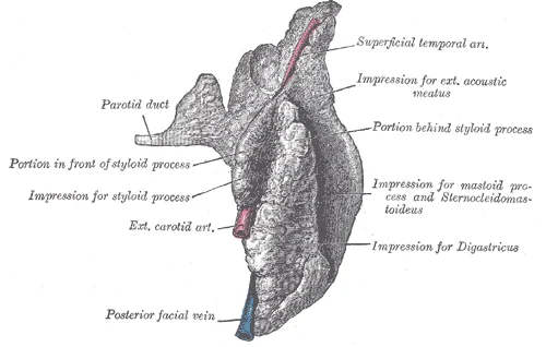
Right parotid gland. Posterior and deep aspects.

Figure 2: Right parotid gland (posterior and deep aspects).
The parotid gland is enclosed within a dense fascial capsule derived from the investing layer of deep cervical fascia. This capsule splits to enclose the gland, forming a superficial and deep layer that fuse at the anterior and posterior borders. [1] [2]
The gland is situated in the parotid bed, lying between the mastoid process, the sternocleidomastoid muscle, and the ramus of the mandible. Anteriorly, the gland overlaps the masseter muscle, which is a key landmark for surgical access. Posteriorly, it rests against the mastoid process and the sternocleidomastoid muscle. The deep part of the gland extends medially into the parapharyngeal space, lying lateral to the styloid process and its associated muscles.
The capsule is thin and adherent to the glandular tissue, preventing the spread of infection or tumour into adjacent spaces. However, it can also restrict the expansion of the gland in cases of inflammation or neoplasia. The relations of the parotid gland are critical for surgical planning, as damage to adjacent structures can lead to significant morbidity.
The following table summarises the key relations of the parotid gland.
| Structure | Relation |
|---|---|
| Masseter Muscle | Anteriorly, overlapped by the superficial lobe |
| Sternocleidomastoid Muscle | Posteriorly, forming the posterior border |
| Mastoid Process | Posteriorly, providing attachment for the capsule |
| Ramus of Mandible | Anteriorly, forming the anterior border |
| Styloid Process | Medially, deep lobe lies lateral to it |
The sympathetic innervation is derived from the superior cervical ganglion, travelling along the external carotid artery to reach the gland.
The lymphatic drainage of the parotid gland is to the preauricular and parotid lymph nodes, which then drain into the deep cervical lymph nodes. These nodes are important in the staging of parotid tumours and the assessment of metastatic disease.
The parotid gland is a complex structure with numerous relations that are critical for its function and for surgical access. Understanding these relations is essential for medical students and surgeons alike.
The external carotid artery is located deep to the gland, giving off branches that supply the parotid tissue. The masseter muscle lies anteriorly and inferiorly, providing a muscular bed for the superficial lobe. The facial nerve trunk emerges from the stylomastoid foramen and enters the posterior aspect of the gland, dividing into its terminal branches within the substance of the gland. These branches fan out to supply the muscles of facial expression. The glandular parenchyma is composed of serous acini arranged in lobules separated by fibrous septa. The intercalated ducts and striated ducts are not individually resolved at this magnification but are part of the internal drainage system leading to the parotid duct. The overall architecture demonstrates the complex relationship between the glandular tissue, the neurovascular structures, and the surrounding musculature and skeletal elements.
Figure 3Gray1025
Long description
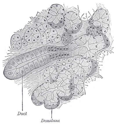
Section of submaxillary gland of kitten. Duct semidiagrammatic. X 200.

Figure 3: Section of submaxillary gland of kitten (duct semidiagrammatic).
The parotid gland is a compound tubuloalveolar gland. It is enclosed by a dense fibrous capsule derived from the investing layer of the deep cervical fascia. This capsule is thick superiorly and laterally but thin inferiorly and medially. The capsule sends septa into the gland, dividing it into lobules. The gland is divided into two lobes by the plane of the facial nerve. The superficial lobe is larger and lies lateral to the facial nerve. The deep lobe is smaller and lies medial to the facial nerve. The two lobes are continuous with each other posteriorly. The parotid duct, also known as Stensen's duct, arises from the anterior border of the superficial lobe. It runs horizontally across the masseter muscle. It then pierces the buccinator muscle to open into the oral cavity opposite the second maxillary molar. It runs vertically downwards. It divides into anterior and posterior divisions. The anterior division joins the facial vein to form the common facial vein. The posterior division joins the posterior auricular vein to form the external jugular vein.
| Structure | Type | Relation to Gland |
|---|---|---|
| Facial Nerve | Nerve | Divides gland into superficial and deep lobes |
| Retromandibular Vein | Vein | Formed within the gland, runs vertically |
| External Carotid Artery | Artery | Passes upwards through the gland |
| Auriculotemporal Nerve | Nerve | Runs upwards, supplies secretomotor fibres |
| Parotid Duct | Duct | Emerges from anterior border of superficial lobe |
This vascular arrangement is critical for understanding both the physiology of salivary secretion and the surgical management of parotid pathology. These branches, along with other smaller vessels, form a rich anastomotic network that perfuses the glandular tissue.
The artery divides into the maxillary artery, which courses anteriorly towards the pterygoid muscles, and the superficial temporal artery, which ascends anterior to the ear. The branching pattern demonstrates how blood is distributed to the superficial and deep lobes of the gland. The vessels are embedded within the connective tissue septa that divide the gland into lobules. This detailed anatomical layout allows for the recognition of these structures on histological sections or during surgical procedures. [2]
The posterior auricular artery runs upwards behind the ear, supplying the posterior aspect of the parotid gland and the scalp. These branches anastomose with the branches from the external carotid artery, ensuring a robust blood supply. The venous drainage follows the arterial pattern, with veins draining into the retromandibular vein, which is formed by the union of the superficial temporal and maxillary veins within the gland.
Understanding this vascular anatomy is essential for surgeons to avoid excessive bleeding during parotidectomy. The proximity of the facial nerve to these vessels also requires careful dissection to prevent iatrogenic injury. The rich anastomotic network means that ligation of one branch may not stop bleeding if collateral circulation is present. Therefore, knowledge of the specific origin, course, and relations of these arteries is vital for safe surgical practice. The external carotid artery's position relative to the mandible and the mastoid process provides key landmarks for identifying the gland's blood supply.
The venous drainage of the parotid gland follows a predictable pattern that mirrors its arterial supply and anatomical boundaries. Understanding this system is critical for surgical planning and interpreting imaging studies, particularly when assessing for deep lobe involvement or vascular invasion by neoplasms.
It arises from the union of several tributaries in the temporal fossa. These veins drain the scalp and the lateral aspect of the forehead. As they descend, they run deep to the zygomatic arch. They join with the maxillary vein to form the retromandibular vein. This convergence typically occurs within the substance of the parotid gland or just posterior to the mandibular ramus. The superficial temporal vein is a key landmark for identifying the upper extent of the parotid bed. [3]
It collects blood from the deep structures of the face, including the infratemporal fossa. It runs anteriorly and medially to meet the superficial temporal vein. The junction of these two veins creates the retromandibular vein. This vessel is crucial as it serves as the main trunk for parotid venous outflow before it divides. Knowledge of the maxillary vein’s course helps in avoiding injury during approaches to the infratemporal region.
Within the gland, it is accompanied by the facial nerve branches. The retromandibular vein typically divides into an anterior and a posterior division. The anterior division joins the facial vein to form the common facial vein. The posterior division joins the posterior auricular vein to form the external jugular vein. This dual termination pattern means that parotid venous drainage can flow into both the internal jugular system via the external jugular and the facial system.
This vein drains the posterior aspect of the parotid gland and the scalp. It is often visible as a superficial structure on the neck. Injury to this vein during parotidectomy can lead to significant bleeding due to its thin walls and fixed position within the fascia.
DefinitionRetromandibular Vein
The main venous trunk of the parotid gland, formed by the union of the superficial temporal and maxillary veins, which divides into anterior and posterior divisions within or below the gland.
| Structure | Formation/Origin | Termination | Key Relation |
|---|---|---|---|
| Superficial Temporal Vein | Tributaries in temporal fossa | Joins maxillary vein | Deep to zygomatic arch |
| Maxillary Vein | Deep facial tributaries | Joins superficial temporal vein | Infratemporal fossa |
| Retromandibular Vein | Union of above two | Divides into anterior/posterior | Within parotid gland |
| External Jugular Vein | Posterior division + posterior auricular | Subclavian vein | Over sternocleidomastoid |
The lymphatic drainage of the parotid gland is a critical pathway for the spread of infection and malignancy, involving both superficial and deep nodal groups. Understanding this anatomy is essential for clinical assessment of head and neck pathology.
Superficial parotid lymph nodes are located within the parotid fascia, superficial to the gland itself. These nodes receive lymph from the lateral aspects of the auricle, the external auditory meatus, and the posterior surface of the parotid gland. They are not always distinct structures but often represent a collection of small nodes embedded in the fatty tissue surrounding the gland.
Deep parotid lymph nodes lie within the substance of the parotid gland, deep to the facial nerve. They receive lymph from the deeper parts of the parotid gland, the medial surface of the tympanic membrane, and the Eustachian tube. These nodes are significant because they can be palpated during examination if enlarged, appearing as a mass within the parotid region.
Preauricular lymph nodes are situated anterior to the tragus of the ear. They drain the scalp anterior to the ear, the upper eyelid, and the lateral aspect of the nose. Lymph from these nodes typically passes to the superficial parotid nodes or directly to the upper deep cervical nodes.
The upper deep cervical lymph nodes, also known as the jugulodigastric nodes, are located along the internal jugular vein, just below the angle of the mandible. They receive lymph from the tonsils, the base of the tongue, and the posterior scalp. The deep parotid nodes often drain into these upper deep cervical nodes, forming a crucial link in the lymphatic chain of the head and neck.
DefinitionClinical Significance
Enlargement of preauricular or deep parotid nodes may indicate pathology in the eye, ear, or parotid gland itself, necessitating thorough clinical examination.
The lymphatic vessels from the parotid region ultimately converge to drain into the upper deep cervical lymph nodes. From there, lymph flows into the jugular trunk, which may join the thoracic duct on the left or the right lymphatic duct on the right, before emptying into the venous system at the junction of the internal jugular and subclavian veins. This pathway is vital for understanding the spread of metastatic disease from primary tumours in the head and neck region.
The parotid gland receives its autonomic innervation via the glossopharyngeal nerve (CN IX). Preganglionic parasympathetic fibres travel through the tympanic branch to form the tympanic plexus. Fibres then continue as the lesser petrosal nerve to reach the otic ganglion. Postganglionic fibres hitchhike on the auriculotemporal nerve to reach the gland. This pathway stimulates serous secretion. Sympathetic fibres arise from the superior cervical ganglion. They follow the external carotid artery branches to the gland. These fibres cause vasoconstriction of the gland’s blood vessels.
Somatic sensation is provided by the auriculotemporal nerve, a branch of the mandibular division of the trigeminal nerve (CN V3). This nerve also carries the postganglionic parasympathetic fibres. The facial nerve (CN VII) does not provide motor supply to the gland itself. Instead, it passes through the gland to supply the muscles of facial expression. The nerve enters the posterior aspect of the gland. It divides within the gland into five terminal branches. These branches emerge from the anterior border of the gland. They are often remembered by the mnemonic 'To Zanzibar By Motor Car'. The branches are temporal, zygomatic, buccal, marginal mandibular, and cervical. [2]
The duct pierces the buccal fat pad to enter the oral cavity. The facial nerve branches lie superficial to the duct in the superficial part of the gland. This anatomical arrangement is critical during surgery. Injury to the facial nerve branches can cause facial paralysis. [3]
Referred pain from the parotid gland is mediated by the auriculotemporal nerve. Patients may perceive pain in the ear or temple region. This occurs because the auriculotemporal nerve also supplies sensation to the external ear and temple. Tumours of the parotid gland can compress these nerves. This leads to pain that is not localised to the gland itself. The secretomotor pathway is essential for understanding gland function. Disruption of the glossopharyngeal pathway leads to dry mouth. Understanding the course of the facial nerve is vital for surgical planning. The nerve’s position within the gland varies. In some cases, the nerve is embedded in the deep lobe. Surgeons must identify the nerve trunk before dissecting the gland. Preservation of the facial nerve is the primary goal of parotid surgery.
The parotid gland is a major salivary gland with a distinct histological architecture that reflects its primary function of serous secretion. Understanding its microscopic structure is fundamental to identifying pathological changes, particularly when distinguishing benign from malignant processes during clinical evaluation. [2]
This capsule is significant clinically, as it restricts the expansion of any swelling within the gland, potentially leading to increased pressure and pain. Within this capsule, the glandular parenchyma is divided into lobules by septa of connective tissue.
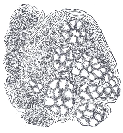
Each lobule contains the secretory units and the ductal system. The secretory units are composed of serous acini. These acini are pyramidal in shape and consist of a single layer of serous cells. The nuclei of these cells are round and located basally, while the cytoplasm is rich in zymogen granules, which appear eosinophilic on standard haematoxylin and eosin staining. These granules contain the inactive enzymes that will be secreted into the lumen. Unlike the submandibular and sublingual glands, the parotid gland is purely serous, meaning it contains no mucous acini.
Figure 4Gray1026
Long description
Human submaxillary gland. (R. Heidenhain.) At the right is a group of mucous alveoli, at the left a group of serous alveoli.

Figure 4: Human submaxillary gland (R.
The ductal system begins with intercalated ducts, which are small, lined by low cuboidal epithelium. These ducts connect the acini to striated ducts. The striated ducts are lined by columnar epithelium with prominent basal striations, which correspond to mitochondrial infoldings involved in ion transport and modification of the primary saliva. The striated ducts eventually merge to form larger excretory ducts. The largest of these is Stensen’s duct, which travels anteriorly across the masseter muscle, pierces the buccinator muscle, and opens into the oral cavity opposite the second maxillary molar.
Accurate histological identification is critical in surgical settings. This highlights the limitations of rapid intraoperative diagnosis and the necessity of final pathological review for definitive management decisions. [2]
For a more detailed exploration of the cellular components and staining characteristics, please refer to the histology page.
The parotid gland is the largest of the major salivary glands, developing from the ectoderm of the stomodeum. It begins as an outgrowth of the oral epithelium during the sixth week of embryonic life. This epithelial bud penetrates the surrounding mesenchyme to form the ductal system and acini. The gland matures in position relative to the mandible and the external acoustic meatus. Understanding its embryological origin helps explain its close relationship with the facial nerve, which develops from the second pharyngeal arch and migrates through the developing gland. [1][2]
This capsule is thick superiorly and laterally, providing structural support. Medially, the capsule is thin and incomplete, allowing the gland to extend into the parapharyngeal space. This anatomical feature is clinically significant because infections or tumours can spread medially with little resistance. The superficial part of the gland lies between the skin and the sternocleidomastoid muscle. The deep part extends anteriorly to the medial pterygoid muscle and posteriorly to the mastoid process.
The parotid duct, also known as Stensen’s duct, arises from the anterior border of the gland. It runs forward over the masseter muscle. At the anterior border of the masseter, it turns medially through the buccal fat pad. The duct pierces the buccinator muscle to open into the oral cavity opposite the second upper molar tooth. This specific opening site is a key landmark for clinical examination. Obstruction of this duct leads to sialolithiasis, causing painful swelling during meals.
The gland is intimately related to several important neurovascular structures. The facial nerve emerges from the stylomastoid foramen and enters the posterior border of the gland. It divides within the gland into its terminal branches. These branches emerge from the anterior and inferior borders of the gland to innervate the muscles of facial expression. The external carotid artery and the retromandibular vein also pass through the substance of the gland. The retromandibular vein divides into anterior and posterior divisions within the gland. The posterior division joins the posterior auricular vein to form the external jugular vein. The anterior division joins the facial vein to form the common facial vein.
The parasympathetic innervation is derived from the glossopharyngeal nerve. Preganglionic fibres travel via the lesser petrosal nerve to the otic ganglion. Postganglionic fibres then hitchhike on the auriculotemporal nerve to reach the gland. This secretomotor pathway is essential for saliva production. Sympathetic fibres arise from the superior cervical ganglion and follow the external carotid artery. They cause vasoconstriction and modify the composition of saliva.
DefinitionParotid Fascia
The dense connective tissue capsule surrounding the parotid gland, derived from the deep cervical fascia, which limits the spread of infection but allows medial extension.
| Structure | Course / Relation | Clinical Significance |
|---|---|---|
| Facial Nerve | Passes through gland substance, dividing into branches | Injury causes facial paralysis; surgical landmark |
| External Carotid Artery | Ascends through posterior part of gland | Source of bleeding in deep lobe surgery |
| Retromandibular Vein | Formed within gland, divides into anterior/posterior parts | Forms external jugular vein posteriorly |
| Auriculotemporal Nerve | Runs with superficial temporal vessels, supplies gland | Carries parasympathetic fibres to gland |
The parotid gland is the largest of the major salivary glands, situated in the parotid bed between the mandibular ramus and the sternocleidomastoid muscle. In radiological anatomy, understanding its spatial relationships is critical for interpreting cross-sectional imaging. This image displays an axial CT view of the head and neck, oriented with the patient’s right side on the left of the screen. The caption for this figure states it shows the parotid gland in axial cross-section, highlighting its lobulated parenchyma and surrounding vascular structures.
The gland is divided into superficial and deep lobes by the plane of the facial nerve. On imaging, the superficial lobe appears as a homogeneous, moderately enhancing structure lateral to the masseter muscle and the mandibular ramus. It lies anterior to the sternocleidomastoid muscle and posterior to the ramus of the mandible. The deep lobe extends medially, occupying the parapharyngeal space. It lies medial to the mandibular ramus and anterior to the sternocleidomastoid muscle. The interface between these two lobes is not a true anatomical capsule but is defined by the course of the facial nerve as it exits the stylomastoid foramen.
Several key structures traverse or relate to the parotid gland. It descends through the gland and exits from its posterior border. The facial nerve (CN VII) exits the skull via the stylomastoid foramen, which lies between the styloid and mastoid processes. It enters the posterior aspect of the parotid gland and divides into its terminal branches. These branches emerge from the anterior border of the gland to innervate the muscles of facial expression.
Ultrasound is particularly useful for evaluating superficial lesions and guiding fine-needle aspiration. It distinguishes cystic from solid components effectively. CT provides excellent detail of bony relationships and deep extension into the parapharyngeal space. MRI offers superior soft-tissue contrast, making it ideal for assessing perineural spread along the facial nerve branches.
Clinical pearlKey Relation
The facial nerve divides the parotid gland into superficial and deep lobes, a landmark essential for surgical and radiological interpretation.
| Structure | Type | Relation to Gland |
|---|---|---|
| Facial Nerve | Nerve | Divides gland into lobes |
| Retromandibular Vein | Vein | Descends through gland |
| External Carotid Artery | Artery | Ascends through posterior part |
| Parotid Duct | Duct | Emerges from anterior border |
The parotid gland is the largest of the salivary glands, situated in the parotid bed between the mastoid process, the sternocleidomastoid muscle, and the ramus of the mandible. Its deep lobe extends into the parapharyngeal space. The gland is enclosed by the parotid fascia, which is continuous with the investing layer of deep cervical fascia. This fascia splits to enclose the gland, forming a strong capsule that limits expansion during inflammation or tumour growth, potentially causing significant pain.
DefinitionParotid Bed
The anatomical space containing the parotid gland, bounded laterally by the skin and platysma, medially by the styloid process and its muscles, posteriorly by the mastoid and sternocleidomastoid, and anteriorly by the ramus of the mandible and masseter muscle.
The contents of the parotid gland are arranged in a specific order from superficial to deep. The facial nerve (CN VII) exits the stylomastoid foramen and enters the posterior aspect of the gland. It divides into its terminal branches within the gland, forming the pes anserinus. These branches emerge from the anterior border of the gland to innervate the muscles of facial expression. It descends behind the mandibular ramus and divides into anterior and posterior divisions.
| Structure | Origin/Source | Course/Relations | Termination/Function |
|---|---|---|---|
| Facial Nerve (CN VII) | Stylomastoid foramen | Divides into temporal, zygomatic, buccal, marginal mandibular, cervical branches | Innervates muscles of facial expression |
| Retromandibular Vein | Union of superficial temporal and maxillary veins | Descends behind mandibular ramus, divides into anterior and posterior divisions | Joins facial vein (anterior) or posterior auricular vein (posterior) to form external jugular vein |
| External Carotid Artery | Common carotid artery | Passes through posterior part of gland, divides into maxillary and superficial temporal arteries | Supplies face and scalp |
Bell palsy is an acute, idiopathic peripheral facial nerve palsy. It results from inflammation and oedema of the facial nerve within the narrow facial canal, particularly at the geniculate ganglion. This compression leads to weakness or paralysis of the muscles of facial expression on the affected side. Patients present with sudden onset of unilateral facial droop, inability to close the eye, and loss of the nasolabial fold. Unlike central lesions, Bell palsy affects the forehead because the upper facial muscles receive bilateral cortical innervation, whereas lower facial muscles receive primarily contralateral innervation. Therefore, a central lesion spares the forehead, while a peripheral lesion like Bell palsy affects the entire half of the face.
The sternocleidomastoid muscle is posterior to the gland. The masseter muscle is anterior.
Exam trapFacial Nerve Injury
Injury to the facial nerve within the parotid gland affects all branches, causing complete hemifacial paralysis. Injury proximal to the stylomastoid foramen may also affect taste (chorda tympani) and lacrimation (greater petrosal nerve).
The parotid gland is the largest of the salivary glands and is classified as a serous gland. It is enclosed within a dense fascial layer known as the parotid sheath, which is a continuation of the deep cervical fascia. This sheath splits into two layers to enclose the gland, creating a potential space for infection or tumour expansion. The gland lies superficial to the masseter muscle and extends deep into the parapharyngeal space.
DefinitionParotid Sheath
A dense fascial enclosure derived from the deep cervical fascia that splits to surround the parotid gland, limiting expansion but facilitating spread of pathology.
The facial nerve (CN VII) exits the skull via the stylomastoid foramen and enters the posterior aspect of the parotid gland. Within the gland, the nerve divides into five terminal branches: temporal, zygomatic, buccal, marginal mandibular, and cervical. These branches emerge from the anterior border of the gland to innervate the muscles of facial expression.
Exam trapFacial Nerve Relations
The facial nerve is superficial to the retromandibular vein and external carotid artery within the parotid gland. Injury during parotidectomy risks facial paralysis.
The retromandibular vein is formed by the union of the superficial temporal vein and the maxillary vein within the parotid gland. It descends through the gland and divides into anterior and posterior divisions. The anterior division joins the facial vein, while the posterior division joins the posterior auricular vein to form the external jugular vein. This vein lies deep to the facial nerve.
The external carotid artery enters the parotid gland posteriorly and divides into the maxillary artery and superficial temporal artery within the substance of the gland. These vessels lie deep to the facial nerve. The great auricular nerve, derived from C2 and C3, provides sensory innervation to the skin overlying the parotid gland and the ear. It is frequently injured during parotid surgery, leading to numbness of the earlobe.
Clinical pearlStensen's Duct
The parotid duct (Stensen's duct) emerges from the anterior border of the gland, pierces the buccinator muscle, and opens into the oral cavity opposite the second maxillary molar.
It opens into the vestibule of the mouth opposite the second upper molar tooth. Obstruction of this duct by sialolithiasis (salivary stones) causes swelling of the gland, particularly during meals. The duct lies superficial to the masseter muscle and deep to the skin and platysma.
DefinitionSialolithiasis
Formation of calcified stones within the salivary ducts, most commonly in the submandibular gland but also occurring in the parotid, causing meal-time swelling and pain.
Tumours of the parotid gland are common, with approximately 80% being benign. Pleomorphic adenoma is the most common benign tumour, typically presenting as a solitary, firm, lobulated, mobile nodule. It occurs most frequently between the fourth and sixth decades of life. The tumour is painless and spares facial nerve function unless it is malignant or very large.
Exam trapBenign Parotid Tumour
Pleomorphic adenoma is the most common benign parotid tumour. It presents as a painless, mobile, firm nodule and typically spares the facial nerve.
Warthin's tumour is the second most common benign tumour. It predominantly affects men after the fifth decade of life and may be bilateral. It is associated with smoking. Malignant tumours, such as mucoepidermoid carcinoma, are less common but more aggressive. They may present with facial nerve paralysis, indicating invasion of the nerve. [2]
Fine needle aspiration biopsy (FNAB) is the diagnostic procedure of choice for parotid masses. It identifies cytological features such as oncocytic cells and lymphocytes. The triad of small oncocytic cells, polymorphic lymphocytes, and a granular proteinaceous background is diagnostic for Warthin's tumour. FNAB is safe and avoids the risk of facial nerve injury associated with open biopsy.
NoteFNAB Diagnosis
FNAB is the preferred diagnostic tool for parotid masses. It avoids facial nerve injury and provides cytological diagnosis, such as the triad of oncocytic cells and lymphocytes for Warthin's tumour.
Surgical excision is the treatment for parotid tumours. Superficial parotidectomy involves removal of the gland superficial to the facial nerve. Total parotidectomy involves removal of the entire gland. Care must be taken to preserve the facial nerve. Post-operative complications include facial nerve weakness, salivary fistula, and Frey's syndrome (gustatory sweating).
Clinical pearlFrey's Syndrome
Gustatory sweating and flushing over the parotid region due to aberrant regeneration of parasympathetic fibres to sweat glands. A complication of parotid surgery.
Infections of the parotid gland, such as acute suppurative parotitis, are usually bacterial, often caused by Staphylococcus aureus. They present with painful swelling, erythema, and purulent discharge from Stensen's duct. Dehydration and poor oral hygiene are risk factors. Treatment involves hydration, antibiotics, and sialogogues. Mumps, a viral infection, causes bilateral parotid swelling but is now rare due to vaccination.
DefinitionAcute Suppurative Parotitis
Bacterial infection of the parotid gland, usually by Staphylococcus aureus, presenting with painful swelling and purulent duct discharge. Risk factors include dehydration and poor oral hygiene.
These fibres travel via the tympanic nerve and lesser petrosal nerve to the otic ganglion. Postganglionic fibres then hitchhike on the auriculotemporal nerve (a branch of V3) to reach the gland. Sympathetic innervation is derived from the superior cervical ganglion, following the external carotid artery.
NoteParasympathetic Innervation
Parasympathetic secretomotor fibres originate from CN IX, synapse in the otic ganglion, and travel via the auriculotemporal nerve to the parotid gland.
Anatomical variations of the facial nerve branches are common. The marginal mandibular branch may cross below the mandible before ascending. Injury to this branch results in asymmetry of the lower lip. The cervical branch innervates the platysma. Knowledge of these variations is essential for safe surgical dissection.
Deep lobe tumours may present with ear pain or hearing loss. Imaging with MRI or CT is useful for assessing deep lobe extension and nerve involvement. Ultrasound is useful for initial evaluation of superficial masses.
NoteDeep Lobe Tumours
Deep lobe parotid tumours may present with ear pain or hearing loss. Imaging with MRI or CT is essential for assessing deep extension and nerve involvement.
Salivary gland tumours are classified by histology. Benign tumours include pleomorphic adenoma and Warthin's tumour. Malignant tumours include mucoepidermoid carcinoma, adenoid cystic carcinoma, and acinic cell carcinoma. Adenoid cystic carcinoma has a propensity for perineural invasion, leading to pain and nerve palsy.
Clinical pearlAdenoid Cystic Carcinoma
A malignant salivary gland tumour with a propensity for perineural invasion, leading to pain and nerve palsy. It has a poor long-term prognosis.
It reaches its adult size by puberty. The gland is composed of acini and ducts. The acini are serous, producing a watery saliva rich in amylase.
DefinitionParotid Development
It is the largest salivary gland and produces serous saliva rich in amylase.
Lymph nodes are present within the parotid gland. They drain the scalp, ear, and face. Metastatic tumours from the skin of the face or scalp may present as parotid masses. Fine needle aspiration can distinguish between primary parotid tumours and metastatic disease.
NoteParotid Lymph Nodes
Lymph nodes within the parotid gland drain the scalp, ear, and face. Metastatic tumours from these areas may present as parotid masses.
The parotid gland is susceptible to radiation damage. Radiation therapy for head and neck cancers can cause xerostomia (dry mouth) and permanent gland damage. The dose of radiation determines the extent of damage. Saliva production may be reduced or abolished. Patients require careful dental care to prevent caries.
Exam trapRadiation Damage
Radiation therapy for head and neck cancers can cause xerostomia and permanent parotid damage. Saliva production may be reduced or abolished, increasing caries risk.
In summary, the parotid gland is a complex structure with important anatomical relations. The facial nerve, retromandibular vein, and external carotid artery are key landmarks. Tumours are common, with pleomorphic adenoma being the most frequent benign lesion. FNAB is the diagnostic procedure of choice. Surgical excision requires careful dissection to preserve the facial nerve.
Clinical pearlKey Landmarks
The facial nerve, retromandibular vein, and external carotid artery are key landmarks in the parotid gland. FNAB is the diagnostic procedure of choice for parotid masses.
Students frequently misidentify the anatomical boundaries of the parotid gland, leading to errors in surgical planning and imaging interpretation. A primary error is confusing the parotid gland with the submandibular gland. The parotid is the largest salivary gland, located anterior to the ear and extending into the neck, whereas the submandibular gland lies beneath the mandible. Another common mistake involves the facial nerve (CN VII). Students often assume the nerve runs superficial to the gland. In reality, the facial nerve emerges from the stylomastoid foramen and passes through the parotid gland, dividing it into superficial and deep lobes. This anatomical relationship is critical; injury to the nerve during parotidectomy results in facial paralysis.
A third pitfall concerns the vascular supply. The external carotid artery enters the posterior aspect of the gland and divides into its terminal branches, the maxillary and superficial temporal arteries, within the gland. Students may overlook the retromandibular vein, which is formed by the union of the superficial temporal and maxillary veins inside the gland. This vein exits the gland and typically divides into anterior and posterior divisions. The anterior division joins the facial vein, while the posterior division joins the posterior auricular vein to form the external jugular vein. Misidentifying these vessels can lead to significant haemorrhage during surgery. [2]
The parotid duct (Stensen’s duct) is another structure often misremembered. It arises from the anterior border of the gland, runs forward across the masseter muscle, pierces the buccinator muscle, and opens into the oral cavity opposite the second upper molar tooth. A frequent error is stating it opens opposite the first molar or into the sublingual caruncle. Additionally, students often confuse the parotid gland with lymph nodes. The parotid gland contains intraglandular lymph nodes, which can become enlarged in infection or malignancy, mimicking a primary glandular pathology. [3]
Finally, the innervation is frequently misunderstood. The glossopharyngeal nerve (CN IX) provides parasympathetic secretomotor fibres to the parotid gland via the otic ganglion. These fibres travel via the lesser petrosal nerve and the auriculotemporal nerve. Students often incorrectly attribute this supply to the facial nerve. The facial nerve provides motor supply to the muscles of facial expression but does not innervate the gland itself. Understanding these distinctions prevents diagnostic and surgical errors.
Exam trapFacial Nerve Course
The facial nerve passes through the parotid gland, not superficial to it. It divides the gland into superficial and deep lobes. Injury causes facial paralysis.
Knowledge of parotid anatomy is essential at the bedside for examining salivary gland disorders. Clinicians palpate the gland anterior to the ear and below the zygomatic arch to detect masses or swelling. Differentiating between parotid and submandibular swelling is a common clinical task. Imaging, particularly MRI and CT, is used to characterise masses. As noted in recent literature, the increasing use of imaging studies leads to more diagnoses of Warthin’s tumours (WTs) as incidentalomas, especially on FDG-PET/CT due to high FDG uptake. This highlights the importance of recognising benign entities to avoid unnecessary intervention. [3]
In the operating theatre, parotidectomy is the standard treatment for parotid masses. The most common procedure is a superficial or partial superficial parotidectomy. The extent of surgery depends on the nature of the tumour. For benign tumours like pleomorphic adenoma, subtotal parotidectomy is often performed, with lymph node resection necessary in only 2.2% of cases. In contrast, full parotidectomy is indicated when there is a conclusive malignant histopathological diagnosis, which occurs in [2][3]
On imaging, the relationship of the mass to the facial nerve is crucial. Radiologists assess whether the nerve is displaced or invaded. For Warthin’s tumours, active surveillance has been proposed due to their benign nature and rare malignant transformation. However, there is no clear consensus on which cases to observe versus operate. Smoking cessation is advised for smokers, as continued smoking increases the risk of metachronous WTs. [3]
In examinations, students are tested on the anatomical relations and surgical landmarks. Questions often focus on the branches of the facial nerve and their order of emergence from the nerve trunk. The five main branches are temporal, zygomatic, buccal, marginal mandibular, and cervical. Understanding the origin, course, and termination of these branches is vital for avoiding iatrogenic injury.
| Structure | Relation to Parotid Gland |
|---|---|
| Facial Nerve (CN VII) | Passes through the gland, dividing it into lobes |
| External Carotid Artery | Enters posterior aspect, divides within gland |
| Retromandibular Vein | Formed within gland from superficial temporal and maxillary veins |
| Glossopharyngeal Nerve (CN IX) | Provides parasympathetic supply via otic ganglion |
| Stensen’s Duct | Arises from anterior border, opens opposite 2nd upper molar |
Parotid Mass Management
- Parotid Mass DetectedHistory, Examination, Imaging
- Question: Malignancy Suspected?
Yes
- Full ParotidectomyBased on histopathology
No
- Superficial ParotidectomyFor benign tumours like pleomorphic adenoma
The parotid gland is the largest salivary gland, located anterior to the ear. The facial nerve passes through the gland, dividing it into superficial and deep lobes. The external carotid artery divides into the maxillary and superficial temporal arteries within the gland. The retromandibular vein is formed inside the gland and exits to form the external jugular vein. The glossopharyngeal nerve provides parasympathetic secretomotor fibres via the otic ganglion. Stensen’s duct opens into the oral cavity opposite the second upper molar tooth. Superficial parotidectomy is the common treatment for benign tumours. Full parotidectomy may be indicated for malignant histopathological diagnoses. [3]
Answer each one in your head first, then open the answer.
Which nerve passes through the parotid gland?
Show answerHide answer
Facial nerve (CN VII)Where does Stensen’s duct open?
Show answerHide answer
Opposite the second upper molar toothWhat artery divides within the parotid gland?
Show answerHide answer
External carotid arteryWhich nerve provides parasympathetic supply to the parotid?
Show answerHide answer
Glossopharyngeal nerve (CN IX)What is the sensitivity of intraoperative frozen section for malignancy?
Show answerHide answer
90.9%
- Betts JG, Young KA, Wise JA, et al. The Mouth, Pharynx, and Esophagus. OpenStax, Anatomy and Physiology 2e (CC BY-NC-SA 4.0). Accessed .
- Maahs GS, Oppermann Pde O, Maahs LG, et al. Parotid gland tumors: a retrospective study of 154 patients. Brazilian journal of otorhinolaryngology 2015;81(3):301-306. doi:10.1016/j.bjorl.2015.03.007. Accessed .
- Quer M, Hernandez-Prera JC, Silver CE, et al. Current Trends and Controversies in the Management of Warthin Tumor of the Parotid Gland. Diagnostics (Basel, Switzerland) 2021;11(8):1467. doi:10.3390/diagnostics11081467. Accessed .
Figure credits
- Figure 1: Henry Vandyke Carter, via Wikimedia Commons, Public domain, Public domain.
- Figure 2: Henry Vandyke Carter, via Wikimedia Commons, Public domain, Public domain.
- Figure 3: Henry Vandyke Carter, via Wikimedia Commons, Public domain, Public domain.
- Figure 4: Henry Vandyke Carter, via Wikimedia Commons, Public domain, Public domain.
Educational content for medical students and doctors. It is not a substitute for clinical judgement or local guidelines.