Temporal and infratemporal fossae
Anatomical spaces lateral to the nasal cavity containing masticatory muscles and autonomic ganglia.
A view of the lateral skull is dominated by the large, rounded brain case above and the upper and lower jaws with their teeth below. Separating these areas is the zygomatic arch, a bony bridge spanning from the cheek to above the ear canal. This arch is formed by the junction of the temporal process of the zygomatic bone anteriorly and the zygomatic process of the temporal bone posteriorly. Above the level of the zygomatic arch lies the temporal fossa, a shallow space on the lateral skull. Below [1]
The temporal bone is subdivided into several regions, including the flattened squamous portion superiorly and the mastoid portion posteriorly. The pterion is a critical landmark where the frontal, parietal, squamous temporal, and greater wing of the sphenoid bones unite. It is the weakest part of the skull, located approximately two finger widths above the zygomatic arch. [1]
It resides within the pterygopalatine fossa, posterior to the maxillary sinus and inferior to the foramen rotundum. This triangular space is bordered by the maxilla anteriorly, the palatine bone medially, and the pterygoid process posteriorly.
DefinitionZygomatic Arch
The bony arch on the side of the skull that spans from the area of the cheek to just above the ear canal.
DefinitionPterion
A small, capital-H-shaped suture line region that unites the frontal bone, parietal bone, squamous portion of the temporal bone, and greater wing of the sphenoid bone.
| Structure | Description |
|---|---|
| Squamous portion | Flattened, upper portion of the temporal bone. |
| Mastoid process | Large prominence projecting inferiorly, serving as a muscle attachment site. |
| Petrous ridge | Prominent, diagonally oriented ridge in the floor of the cranial cavity. |
The zygomatic arch is the most prominent feature, formed by the zygomatic process of the temporal bone and the temporal process of the zygomatic bone. This arch serves as the superior boundary of the infratemporal fossa and the inferior boundary of the temporal fossa. Anterior to the zygomatic arch lies the zygomatic bone, which forms the lateral wall of the orbit and the lateral-inferior margins of the anterior orbital opening. Superior to the zygomatic arch is the temporal fossa, a shallow depression on the lateral aspect of the skull. The floor of this fossa is formed by parts of the frontal, parietal, temporal, and sphenoid bones. The parietal bone forms most of the upper lateral side of the skull, bounded anteriorly by the frontal bone and inferiorly by the temporal bone. The temporal bone itself forms the lower lateral side of the skull. Inferior and deep to the zygomatic arch is the infratemporal fossa. The surface anatomy is critical for identifying the location of the underlying structures, including the muscles of mastication and the maxillary artery. The skin overlying these regions is thin and adherent to the underlying fascia. Palpation of the zygomatic arch is straightforward, while the temporalis muscle belly can be felt contracting during clenching. The infratemporal fossa is deeper and requires knowledge of the bony landmarks to appreciate its extent. The relationship between the zygomatic arch and the mandibular condyle is key for understanding temporomandibular joint function. The surface markings guide clinicians to the deeper neurovascular structures, such as the maxillary nerve and artery, which pass through the infratemporal fossa. Understanding these surface landmarks allows for accurate physical examination and procedural planning. The temporal fossa contains the temporalis muscle, which originates from the temporal fossa and inserts onto the coronoid process of the mandible. The infratemporal fossa contains the lateral and medial pterygoid muscles, the maxillary artery, and the mandibular nerve. These structures are protected by the dense bone of the skull but are accessible via specific approaches. The surface anatomy provides the roadmap for navigating these complex regions. [1]
Figure 1Gray137
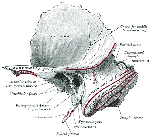
Long description
Left temporal bone. Outer surface. Labelled illustration from Wikimedia Commons, used under its open licence.

Figure 1: Left temporal bone (outer surface).
Figure 2Gray382
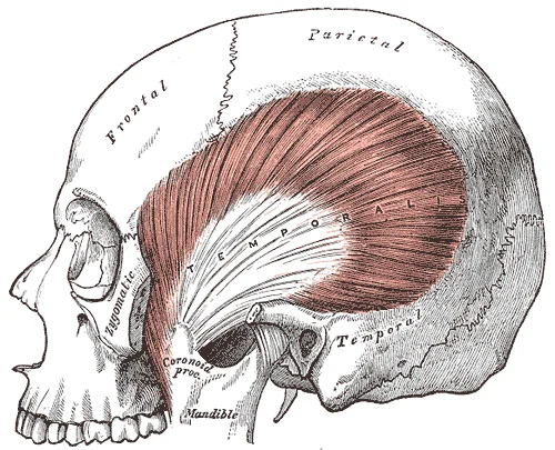
Long description
The Temporalis; the zygomatic arch and Masseter have been removed.

Figure 2: The Temporalis; the zygomatic arch and Masseter have been removed.
The temporal and infratemporal fossae are complex anatomical spaces defined by the surrounding skeletal architecture of the skull base and face. Understanding these fossae requires a precise knowledge of the bones that form their boundaries. The cranium is subdivided into the facial bones and the brain case. The temporal fossa, located on the lateral aspect of the skull, is primarily bounded by the temporal lines on the frontal, parietal, and temporal bones. The infratemporal fossa lies inferior and medial to the zygomatic arch and lateral to the lateral pterygoid plate. [1]
Figure 3705 Lateral View of Skull-01
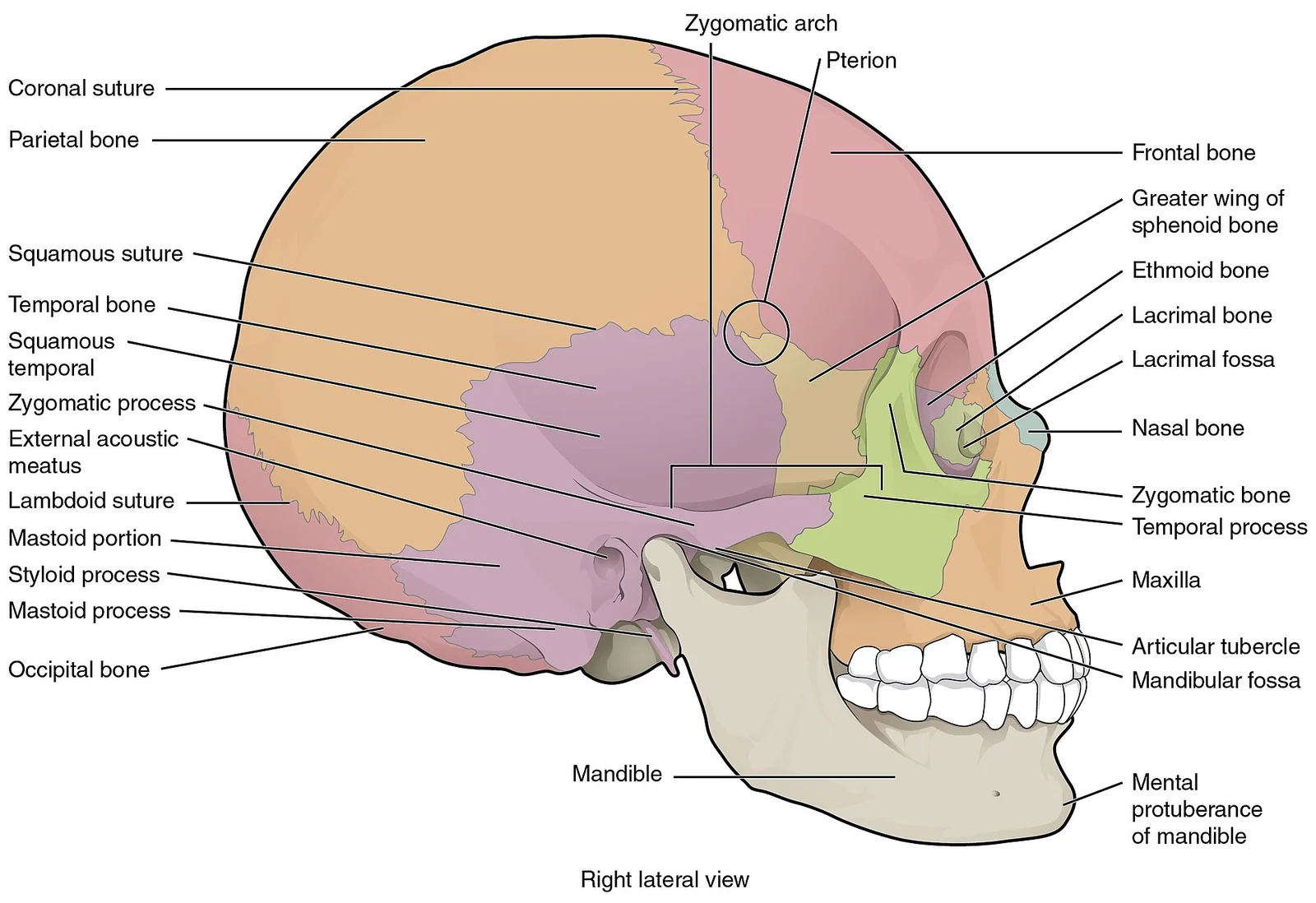
Long description
The lateral skull shows the large rounded brain case, zygomatic arch, and the upper and lower jaws. The zygomatic arch is formed jointly by the zygomatic process of the temporal bone and the temporal process of the zygomatic bone. The shallow space above the zygomatic arch is the temporal fossa. The

Figure 3: The lateral skull shows the large rounded brain case, zygomatic arch, and the upper and lower jaws.
The sphenoid bone is central to this region. The right and left medial pterygoid plates form the posterior, lateral walls of the nasal cavity. The somewhat larger lateral pterygoid plates serve as attachment sites for chewing muscles that fill the infratemporal space and act on the mandible. [1]
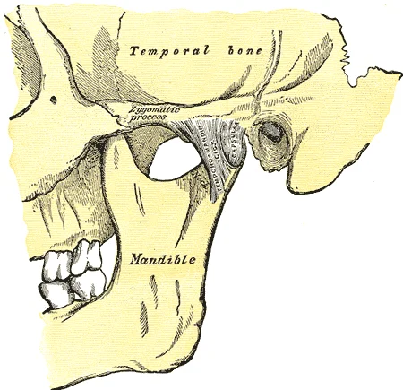
The mandible also contributes key landmarks. The ramus on each side of the mandible has two upward-going bony projections. The more anterior projection is the flattened coronoid process of the mandible, which provides attachment for one of the biting muscles. The posterior projection is the condylar process of the mandible, which is topped by the oval-shaped condyle. The condyle of the mandible articulates with the mandibular fossa and articular tubercle of the temporal bone. Together these articulations form the temporomandibular joint. The broad U-shaped curve located between the coronoid and condylar processes is the mandibular notch. [1]
Figure 4Gray310
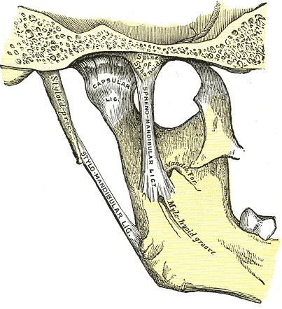
Long description
Articulation of the mandible. Medial aspect.

Figure 4: Articulation of the mandible (medial aspect).
Figure 5Gray309
Long description
Articulation of the mandible. Lateral aspect.

Figure 5: Articulation of the mandible (lateral aspect).
The zygomatic bone forms the lateral wall of the orbit and contributes to the temporal fossa boundary. The maxilla forms the upper jaw and contributes to the floor of the orbit and the lateral wall of the nasal cavity. The palatine bone contributes small areas to the lateral walls of the nasal cavity and the medial wall of each orbit. The largest region of each palatine bone is the horizontal plate. The plates from the right and left palatine bones join together at the midline to form the posterior quarter of the hard palate. [1]
DefinitionPterygoid Plates
The medial and lateral pterygoid plates are vertical bony projections of the sphenoid bone that serve as attachment sites for muscles of mastication.
The infratemporal fossa is bounded laterally by the ramus of the mandible and the zygomatic arch. Medially, it is bounded by the lateral pterygoid plate of the sphenoid bone. Superiorly, it is bounded by the infratemporal surface of the greater wing of the sphenoid bone. Anteriorly, it is bounded by the posterior surface of the maxilla. Posteriorly, it is bounded by the styloid process and its associated muscles. The temporal fossa is bounded superiorly by the temporal lines. Anteriorly, it is bounded by the frontal and zygomatic bones. Inferiorly, it is bounded by the zygomatic arch. Medially, it is bounded by the frontal, parietal, temporal, and greater wing of the sphenoid bones. Posteriorly, it is bounded by the temporal and occipital bones. [1]
Figure 6Gray141
Long description
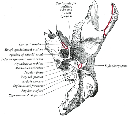
Left temporal bone. Inferior surface. Labelled illustration from Wikimedia Commons, used under its open licence.

Figure 6: Left temporal bone (inferior surface).
Figure 7Gray139
Long description
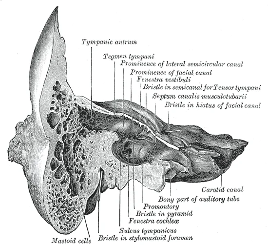
Coronal section of right temporal bone. Labelled illustration from Wikimedia Commons, used under its open licence.

Figure 7: Coronal section of right temporal bone.
Figure 8Gray138
Long description
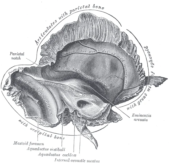
Left temporal bone. Inner surface. Labelled illustration from Wikimedia Commons, used under its open licence.

Figure 8: Left temporal bone (inner surface).
| Structure | Location | Relation |
|---|---|---|
| Lateral Pterygoid Plate | Sphenoid Bone | Medial boundary of infratemporal fossa |
| Medial Pterygoid Plate | Sphenoid Bone | Posterior lateral wall of nasal cavity |
| Coronoid Process | Mandible | Anterior projection of ramus |
| Condylar Process | Mandible | Posterior projection of ramus |
| Mandibular Notch | Mandible | Between coronoid and condylar processes |
| Temporal Lines | Frontal, Parietal, Temporal | Superior boundary of temporal fossa |
The temporal and infratemporal fossae are anatomical spaces defined by the surrounding bony architecture rather than containing intrinsic synovial joints within their cavities. The primary articulation relevant to this region is the temporomandibular joint (TMJ), which connects the mandible to the temporal bone. This joint allows for the complex movements required for mastication and speech.
The articular eminence lies anterior to the condyle. The joint capsule encloses the articulation. The sphenoid bone forms the anterior boundary, with the greater wing extending laterally.
The TMJ is a synovial joint. It consists of the mandibular condyle and the mandibular fossa of the temporal bone. An articular disc separates the two bony surfaces. The joint is supported by ligaments, including the lateral ligament, which reinforces the capsule laterally. Movements include depression, elevation, protrusion, retrusion, and lateral excursion.
It is located in the upper neck near the level of the inferior mandible. It serves as the base for the tongue above, and is attached to the larynx below and the pharynx posteriorly. Movements of the hyoid are coordinated with movements of the tongue, larynx, and pharynx during swallowing and speaking. [1]
Finally, students may overlook the depth of the posterior cranial fossa. It contains the cerebellum of the brain[1]. The posterior fossa is bounded anteriorly by the petrous ridges, while the occipital bone forms the floor and posterior wall[1]. It is divided at the midline by the large foramen magnum (“great aperture”), the opening that provides for passage of the spinal
DefinitionTMJ
The temporomandibular joint is the synovial articulation between the mandibular condyle and the temporal bone, allowing complex jaw movements.
| Structure | Type | Relation |
|---|---|---|
| Mandibular Condyle | Bone | Articulates with temporal bone |
| Hyoid Bone | Bone | Suspended by muscles, no bony joint |
| Sphenoid Greater Wing | Bone | Forms anterior floor of middle cranial fossa |
The rounded depression in the floor of the sella turcica is the hypophyseal (pituitary) fossa, which houses the pea-sized pituitary (hypophyseal) gland. This structure is central but not part of the fossae joints.
Exam trapHyoid Joint
The hyoid bone does not articulate with any other bone; it is suspended by muscular attachments.
The temporal and infratemporal fossae are deep anatomical spaces located lateral to the cranial cavity. They are bounded by complex bony structures and covered by strong fascial layers that compartmentalise the contained muscles and neurovascular bundles. Understanding these compartments is essential for interpreting surgical approaches and the spread of infection.
The temporalis muscle occupies the temporal fossa, covered by the temporal fascia. This fascia is thick and strong, attaching superiorly to the superior temporal line and inferiorly to the zygomatic arch.
The temporal fascia encloses the temporalis muscle. It splits to attach to the zygomatic arch, creating a compartment for the temporalis muscle. The infratemporal fascia covers the lateral pterygoid muscle. These fascial layers provide attachment points for muscles and protect neurovascular structures. The compartments are separated by bony landmarks such as the zygomatic arch and the lateral pterygoid plate.
DefinitionTemporal Fascia
A strong fibrous layer covering the temporalis muscle, attaching to the zygomatic arch and temporal lines.
| Structure | Attachment |
|---|---|
| Temporal Fascia | Superior temporal line to zygomatic arch |
| Pterygoid Fascia | Lateral pterygoid plate to zygomatic arch |
The relationship between these fascial compartments determines the spread of pathology. Infections in the temporal fossa may track into the infratemporal fossa if the fascia is breached. Surgical access requires careful incision through these layers to avoid damaging the underlying nerves and vessels.
Figure 9Gray383
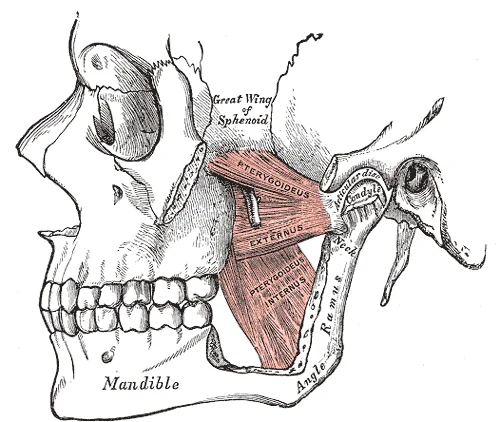
Long description
The Pterygoidei; the zygomatic arch and a portion of the ramus of the mandible have been removed.

Figure 9: The Pterygoidei; the zygomatic arch and a portion of the ramus of the mandible have been removed.
Figure 10Gray311
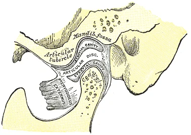
Long description
Sagittal section of the articulation of the mandible.

Figure 10: Sagittal section of the articulation of the mandible.
The masseter is lateral, while the medial and lateral pterygoids occupy the infratemporal fossa. The maxillary artery courses through the infratemporal fossa. The temporomandibular joint (TMJ) is indicated laterally.
The muscles of mastication are innervated by the mandibular division of the trigeminal nerve (CN V3). They are grouped into four main muscles: the temporalis, masseter, medial pterygoid, and lateral pterygoid. These muscles act on the mandible to facilitate chewing and jaw movement.
| Muscle | Origin | Insertion | Action | Nerve Supply |
|---|---|---|---|---|
| Temporalis | Temporal fossa | Coronoid process of mandible | Elevates and retracts mandible | Mandibular nerve (V3) |
| Masseter | Zygomatic arch | Lateral surface of ramus and angle of mandible | Elevates mandible | Mandibular nerve (V3) |
| Medial Pterygoid | Medial surface of lateral pterygoid plate | Medial surface of ramus and angle of mandible | Elevates and protrudes mandible | Mandibular nerve (V3) |
| Lateral Pterygoid | Lateral pterygoid plate | Neck of mandible and TMJ capsule | Protrudes and depresses mandible | Mandibular nerve (V3) |
It elevates and retracts the mandible. The masseter originates from the zygomatic arch and inserts on the lateral surface of the ramus and angle of the mandible. It primarily elevates the mandible. The medial pterygoid originates from the medial surface of the lateral pterygoid plate and inserts on the medial surface of the ramus and angle. It elevates and protrudes the mandible. It protrudes and depresses the mandible.
The infratemporal fossa contains these muscles, along with the maxillary artery and the mandibular nerve. The pterygoid venous plexus is also located here. Infections can spread from the teeth to this plexus. Nerve blocks of the mandibular nerve are performed in this region. The spread of infection can follow the fascial planes of these muscles.
Figure 11Deep temporal arteries
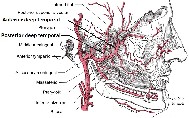
Long description
Branches of the maxillary artery. Labelled illustration from Wikimedia Commons, used under its open licence.

Figure 11: Branches of the maxillary artery.
The arterial supply of the temporal and infratemporal fossae is dominated by the maxillary artery, a terminal branch of the external carotid artery. The maxillary artery enters the infratemporal fossa through the pterygomaxillary fissure, lying deep to the lateral pterygoid muscle and posterior to the lateral pterygoid plate. From its origin it divides into three principal segments: the mandibular, pterygoid, and pterygopalatine portions. Each segment gives rise to a series of smaller branches that nourish the deep structures of the skull base, the mandible, and the surrounding soft tissues.
Key branches include the inferior alveolar artery, which accompanies the inferior alveolar nerve through the mandibular foramen into the mandibular canal, and the deep auricular artery, which supplies the temporomandibular joint capsule. The pterygoid segment gives rise to the lateral and medial pterygoid arteries, which run along the respective pterygoid muscles, and the ascending palatine artery, which ascends to the soft palate. The pterygopalatine segment supplies the nasal cavity, the palate, and the pharynx via the sphenopalatine artery and the descending palatine artery. The sphenopalatine artery, the terminal branch of the maxillary artery, enters the nasal cavity through the sphenopalatine foramen and gives off the posterior superior nasal branches that supply the posterior nasal septum and lateral walls.
The temporal fossa receives its arterial supply primarily from branches of the superficial temporal artery, a terminal branch of the external carotid that runs superficially over the temporalis muscle. The middle meningeal artery, a branch of the maxillary artery, enters the cranial cavity through the foramen spinosum and supplies the dura mater of the middle cranial fossa, including the temporal lobe.
The following table summarises the major branches of the maxillary artery and their primary territories:
| Branch | Territory supplied |
|---|---|
| Inferior alveolar | Mandibular canal, lower teeth |
| Deep auricular | TMJ capsule |
| Lateral pterygoid | Lateral pterygoid muscle |
| Medial pterygoid | Medial pterygoid muscle |
| Ascending palatine | Soft palate |
| Sphenopalatine | Nasal cavity |
| Descending palatine | Hard palate, tonsillar fossa |
The venous drainage of the temporal and infratemporal fossae is primarily mediated by the pterygoid venous plexus. This network is situated deep within the infratemporal fossa, lying between the lateral pterygoid muscle and the temporalis muscle. It serves as a crucial communication channel between the superficial veins of the face and the deeper intracranial venous sinuses.
Superiorly, it relates to the middle meningeal vein, which passes through the foramen spinosum to drain into the cavernous sinus. Anteriorly, it connects with the deep facial vein, which descends to join the facial vein. Posteriorly, it communicates with the maxillary vein, which contributes to the formation of the retromandibular vein. The plexus is embedded in the loose connective tissue of the infratemporal fossa, allowing it to expand and contract with changes in pressure.
The retromandibular vein then divides into anterior and posterior divisions. The anterior division joins the facial vein to form the common facial vein, which drains into the internal jugular vein. The posterior division joins the posterior auricular vein to form the external jugular vein.
DefinitionPterygoid Venous Plexus
A network of veins located in the infratemporal fossa that connects the facial vein with the cavernous sinus and the maxillary vein.
| Structure | Connection | Direction |
|---|---|---|
| Middle Meningeal Vein | Cavernous Sinus | Intra- to Extracranial |
| Deep Facial Vein | Facial Vein | Extracranial |
| Maxillary Vein | Retromandibular Vein | Outflow |
| Superior Temporal Vein | Temporalis Muscle | Inflow |
The clinical significance of this drainage pattern lies in its potential for spreading infection. Because the pterygoid plexus communicates directly with the cavernous sinus via the emissary veins, infections from the face or teeth can spread intracranially. This is particularly dangerous as the cavernous sinus contains cranial nerves III, IV, V1, V2, and VI, as well as the internal carotid artery.
Exam trapInfection Spread
Infections in the 'danger triangle' of the face can spread to the cavernous sinus via the pterygoid plexus, leading to cavernous sinus thrombosis.
Several cranial nerves from the brain exit the skull via this opening. It is also the exit point through the base of the skull for all the venous return blood leaving the brain. The venous structures that carry blood inside the skull form large, curved grooves on the inner walls of the posterior cranial fossa, which terminate at each jugular foramen. [1]
The nerves of the temporal and infratemporal fossae are predominantly branches of the mandibular division of the trigeminal nerve (CN V3), which provides both motor supply to the muscles of mastication and cutaneous sensation to the lower face. The main trunk of CN V3 emerges from the foramen ovale and immediately divides into anterior and posterior divisions. The buccal nerve is sensory, running anteriorly between the two heads of the lateral pterygoid muscle to supply the skin of the cheek and the buccal mucosa. The posterior division is primarily sensory, containing the auriculotemporal, lingual, and inferior alveolar nerves. The lingual nerve runs inferior to the lateral pterygoid muscle, carrying general sensation from the anterior two-thirds of the tongue. The inferior alveolar nerve enters the mandibular foramen on the medial surface of the ramus, running within the mandibular canal to supply the lower teeth before emerging as the mental nerve. [1]
DefinitionMandibular Nerve
The third division of the trigeminal nerve, providing motor innervation to muscles of mastication and sensory innervation to the lower face, tongue, and teeth.
| Nerve | Origin | Primary Supply |
|---|---|---|
| Auriculotemporal | Posterior division of CN V3 | Skin of temple, external ear, temporomandibular joint |
| Lingual | Posterior division of CN V3 | Anterior two-thirds of tongue (general sensation) |
| Inferior Alveolar | Posterior division of CN V3 | Lower teeth, chin (via mental nerve) |
| Buccal | Anterior division of CN V3 | Skin of cheek, buccal mucosa |
| Deep Temporal | Anterior division of CN V3 | Temporalis muscle (motor) |
| Nerve to Lateral Pterygoid | Anterior division of CN V3 | Lateral pterygoid muscle (motor) |
| Nerve | Origin | Primary Supply |
|---|---|---|
| Auriculotemporal | Posterior division of CN V3 | Skin of temple, external ear, temporomandibular joint |
| Lingual | Posterior division of CN V3 | Anterior two-thirds of tongue (general sensation) |
| Inferior Alveolar | Posterior division of CN V3 | Lower teeth, chin (via mental nerve) |
| Buccal | Anterior division of CN V3 | Skin of cheek, buccal mucosa |
| Deep Temporal | Anterior division of CN V3 | Temporalis muscle (motor) |
| Nerve to Lateral Pterygoid | Anterior division of CN V3 | Lateral pterygoid muscle (motor) |
Lesions of these nerves result in specific sensory deficits or motor weakness. For instance, damage to the auriculotemporal nerve may cause loss of sensation over the temple and ear, while injury to the inferior alveolar nerve results in anaesthesia of the lower lip and chin. Understanding these pathways is essential for localising lesions in clinical practice.
The lymphatic drainage of the temporal and infratemporal fossae follows a predictable pattern that is critical for understanding the spread of infection or malignancy in the head and neck region. The superficial tissues of the temporal region, including the skin and subcutaneous tissue of the temple, drain primarily into the preauricular and superficial parotid lymph nodes. These nodes are situated anterior to the ear and within the parotid gland parenchyma. From these initial stations, efferent vessels pass deep to drain into the deep cervical lymph nodes. In this lateral view, the preauricular nodes are located just anterior to the tragus of the ear. The superficial parotid nodes lie within the superficial layer of the parotid fascia. The efferent vessels from these nodes descend to join the upper deep cervical nodes, which are arranged along the internal jugular vein.
DefinitionPreauricular Nodes
Lymph nodes located anterior to the ear that receive drainage from the lateral scalp, eyelids, and external acoustic meatus.
The infratemporal fossa contains the pterygoid muscles, the maxillary artery, and the mandibular nerve. Lymphatic vessels from the infratemporal fossa drain into the deep cervical lymph nodes, specifically those along the internal jugular vein. This direct drainage bypasses the superficial nodal groups, highlighting the importance of deep cervical examination in patients with pathology in this region.
| Structure | Primary Drainage | Secondary Drainage |
|---|---|---|
| Temporal Skin | Preauricular Nodes | Deep Cervical Nodes |
| Temporalis Muscle | Deep Parotid Nodes | Deep Cervical Nodes |
| Infratemporal Fossa | Deep Cervical Nodes | Lower Deep Cervical Nodes |
Clinical correlation is essential. Infection or tumour in the temporal region may present with enlarged preauricular nodes, while pathology in the infratemporal fossa may present with deep cervical lymphadenopathy. Understanding these pathways allows for accurate staging of head and neck cancers and appropriate management of infections. The deep cervical nodes are the final common pathway for most head and neck lymphatic drainage, making them a critical site for examination.
The temporal and infratemporal fossae are complex anatomical regions situated lateral to the skull base. This view demonstrates the lateral aspect of the skull, showing the temporal fossa superiorly and the infratemporal fossa inferiorly. The temporal fossa is bounded superiorly by the temporal lines and inferiorly by the zygomatic arch. Its floor is formed by parts of the frontal, parietal, temporal, and sphenoid bones. The infratemporal fossa lies deep to the ramus of the mandible and the zygomatic arch. It communicates with the temporal fossa through the gap between the zygomatic arch and the skull base.
DefinitionTemporal Fossa
A shallow depression on the lateral aspect of the skull, bounded by the temporal lines and zygomatic arch.
The contents of the temporal fossa include the temporalis muscle, which originates from the temporal fossa and inserts onto the coronoid process of the mandible.
| Muscle | Origin | Insertion | Action | Nerve Supply |
|---|---|---|---|---|
| Lateral Pterygoid | Greater wing of sphenoid; lateral pterygoid plate | Neck of mandible; articular disc | Protrusion; depression of mandible | Mandibular nerve |
| Medial Pterygoid | Lateral surface of lateral pterygoid plate | Medial surface of ramus of mandible | Elevation; protrusion of mandible | Mandibular nerve |
They serve to reduce bone mass and thus lighten the skull, and they also add resonance to the voice. This second feature is most obvious when you have a cold or sinus congestion. These produce swelling of the mucosa and excess mucus production, which can obstruct the narrow passage [1]
The temporal and infratemporal fossae are deep anatomical spaces critical for neurovascular supply to the head. Understanding their radiological appearance is essential for accurate diagnosis and intervention.
X-ray imaging provides limited detail of these fossae due to overlapping structures. It is primarily used to detect gross bony abnormalities or fractures involving the zygomatic arch or sphenoid bone.
Computed tomography (CT) is the modality of choice for evaluating bony anatomy. It offers high-resolution images of the skull base, mandible, and sphenoid sinus. CT is particularly useful for identifying fractures, tumours, or inflammatory changes affecting the bony boundaries of the fossae. It also visualises the course of the maxillary artery and its branches.
Magnetic resonance imaging (MRI) excels in soft tissue contrast. It is superior for visualising the contents of the infratemporal fossa, including the pterygoid muscles, the mandibular nerve, and the maxillary artery. MRI is essential for evaluating soft tissue masses, inflammation, or nerve compression. It can distinguish between muscle, fat, and neural structures with high precision.
Ultrasound has a limited role in imaging these deep fossae. It is primarily used for superficial structures such as the temporomandibular joint. Ultrasound can guide needle placement for nerve blocks in the infratemporal fossa, providing real-time visualisation of the needle tip relative to vascular and neural structures. [3]
Clinical pearlImaging Choice
CT is best for bone; MRI is best for soft tissue and nerves.
The temporal and infratemporal fossae are critical anatomical spaces housing muscles of mastication, the maxillary artery, and the mandibular nerve. Clinical assessment often relies on surface landmarks to identify deep structures. The zygomatic arch serves as a key landmark for the coronoid process of the mandible and the insertion of the temporalis tendon. Palpation here helps assess temporomandibular joint function and muscle hypertrophy.
Injuries to these fossae typically result from high-impact trauma, such as motor vehicle accidents or falls. Fractures of the zygomaticomaxillary complex often extend into the infratemporal fossa, potentially damaging the maxillary artery or its branches. Such injuries may present with trismus due to haematoma formation or direct muscle involvement. Nerve palsies are a significant concern. The mandibular division of the trigeminal nerve (V3) exits the skull via the foramen ovale and courses through the infratemporal fossa. Compression or transection during surgical approaches or trauma can lead to sensory loss in the lower face and motor deficits in the muscles of mastication.
Procedures involving these spaces require precise anatomical knowledge.
Surface landmarks also guide surgical access. The lateral pterygoid plate forms the medial boundary of the infratemporal fossa. The masseter muscle covers the lateral aspect of the ramus of the mandible. Understanding these relations is vital for avoiding iatrogenic injury.
Clinical pearlClinical Pearl
Always consider the proximity of the maxillary artery to the lateral pterygoid muscle during deep facial procedures.
| Muscle | Origin | Insertion | Action | Nerve Supply |
|---|---|---|---|---|
| Temporalis | Temporal fossa | Coronoid process | Elevates mandible | Mandibular nerve (V3) |
| Masseter | Zygomatic arch | Mandible angle | Elevates mandible | Mandibular nerve (V3) |
| Medial Pterygoid | Medial pterygoid plate | Mandible angle | Elevates mandible | Mandibular nerve (V3) |
| Lateral Pterygoid | Lateral pterygoid plate | Mandibular condyle | Protrudes mandible | Mandibular nerve (V3) |
The temporal fossa is a shallow depression on the lateral aspect of the skull, bounded superiorly by the temporal lines and inferiorly by the zygomatic arch. Its floor is formed by parts of the frontal, parietal, temporal, and sphenoid bones. The fossa contains the temporalis muscle, which is a major muscle of mastication. The infratemporal fossa lies deep and inferior to the temporal fossa, separated by the lateral pterygoid muscle. It contains the mandibular nerve, maxillary artery, pterygoid muscles, and the pterygoid venous plexus.
DefinitionTemporalis Muscle
A fan-shaped muscle originating from the temporal fossa and inserting onto the coronoid process of the mandible, responsible for elevating and retracting the jaw.
It gives off several branches including the middle meningeal artery, which enters the cranial cavity via the foramen spinosum. This artery is clinically significant as its rupture can lead to an epidural hematoma. The mandibular nerve (V3) exits the skull via the foramen ovale and enters the infratemporal fossa, providing sensory and motor innervation to the lower face and muscles of mastication.
| Muscle | Origin | Insertion | Action | Nerve Supply |
|---|---|---|---|---|
| Temporalis | Temporal fossa | Coronoid process | Elevates and retracts mandible | Anterior trunk of V3 |
| Masseter | Zygomatic arch | Ramus of mandible | Elevates mandible | Masseteric nerve (V3) |
| Medial Pterygoid | Pterygoid plate | Medial surface of ramus | Elevates and protrudes mandible | Medial pterygoid nerve (V3) |
| Lateral Pterygoid | Pterygoid plate | Neck of mandible | Protrudes and depresses mandible | Lateral pterygoid nerve (V3) |
The sphenomandibular ligament, a remnant of Meckel's cartilage, runs between the spine of the sphenoid and the mandibular foramen, providing support to the temporomandibular joint.
Exam trapArterial Branches
Do not confuse the middle meningeal artery (branch of maxillary) with the anterior meningeal artery (branch of anterior ethmoidal). The former is the primary blood supply to the dura mater in the middle cranial fossa.
DefinitionMandibular Foramen
Located on the medial surface of the ramus of the mandible, this foramen allows the entry of the inferior alveolar nerve and artery into the mandibular canal.
Clinical pearlSurgical Approach
Access to the infratemporal fossa is often required for removal of tumors or fractures of the mandibular condyle. Care must be taken to avoid injury to the maxillary artery and mandibular nerve.
NoteLymphatic Drainage
Lymph from the temporal and infratemporal regions drains into the deep cervical lymph nodes. Infection in these areas can lead to swelling and tenderness in the neck.
Exam trapJoint Anatomy
The temporomandibular joint (TMJ) is a synovial joint with an articular disc. The disc is attached anteriorly to the joint capsule and posteriorly to the retrodiscal tissue, which is highly vascular and innervated.
Clinical pearlClinical Significance
Trigeminal neuralgia can present with pain in the distribution of V3, affecting the temporal and infratemporal regions. Diagnosis is clinical, based on the characteristic sharp, shooting pain.
Students frequently confuse the bony boundaries of the nasal cavity with the deeper infratemporal fossa. A primary error is assuming the nasal septum is entirely cartilaginous. The anterior nasal septum is formed by septal cartilage, a flexible plate that fills the gap between the perpendicular plate of the ethmoid and vomer bones[1]. However, the posterior and inferior parts are bony. The upper portion of the septum is formed by the perpendicular plate of the ethmoid bone[1] The vomer is best seen when looking from behind into the posterior openings of the nasal cavity
Another frequent mistake involves the mandible's development and anatomy. The mandible forms the lower jaw and is the only moveable bone of the skull[1]. At the time of birth, the mandible consists of paired right and left bones, but these fuse together during the first year to form the single U-shaped mandible of the adult skull[1]. Students often misidentify the angle of the mandible. The outside margin of the mandible, where the body and ramus come together is called the angle of the mandible.
A third pitfall relates to the sphenopalatine ganglion (SPG) block technique. The SPG is a critical integration center between trigeminal nociceptive signaling and cranial autonomic responses. It is closely associated with the trigeminal-autonomic reflex, a loop in which trigeminal sensory input triggers parasympathetic output, leading to symptoms such as lacrimation, rhinorrhea, and nasal congestion. Students often forget that this reflex explains the autonomic symptoms in cluster headaches. The parasympathetic activity is blocked which inhibits the cerebral vasodilatation. The vasodilatation occurs to restore intracranial volume. Understanding this mechanism prevents the error of thinking the block is purely analgesic rather than anti-inflammatory and anti-vasodilatory. [3] [2]
Finally, students may overlook the depth of the posterior cranial fossa. It contains the cerebellum of the brain[1]. The posterior fossa is bounded anteriorly by the petrous ridges, while the occipital bone forms the floor and posterior wall[1]. It is divided at the midline by the large foramen magnum (“great aperture”), the opening that provides for passage of the spinal
This knowledge is applied at the bedside for managing severe headaches. The approaches practiced by pain physicians are not only invasive, but also necessary training for use of fluoroscopy, the operation room, and sedation in some patients to facilitate the procedure. The SPG block practiced for PDPH is simple, and therefore, it can be done bedside in wards, or in the out-patient department, and does not need fluoroscopy or an operating room. This distinction is vital for triage. Relief is ofte [2]
In the operating theatre, anatomical precision is required for endoscopic sinus surgery. Surgeons must navigate the sphenopalatine foramen to access the SPG. This method targets the sphenopalatine foramen, approximating the SPG by capillary absorption and diffusion. The safety profile was acceptable, with only transient sensory disturbances reported. This suggests that selective patient screening may improve success rates, and targeting multiple cranial pain pathways may be necessary for complex facial pain syndromes. Surgeons must avoid damaging the trigeminal-autonomic reflex loop to prevent unintended autonomic symptoms. [3]
The temporalis muscle occupies the temporal fossa, covered by the temporal fascia. This fascia is thick and strong, attaching superiorly to the superior temporal line and inferiorly to the zygomatic arch.
In examinations, students must demonstrate knowledge of the skull's composition. In the adult, the skull consists of 22 individual bones, 21 of which are immobile and united into a single unit[1]. The 22nd bone is the mandible (lower jaw), which is the only moveable bone of the skull[1]. Questions often test the origin and insertion of muscles in the infratemporal fossa, such as the lateral pterygoid. Understanding the relations of the mandibular ramus is key. Each side of the mandible consists of a horizontal body and posteriorly, a vertically oriented ramus of the mandible (ramus = “branch”)[1]. This structural knowledge underpins the understanding of nerve blocks and surgical approaches to the maxillofacial region.
The mandible is the only moveable bone of the adult skull, consisting of 22 bones total. The nasal septum comprises the perpendicular plate of the ethmoid, the vomer, and septal cartilage. The vomer forms the posterior-inferior part of the nasal septum and is best viewed from behind. The sphenopalatine ganglion integrates trigeminal nociceptive signaling with cranial autonomic responses. SPG blocks can be performed bedside without fluoroscopy for certain headache types. The posterior cranial fossa is bounded anteriorly by the petrous ridges and contains the cerebellum. Septal cartilage is flexible and separates the nostrils but is absent in dry skulls. [1] The angle of the mandible is the junction of the body and ramus.
Answer each one in your head first, then open the answer.
Which bone forms the posterior-inferior part of the nasal septum?
Show answerHide answer
VomerWhat is the only moveable bone of the adult skull?
Show answerHide answer
MandibleWhich structure separates the right and left nostrils?
Show answerHide answer
Septal cartilageWhat reflex loop involves the sphenopalatine ganglion?
Show answerHide answer
Trigeminal-autonomic reflexWhere is the posterior cranial fossa bounded anteriorly?
Show answerHide answer
Petrous ridges
- Betts JG, Young KA, Wise JA, et al. The Skull. OpenStax, Anatomy and Physiology 2e (CC BY-NC-SA 4.0). Accessed .
- Nair AS, Rayani BK. Sphenopalatine ganglion block for relieving postdural puncture headache: technique and mechanism of action of block with a narrative review of efficacy. The Korean journal of pain 2017. Accessed .
- Bautista AL, Coyne K, Bautista A, Abd-Elsayed A. Sphenopalatine Ganglion Blocks in Headache Management: A Review. Brain sciences 2025. Accessed .
Figure credits
- Figure 1: Henry Vandyke Carter, via Wikimedia Commons, Public domain, Public domain.
- Figure 2: Henry Vandyke Carter, via Wikimedia Commons, Public domain, Public domain.
- Figure 3: OpenStax College, via Wikimedia Commons, CC BY 3.0, CC BY 3.0. resized.
- Figure 4: Henry Vandyke Carter, via Wikimedia Commons, Public domain, Public domain.
- Figure 5: Henry Vandyke Carter, via Wikimedia Commons, Public domain, Public domain.
- Figure 6: Henry Vandyke Carter, via Wikimedia Commons, Public domain, Public domain.
- Figure 7: Henry Vandyke Carter, via Wikimedia Commons, Public domain, Public domain.
- Figure 8: Henry Vandyke Carter, via Wikimedia Commons, Public domain, Public domain.
- Figure 9: Henry Vandyke Carter, via Wikimedia Commons, Public domain, Public domain.
- Figure 10: Henry Vandyke Carter, via Wikimedia Commons, Public domain, Public domain.
- Figure 11: Mikael Häggström, via Wikimedia Commons, Public domain, Public domain.
Educational content for medical students and doctors. It is not a substitute for clinical judgement or local guidelines.