Pterygopalatine fossa
A deep skull space housing the maxillary nerve and pterygoid muscles, bounded by sphenoid and palatine bones.
The pterygopalatine fossa is a small, irregular space situated deep to the maxilla, posterior to the maxillary sinus, and medial to the infratemporal fossa. The region is not a single open cavity but a complex arrangement of bony plates and foramina that connect the orbit, nasal cavity, and infratemporal fossa. Understanding its boundaries is essential for interpreting imaging and planning surgical approaches to the midface. [2]
The anterior wall is formed by the posterior surface of the maxilla, specifically the area behind the maxillary sinus. The posterior wall is the pterygoid process of the sphenoid bone, which separates the fossa from the infratemporal fossa laterally. The superior boundary is the floor of the middle cranial fossa, marked by the greater wing of the sphenoid bone. [1]
The fossa communicates with adjacent spaces through specific foramina. The foramen rotundum transmits the maxillary nerve from the middle cranial fossa into the pterygopalatine fossa. The pterygopalatine canal transmits the greater and lesser palatine nerves to the palate. These pathways are critical for the spread of infection or tumor from the nasal cavity or nasopharynx into the central nervous system. [2]
The pterygopalatine fossa is a central station in the trigeminal nerve pathway. It is often the site of perineural spread from cancers of the palate, cheek, maxillary sinus, and nasopharynx. Direct invasion can occur in tumors such as juvenile nasopharyngeal angiofibroma and nasopharyngeal carcinoma. [2]
| Structure | Boundary or Opening | Content |
|---|---|---|
| Anterior Wall | Posterior Maxilla | Maxillary Sinus |
| Posterior Wall | Pterygoid Process | Infratemporal Fossa |
| Superior Wall | Sphenoid Greater Wing | Middle Cranial Fossa |
| Inferior Wall | Hard Palate | Nasal Cavity |
| Foramen Rotundum | Anterior | Maxillary Nerve |
| Pterygopalatine Canal | Inferior | Palatine Nerves |
The pterygopalatine fossa is a small, pyramidal space located deep within the facial skeleton. It sits posterior to the maxilla and anterior to the pterygoid process of the sphenoid bone. To locate it clinically, one must understand the surrounding bony landmarks. The zygomatic arch forms a critical horizontal divider on the lateral skull. It consists of the temporal process of the zygomatic bone anteriorly and the zygomatic process of the temporal bone posteriorly. The temporal fossa lies above this arch, while the infratemporal fossa lies below it. The pterygopalatine fossa is situated medial to the infratemporal fossa. [1] [3]
Palpable landmarks guide the surgeon to this deep region. Specifically, the posterior surface of the maxilla faces the fossa. The sphenoid bone forms the posterior boundary. The greater wings of the sphenoid bone extend laterally from the sella turcica. On the outside of the lateral skull, the greater wing forms a rectangular area immediately anterior to the squamous portion of the temporal bone. This area is easily palpable.
Anteriorly, the maxilla forms the front wall. The roof also includes the sphenoid bone.
The zygomatic arch consists of two parts. The sphenoid bone is posterior. The foramina transmitting nerves are marked.
The lacrimal bone is a small, rectangular bone forming the anterior, medial wall of the orbit. It lies anterior to the pterygopalatine fossa. The lacrimal fossa is a shallow depression on this bone. The nasolacrimal canal extends inferiorly from this fossa. This canal drains tears into the nasal cavity. The pterygopalatine fossa is deep to these structures. [1]
The hypophyseal fossa is a rounded depression in the floor of the sella turcica. It houses the pituitary gland. This structure is superior and medial to the pterygopalatine fossa. The greater wings of the sphenoid bone form the anterior floor of the middle cranial fossa. These wings are lateral to the sella turcica.
Figure 1709 Sphenoid Bone
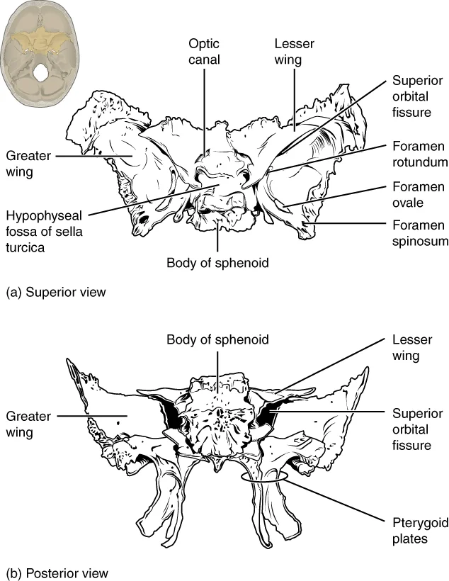
Long description
Shown in isolation in (a) superior and (b) posterior views, the sphenoid bone is a single midline bone that forms the anterior walls and floor of the middle cranial fossa. It has a pair of lesser wings and a pair of greater wings. The sella turcica surrounds the hypophyseal fossa. Projecting downwar

Figure 1: Shown in isolation in (a) superior and (b) posterior views, the sphenoid bone is a single midline bone that forms the anterior walls and floor of the middle cranial fossa.
Understanding these landmarks allows precise localization. The pterygopalatine fossa is a key hub for neurovascular structures. It connects the nasal cavity, orbit, and cranial cavity. Surgeons must identify the zygomatic arch and the maxilla to access this space. The infratemporal fossa provides a lateral approach. The nasal cavity provides a medial approach. The orbit provides a superior approach.
The pterygopalatine fossa is a small, inverted pyramid-shaped space located deep within the facial skeleton. It serves as a critical crossroads for neurovascular structures passing between the middle cranial fossa and the oral and nasal cavities. Understanding its bony boundaries is essential for interpreting imaging and performing surgical approaches. [1]
The anterior boundary is formed by the posterior surface of the maxilla. Specifically, the pterygopalatine fossa lies posterior to the maxillary sinus.
The maxilla contributes significantly to the anterior and lateral walls. The palatine bone forms the medial wall and part of the inferior boundary. The sphenoid bone forms the posterior wall. The body of the sphenoid bone articulates with the palatine bone medially and the maxilla anteriorly. The pterygoid process of the sphenoid bone provides the posterior boundary.
The sphenoid bone is one of the eight bones forming the brain case. It is an unpaired bone located at the base of the skull. The sphenoid bone articulates with the frontal, parietal, temporal, and occipital bones. In the context of the pterygopalatine fossa, the sphenoid bone's pterygoid process is the key posterior landmark. The maxilla is a paired facial bone that forms the upper jaw. It also contributes to the floor of the orbit and the lateral wall of the nasal cavity. The palatine bone is a [1]
The bony communications of the pterygopalatine fossa are numerous. These include the sphenopalatine foramen, which connects to the nasal cavity, and the pterygomaxillary fissure, which connects to the infratemporal fossa. High-resolution computed tomography is the modality of choice for evaluating these bony communications. Typically, very thin axial sections are obtained on a multidetector CT scanner, parallel to the lower borders of the upper teeth, starting from the hard-palate to the mid-hyp
DefinitionPterygopalatine Fossa
An inverted pyramid-shaped space bounded by the junction of the maxilla, palatine, and sphenoid bones. [2]
| Boundary | Bone |
|---|---|
| Anterior | Maxilla |
| Posterior | Sphenoid (Pterygoid Process) |
| Medial | Palatine (Perpendicular Plate) |
| Lateral | Pterygomaxillary Fissure |
The pterygopalatine fossa (PPF) is a small, pyramid-shaped space situated deep within the skull. It serves as a critical crossroads for neurovascular structures connecting the cranial cavity, the orbit, the nasal cavity, and the infratemporal fossa. Understanding its boundaries is essential for localising pathology and planning surgical approaches.
Anteriorly, it is bounded by the posterior wall of the maxillary sinus. Posteriorly, the pterygoid plates of the sphenoid bone define the limit. Laterally, the fossa communicates with the infratemporal fossa via the pterygomaxillary fissure.
The contents of the fossa are organised around the pterygopalatine ganglion. This ganglion hangs from the maxillary nerve (V2) within the fossa. It receives parasympathetic preganglionic fibres from the facial nerve via the greater petrosal nerve. These fibres travel through the Vidian nerve, which passes through the Vidian canal. The Vidian canal traverses the body of the sphenoid bone posterolaterally. The ganglion provides postganglionic fibres that supply the lacrimal gland and the mucosa of [2]
The maxillary nerve (V2) enters the pterygopalatine fossa through the foramen rotundum. This foramen is located in the posterosuperior aspect of the fossa. V2 continues as the infraorbital nerve, which exits via the inferior orbital fissure. The inferior orbital fissure also transmits the zygomatic branch of V2. This fissure connects the PPF to the orbit. The pterygopalatine ganglion sends ascending branches through the inferior orbital fissure to reach the orbit. [2] [1]
Communications with other spaces are vital for the spread of disease. The pterygopalatine fossa communicates with the nasal cavity via the sphenopalatine foramen. This foramen is located just anterior to the opening of the Vidian canal. Nasopharyngeal carcinoma can spread anteriorly into the nasal cavity and then into the PPF through this foramen. From the PPF, tumours can extend into the orbit via the inferior orbital fissure or into the masticator space via the pterygomaxillary fissure. Such e [2]
Clinical pearlKey Communication
The pterygopalatine fossa acts as a central hub. Its connections allow pathology to spread rapidly to the orbit, infratemporal fossa, and middle cranial fossa.
The pterygopalatine fossa is a small, pyramid-shaped space located deep within the skull, situated between the maxilla anteriorly, the pterygoid process of the sphenoid posteriorly, and the palatine bone inferiorly. It serves as a critical crossroads for neurovascular structures entering and exiting the face and orbit. While the fossa itself does not contain intrinsic muscles that originate and insert within its boundaries, it is intimately related to several key muscles of mastication and facial expression that define its anatomical limits. [1]
The central structure is the pterygopalatine fossa itself, appearing as a narrow vertical cleft. Anterior to this fossa lies the posterior wall of the maxillary sinus.
The muscles surrounding the pterygopalatine fossa are essential for understanding its clinical relevance, particularly in surgical approaches and pathological spread. The lateral pterygoid muscle, for instance, is often involved in pathologies extending from the infratemporal fossa into the pterygopalatine fossa. Similarly, the medial pterygoid muscle provides attachment points that help stabilize the mandible during mastication. Understanding the origin, insertion, action, and nerve supply of these muscles is crucial for medical students. [3]
| Muscle | Origin | Insertion | Action | Nerve Supply |
|---|---|---|---|---|
| Lateral Pterygoid | Lateral surface of lateral pterygoid plate and greater wing of sphenoid | Neck of mandible and articular disc | Protrudes and depresses mandible; aids in lateral movements | Mandibular nerve (V3) |
| Medial Pterygoid | Medial surface of lateral pterygoid plate and pyramidal process of palatine bone | Medial surface of ramus and angle of mandible | Elevates and protrudes mandible; aids in lateral movements | Mandibular nerve (V3) |
These muscles are innervated by the mandibular division of the trigeminal nerve (V3), which passes through the foramen ovale and enters the infratemporal fossa before branching to supply the muscles of mastication. The close anatomical relationship between these muscles and the pterygopalatine fossa means that any pathology affecting the fossa, such as tumors or infections, can potentially involve these muscles, leading to functional deficits. [1]
The arterial supply to the pterygopalatine fossa is derived primarily from the terminal part of the maxillary artery. This vessel enters the fossa through the pterygomaxillary fissure, a narrow gap between the pterygoid process of the sphenoid bone and the maxilla. Once within the fossa, the maxillary artery gives rise to several key branches that supply the surrounding structures and the fossa contents itself.
The sphenopalatine artery supplies the nasal cavity and the lateral nasal wall. Other branches, such as the posterior superior alveolar artery, descend anteriorly to supply the maxillary teeth and gingiva. This canal opens as the greater palatine foramen in the hard palate. These canals house the corresponding palatine nerves and arteries, which serve as efferent branches from the pterygopalatine ganglion to the palate. Mild post-contrast enhancement is normal due to small emissary veins. [1]
DefinitionSphenopalatine Artery
The largest branch of the maxillary artery, entering the nasal cavity via the sphenopalatine foramen to supply the nasal mucosa.
The anatomical relationships are critical for surgical access and understanding pathology. The maxillary sinus lies anteriorly. Tumours of the maxillary sinus, such as squamous cell carcinoma, may directly involve the pterygopalatine fossa through the posterior wall of the sinus. Perineural spread via the posterior superior alveolar nerve or the infraorbital nerve, both branches of the maxillary nerve (V2), is another route of tumour extension.
| Vessel | Origin | Course | Territory |
|---|---|---|---|
| Sphenopalatine Artery | Maxillary Artery | Through sphenopalatine foramen | Nasal cavity, lateral wall |
| Greater Palatine Artery | Maxillary Artery | Through greater palatine canal | Hard palate |
| Lesser Palatine Artery | Maxillary Artery | Through lesser palatine canal | Soft palate |
| Posterior Superior Alveolar Artery | Maxillary Artery | Descends anteriorly | Maxillary teeth, gingiva |
Infection can also spread to this region. Abscess formation in the pterygopalatine fossa may occur secondary to dental or periodontal disease. Clinicians should suspect this whenever patients with sinusitis present with symptoms related to any of the V2 branches.
The venous drainage of the pterygopalatine fossa is a critical pathway for blood return from the deep facial structures, connecting directly to the cavernous sinus. This route is clinically significant because it provides a potential conduit for the spread of infection from the face to the intracranial cavity. Understanding this drainage requires an appreciation of the pterygoid plexus and its communications.
The pterygoid plexus is a network of veins located between the temporalis and lateral pterygoid muscles. It receives blood from the deep structures of the face, including the maxillary teeth and the pterygopalatine fossa itself. The plexus is valveless, which allows for retrograde flow under certain conditions. This anatomical feature is crucial for understanding the spread of infection. [1]
The vein then courses anteriorly to join the superficial temporal vein, forming the retromandibular vein. This junction usually occurs within the parotid gland. The maxillary vein serves as the primary efferent vessel from the pterygoid plexus.
A key relationship is the connection between the pterygoid plexus and the cavernous sinus. Because these veins are valveless, blood can flow in either direction. This bidirectional flow explains how infections from the 'danger triangle' of the face can lead to cavernous sinus thrombosis.
The valveless nature of these veins is the primary clinical concern. Infection from the upper lip or nose can travel retrograde through the facial vein, into the angular vein, and then into the ophthalmic veins, reaching the cavernous sinus. Alternatively, infection from the pterygopalatine fossa can drain directly into the pterygoid plexus and subsequently into the cavernous sinus via the emissary veins. This pathway bypasses the heart's filtration mechanisms, allowing pathogens to reach the brain directly.
Clinical pearlValveless Veins
The pterygoid plexus and associated emissary veins are valveless, allowing retrograde spread of infection from the face to the cavernous sinus.
| Structure | Connection | Clinical Relevance |
|---|---|---|
| Pterygoid Plexus | Maxillary Vein | Primary drainage route |
| Pterygoid Plexus | Cavernous Sinus | Via emissary veins |
| Facial Vein | Angular Vein | Danger triangle spread |
The pterygopalatine fossa is a deep, pyramid-shaped space located posterior to the maxillary sinus and medial to the infratemporal fossa. It serves as a critical conduit for neurovascular structures passing between the cranial cavity and the face. Understanding its innervation requires tracing the pathways of the maxillary nerve and the pterygopalatine ganglion.
Communications with other spaces are vital for the spread of disease. The pterygopalatine fossa communicates with the nasal cavity via the sphenopalatine foramen. This foramen is located just anterior to the opening of the Vidian canal. Nasopharyngeal carcinoma can spread anteriorly into the nasal cavity and then into the PPF through this foramen. From the PPF, tumours can extend into the orbit via the inferior orbital fissure or into the masticator space via the pterygomaxillary fissure. Such e
DefinitionPterygopalatine Ganglion
A parasympathetic ganglion suspended in the pterygopalatine fossa, receiving input from the facial nerve via the greater petrosal nerve and distributing fibres to the lacrimal gland and mucosal glands of the nose and palate.
The motor supply to the muscles of mastication does not originate in the pterygopalatine fossa. The muscles of mastication, including the masseter, temporalis, and pterygoids, are innervated by the mandibular division of the trigeminal nerve (V3). The masseter arises from the zygomatic arch and inserts into the ramus and angle of the mandible. The temporalis arises from the temporal fossa and inserts into the coronoid process. The lateral pterygoid arises from the lateral pterygoid plate and inserts into the neck of the mandible and the articular disc. The medial pterygoid arises from the medi
The cutaneous supply to the face in this region is provided by branches of V2. The infraorbital nerve, a terminal branch of V2, exits the infraorbital foramen. It supplies the skin of the lower eyelid, the side of the nose, and the upper lip. The zygomaticofacial nerve supplies the skin over the prominence of the cheek. The greater palatine nerve supplies the gingiva and mucosa of the hard palate. The nasopalatine nerve supplies the mucosa of the anterior nasal septum.
| Structure | Origin | Course | Termination | Supply |
|---|---|---|---|---|
| Maxillary Nerve (V2) | Trigeminal ganglion | Through foramen rotundum | Infraorbital foramen | Sensory to mid-face |
| Pterygopalatine Ganglion | Greater petrosal nerve | Suspended in fossa | Multiple branches | Parasympathetic to glands |
| Greater Palatine Nerve | Pterygopalatine ganglion | Through greater palatine foramen | Incisive foramen | Palatal mucosa and gingiva |
| Zygomatic Nerve | Maxillary nerve | Through lateral wall | Zygomatic bone | Sensory to cheek and temple |
Exam trapMotor Supply
Do not attribute motor supply to the pterygopalatine fossa. It contains only sensory and parasympathetic fibres. Motor supply to the face and jaw comes from V3 and the facial nerve.
Lesions in this area can compress V2 or the ganglion. This leads to sensory loss in the distribution of V2. Patients may report numbness in the upper lip, cheek, and nose. Parasympathetic disruption can cause dry eyes and dry mouth. Understanding the precise anatomy allows for accurate localisation of pathology.
It serves as a critical junction for neurovascular structures, acting as a conduit between the nasal cavity, the orbit, and the intracranial space. Understanding its lymphatic drainage is essential for comprehending the spread of infection and malignancy in this region.
The lymphatic drainage of the pterygopalatine fossa is not isolated; it is part of a complex network that includes the nasal cavity and the orbit. This dual drainage pattern explains why infections in the pterygopalatine region can present with swelling in either the neck or the deep throat. The nerve travels with the vessels, ensuring that any inflammatory process in the fossa is perceived as pain in the face or throat.
Clinically, the lymphatic drainage of the pterygopalatine fossa is relevant in the context of nasopharyngeal carcinoma. Medical students must understand that the pterygopalatine fossa is a "silent" space, meaning that pathology here often presents with referred symptoms rather than local signs. The lymphatic system in this area is therefore a key factor in the clinical presentation of deep facial infections and malignancies.
The pterygopalatine fossa (PPF) is a small, pyramid-shaped space situated deep in the face. It acts as a ‘central station’ in the deep face where various neurovascular crossroads meet; it acts as a relay terminus for the spread of tumour, infection, and inflammation from one deep neck space to another. Because of this role, pathologies in adjacent spaces can extend to the PPF by direct spread or via neurovascular communications. The presence of disease within the PPF has serious treatment and prognostic [2]
NoteClinical Relevance
Review all anatomical connections of the PPF to avoid missing out on subtle disease lurking in a blind spot. [2]
The boundaries of the pterygopalatine fossa are formed by several bones. Anteriorly, it is bounded by the posterior surface of the maxilla. Posteriorly, it is limited by the pterygoid process of the sphenoid bone. Medially, it opens into the nasal cavity via the sphenopalatine foramen. Laterally, it communicates with the infratemporal fossa through the pterygomaxillary fissure. Superiorly, it is closed by the greater wing of the sphenoid and the inferior orbital fissure. Inferiorly, it tapers to a point near the palatine bone.
The main contents include the maxillary nerve (V2), the pterygopalatine ganglion, and the terminal part of the maxillary artery. The maxillary nerve enters the fossa through the foramen rotundum. It lies lateral to the pterygopalatine ganglion. The ganglion is suspended from the nerve by two roots. The maxillary artery enters from the infratemporal fossa and gives off branches within the fossa. These branches include the posterior superior alveolar artery, the infraorbital artery, the descending palatine artery, and the sphenopalatine artery.
The pterygopalatine fossa communicates with several surrounding spaces. It connects to the nasal cavity via the sphenopalatine foramen. It links to the orbit through the inferior orbital fissure. It communicates with the infratemporal fossa via the pterygomaxillary fissure. It also connects to the oral cavity through the greater and lesser palatine canals. These communications allow for the spread of pathology and provide routes for surgical access.
The maxilla forms the anterior wall. The pterygoid process of the sphenoid forms the posterior wall. The pterygopalatine ganglion hangs below the nerve. The sphenopalatine foramen is located medially, leading to the nasal cavity. The inferior orbital fissure is superior, leading to the orbit. The pterygomaxillary fissure is lateral, leading to the infratemporal fossa. The greater palatine canal is inferior, leading to the palate.
Understanding the pterygopalatine fossa (PPF) requires integrating cross-sectional imaging modalities, as each provides distinct information about its bony confines and soft-tissue contents. The PPF is a small, pyramidal space located deep within the face, lateral to the nasal cavity and inferior to the sphenoid bone. Its identification on imaging is critical for diagnosing pathologies such as tumours, infections, or neural spread.
Computed tomography is the primary modality for delineating the bony architecture of the pterygopalatine fossa. It provides high-resolution detail of the fossa’s walls, including the posterior wall formed by the pterygoid process of the sphenoid and the anterior wall formed by the maxilla. CT is particularly useful for detecting bony erosion or destruction, which is a key sign of aggressive pathology. While CT is useful for studying bony anatomy, magnetic resonance imaging (MRI) is excellent for evaluating pathologies of the PPF, especially perineural tumour spread (PNS). Osseous abnormalities are best seen on CT whilst replacement of fat is best seen on T1W MR images. [2]
MRI is superior for evaluating soft-tissue structures within the PPF, including the pterygopalatine ganglion, the maxillary nerve (V2), and the internal maxillary artery. The fossa is normally filled with fat, which appears hyperintense on T1-weighted images. Obliteration or replacement of the fat with soft tissue, widening of the PPF, and bony erosion of its walls are key imaging findings of its pathological involvement. High-resolution, thin-section (2–3 mm), small field-of-view MRI using a he [2]
Plain X-rays have limited utility in visualising the deep structures of the pterygopalatine fossa due to overlapping bony structures. Ultrasound is generally not used for direct imaging of the PPF due to its deep location and bony obstruction, though it may be useful for superficial extensions or lymph node assessment in adjacent regions.
The anterior boundary is the posterior wall of the maxillary sinus. The posterior boundary is the pterygoid process of the sphenoid bone. The lateral boundary is the pterygomaxillary fissure, which communicates with the infratemporal fossa. The contents include the maxillary nerve (V2), the pterygopalatine ganglion, and the terminal part of the internal maxillary artery. On MRI, this fat is hyperintense on T1-weighted images. Pathological changes such as tumour infiltration will replace this fat, appearing as soft-tissue density on CT or isointense/hypointense signal on T1-weighted MRI.
The pterygopalatine fossa is a small, pyramid-shaped space located deep within the facial skeleton, serving as a critical crossroads for neurovascular structures. Understanding these bony landmarks is essential for surgical approaches and interpreting imaging.
DefinitionPterygopalatine Fossa
A deep facial space situated between the maxilla, sphenoid, and palatine bones, containing the maxillary artery branches and the maxillary nerve.
Injuries to this region are rare due to its protected location but can occur during severe midface trauma or iatrogenically during sinus surgery. Damage to the structures within the fossa often results in specific nerve palsies. The maxillary nerve (V2) passes through the foramen rotundum to enter the fossa. Compression or transection here leads to sensory loss over the mid-face, including the lower eyelid, side of the nose, and upper lip. The greater and lesser palatine nerves, which exit via the greater and lesser palatine foramina respectively, may also be affected, causing palatal anaesthesia.
| Structure | Course/Relation | Clinical Consequence of Lesion |
|---|---|---|
| Maxillary Nerve (V2) | Enters via foramen rotundum | Sensory loss in mid-face distribution |
| Greater Palatine Nerve | Exits via greater palatine foramen | Anaesthesia of hard palate |
| Lesser Palatine Nerve | Exits via lesser palatine foramen | Anaesthesia of soft palate |
| Maxillary Artery | Branches within fossa | Risk of significant haemorrhage |
Procedures involving the pterygopalatine fossa are primarily diagnostic or therapeutic for intractable facial pain or chronic rhinosinusitis. The fossa is accessed via the maxillary sinus or through the pterygomaxillary fissure. A key surface landmark for identifying the general area is the maxillary tuberosity, which overlies the posterior aspect of the maxilla. However, direct surface landmarks are limited due to the depth of the fossa.
The central structure is the fossa itself, bounded laterally by the pterygomaxillary fissure. Posteriorly, the pterygoid process of the sphenoid forms the boundary.
Lesions such as juvenile nasopharyngeal angiofibroma (JNA) often originate in the nasopharynx but extend into the pterygopalatine fossa. This distinction is crucial for determining whether endoscopic or open surgical resection is appropriate. Endoscopic excision is now standard for lesions confined to or extending into the pterygopalatine fossa, offering high complete resection rates with minimal morbidity. In contrast, lesions extending beyond this space into the infratemporal fossa or intracranially require more complex external approaches.
The pterygopalatine fossa (PPF) serves as a central communication hub in the deep face. It is a small, inverted pyramid-shaped space located deep to the maxilla. The PPF connects with the infratemporal fossa, nasal cavity, orbit, and cranial cavity via several foramina. Understanding its boundaries is essential for interpreting spread of pathology.
Exam trapPPF Spread
Sarcomas of the masticator space can directly extend medially via the pterygomaxillary fissure and infiltrate the PPF, which then acts as a highway for further intracranial spread of disease.
Mesenchymal tumours such as rhabdomyosarcoma and fibrosarcoma are common primary tumours of the masticator space. These sarcomas can directly extend medially via the pterygomaxillary fissure and infiltrate the PPF. This infiltration allows for further intracranial spread of disease. Recognising this pathway is vital for staging and surgical planning. [2]
The PPF contains the maxillary nerve (V2), the terminal part of the maxillary artery, and the nerve of the pterygoid canal. It is bounded laterally by the pterygomaxillary fissure, medially by the perpendicular plate of the palatine bone, anteriorly by the posterior maxillary wall, posteriorly by the pterygoid process, and superiorly by the sphenoid bone.
Clinical pearlPPF Contents
The PPF contains the maxillary nerve (V2), the terminal part of the maxillary artery, and the nerve of the pterygoid canal.
Lesions in the PPF can cause facial pain, nasal obstruction, or ophthalmological symptoms due to nerve compression. Imaging is crucial for diagnosis. CT and MRI are the modalities of choice.
Exam trapImaging Choice
CT and MRI are the modalities of choice for diagnosing PPF lesions.
DefinitionPPF Intervention
Radiofrequency ablation or glycerol rhizotomy may be performed in the PPF for trigeminal neuralgia.
The sphenopalatine artery, a branch of the maxillary artery, enters the nasal cavity through the sphenopalatine foramen from the PPF. It is a common source of posterior epistaxis.
Clinical pearlSphenopalatine Artery
The sphenopalatine artery, a branch of the maxillary artery, enters the nasal cavity through the sphenopalatine foramen from the PPF and is a common source of posterior epistaxis.
The PPF is also involved in the spread of nasopharyngeal carcinoma.
NoteNPC Spread
The PPF is involved in the spread of nasopharyngeal carcinoma.
The pterygoid venous plexus communicates with the PPF via emissary veins.
DefinitionVenous Communication
The pterygoid venous plexus communicates with the PPF via emissary veins.
The PPF is bounded posteriorly by the pterygoid process of the sphenoid bone.
DefinitionPosterior Boundary
The PPF is bounded posteriorly by the pterygoid process of the sphenoid bone.
The PPF is bounded laterally by the pterygomaxillary fissure.
DefinitionLateral Boundary
The PPF is bounded laterally by the pterygomaxillary fissure.
The PPF contains the pterygopalatine ganglion.
Clinical pearlGanglion
The PPF contains the pterygopalatine ganglion.
The PPF is a site for biopsy of deep facial masses.
NoteBiopsy Site
The PPF is a site for biopsy of deep facial masses.
The PPF is involved in the spread of fungal sinusitis.
DefinitionFungal Spread
The PPF is involved in the spread of fungal sinusitis.
The PPF is a key landmark for endoscopic sinus surgery.
Clinical pearlSurgical Landmark
The PPF is a key landmark for endoscopic sinus surgery.
The PPF is bounded anteriorly by the posterior maxillary wall.
DefinitionAnterior Wall
The PPF is bounded anteriorly by the posterior maxillary wall.
The PPF is bounded posteriorly by the pterygoid process.
Clinical pearlPosterior Wall
The PPF is bounded posteriorly by the pterygoid process.
The PPF is bounded laterally by the pterygomaxillary fissure.
Clinical pearlLateral Wall
The PPF is bounded laterally by the pterygomaxillary fissure.
The PPF is bounded superiorly by the sphenoid bone.
DefinitionSuperior Wall
The PPF is bounded superiorly by the sphenoid bone.
The PPF contains the maxillary nerve.
DefinitionNerve
The PPF contains the maxillary nerve.
The PPF contains the maxillary artery.
Clinical pearlArtery
The PPF contains the maxillary artery.
The PPF contains the nerve of the pterygoid canal.
DefinitionCanal Nerve
The PPF contains the nerve of the pterygoid canal.
The PPF contains the pterygopalatine ganglion.
Clinical pearlGanglion
The PPF contains the pterygopalatine ganglion.
The PPF is involved in the spread of nasopharyngeal carcinoma.
Clinical pearlNPC Spread
The PPF is involved in the spread of nasopharyngeal carcinoma.
The PPF is involved in the spread of fungal sinusitis.
DefinitionFungal Spread
The PPF is involved in the spread of fungal sinusitis.
The PPF is a key landmark for endoscopic sinus surgery.
Clinical pearlSurgical Landmark
The PPF is a key landmark for endoscopic sinus surgery.
The PPF is bounded anteriorly by the posterior maxillary wall.
DefinitionAnterior Wall
The PPF is bounded anteriorly by the posterior maxillary wall.
The PPF is bounded posteriorly by the pterygoid process.
Clinical pearlPosterior Wall
The PPF is bounded posteriorly by the pterygoid process.
The PPF is bounded laterally by the pterygomaxillary fissure.
Clinical pearlLateral Wall
The PPF is bounded laterally by the pterygomaxillary fissure.
The PPF is bounded superiorly by the sphenoid bone.
DefinitionSuperior Wall
The PPF is bounded superiorly by the sphenoid bone.
The PPF contains the maxillary nerve.
DefinitionNerve
The PPF contains the maxillary nerve.
The PPF contains the maxillary artery.
Clinical pearlArtery
The PPF contains the maxillary artery.
The PPF contains the nerve of the pterygoid canal.
DefinitionCanal Nerve
The PPF contains the nerve of the pterygoid canal.
The PPF contains the pterygopalatine ganglion.
Clinical pearlGanglion
The PPF contains the pterygopalatine ganglion.
The PPF is involved in the spread of nasopharyngeal carcinoma.
Clinical pearlNPC Spread
The PPF is involved in the spread of nasopharyngeal carcinoma.
The PPF is involved in the spread of fungal sinusitis.
DefinitionFungal Spread
The PPF is involved in the spread of fungal sinusitis.
The PPF is a key landmark for endoscopic sinus surgery.
Clinical pearlSurgical Landmark
The PPF is a key landmark for endoscopic sinus surgery.
Students frequently confuse the anatomical boundaries of the pterygopalatine fossa with adjacent structures, leading to errors in identifying its contents and communications. A primary error is assuming the fossa is a simple open space rather than a complex, wedge-shaped depression located deep within the skull. It is bounded anteriorly by the posterior surface of the maxilla, posteriorly by the pterygoid process of the sphenoid bone, and medially by the perpendicular plate of the palatine bone. Confusing these bony landmarks often results in misidentifying the path of neurovascular structures. For instance, the maxillary nerve (V2) and the third part of the maxillary artery enter the fossa through the foramen rotundum and pterygomaxillary fissure, respectively. Misunderstanding these entry points leads to incorrect tracing of their courses.
Another frequent mistake involves the innervation of the nasal cavity and palate. Students often attribute all parasympathetic secretomotor fibres to the facial nerve directly, forgetting that these fibres hitchhike via the nerve of the pterygoid canal. The greater petrosal nerve joins the deep petrosal nerve to form the nerve of the pterygoid canal, which carries preganglionic parasympathetic fibres to the pterygopalatine ganglion. From there, postganglionic fibres distribute to the lacrimal gland, nasal mucosa, and palatine mucosa. Failing to recognise this relay station causes errors in localising lesions causing dry eye or rhinorrhoea. [2]
A third pitfall is overlooking the clinical significance of the fossa’s communications. It connects anteriorly to the nasal cavity via the sphenopalatine foramen, laterally to the infratemporal fossa via the pterygomaxillary fissure, superiorly to the middle cranial fossa via the foramen rotundum, and inferiorly to the oral cavity via the greater palatine canal. Ignoring these pathways prevents understanding how infections or tumours spread. For example, a tumour in the maxillary sinus can extend posteriorly into the pterygopalatine fossa, potentially compressing the maxillary nerve and causing facial pain or numbness.
Furthermore, students often misidentify the branches of the maxillary artery within the fossa. The third part of the maxillary artery gives off several branches, including the posterior superior alveolar artery, infraorbital artery, descending palatine artery, and artery of the pterygoid canal. Confusing these with branches from the external carotid artery in the infratemporal fossa leads to errors in surgical planning for epistaxis or tumour resection. The descending palatine artery, for instance, divides into the greater and lesser palatine arteries, which supply the hard and soft palates respectively.
Finally, a common error is neglecting the relationship between the pterygopalatine ganglion and the maxillary nerve. The ganglion is suspended from the maxillary nerve by two roots but contains no sensory fibres. It is purely parasympathetic and sensory fibres pass through it without synapsing. Assuming the ganglion provides sensory innervation to the palate or nose is incorrect; these sensations are carried by the palatine nerves, which are branches of V2. Recognising this distinction is crucial for interpreting neurological deficits.
Knowledge of the pterygopalatine fossa is essential at the bedside for diagnosing conditions affecting the midface. Clinicians use this anatomy to localise the source of facial pain or numbness. For example, trigeminal neuralgia involving the maxillary division may present with pain in the cheek or upper teeth, suggesting pathology near the foramen rotundum or within the fossa itself. Understanding the course of the maxillary nerve helps in planning nerve blocks for pain management.
In the operating theatre, surgeons must navigate the pterygopalatine fossa during procedures such as endoscopic sinus surgery or tumour resection. The fossa serves as a critical corridor for accessing the posterior nasal cavity and maxillary sinus. Surgeons often perform sphenopalatine artery ligation to control severe epistaxis. This procedure requires precise identification of the sphenopalatine foramen, which leads into the fossa, and careful dissection to avoid damaging the maxillary nerve or artery.
On imaging, particularly CT and MRI, the pterygopalatine fossa is a key landmark for evaluating midface pathology. Radiologists look for asymmetry, mass effect, or bone erosion in this region to detect tumours such as nasopharyngeal carcinoma or benign lesions like pleomorphic adenomas. The fossa’s distinct bony boundaries make it easily identifiable on axial and coronal images. Abnormal enhancement or expansion of the fossa can indicate malignancy or infection, guiding further diagnostic workup.
In examinations, medical students are tested on the contents and communications of the pterygopalatine fossa. Questions often ask for the branches of the maxillary nerve and artery within the fossa or the pathway of parasympathetic fibres to the lacrimal gland. Students must demonstrate an understanding of how structures pass through the various foramina and fissures. For instance, knowing that the greater palatine nerve exits the fossa via the greater palatine foramen to supply the hard palate is a common exam point.
DefinitionPterygopalatine Fossa
A deep, wedge-shaped space located between the maxilla, sphenoid, and palatine bones, serving as a conduit for neurovascular structures to the midface.
| Structure | Type | Key Relations/Pathway |
|---|---|---|
| Maxillary Nerve (V2) | Sensory | Enters via foramen rotundum; gives off zygomatic, ganglionic, and nasal branches. |
| Maxillary Artery (3rd Part) | Vascular | Enters via pterygomaxillary fissure; gives off posterior superior alveolar, infraorbital, and descending palatine arteries. |
| Pterygopalatine Ganglion | Parasympathetic | Suspended from V2; receives preganglionic fibres via nerve of pterygoid canal; distributes to lacrimal and nasal glands. |
| Greater Palatine Nerve | Sensory | Branch of V2; exits via greater palatine canal to supply hard palate. |
| Sphenopalatine Artery | Vascular | Branch of maxillary artery; exits via sphenopalatine foramen to supply nasal cavity. |
Parasympathetic Pathway to Lacrimal Gland
- Start: Superior Salivatory NucleusPreganglionic parasympathetic fibres originate here.
- Facial Nerve (CN VII)Fibres travel via the greater petrosal nerve.
- Question: Join Deep Petrosal Nerve?
Yes
- Form Nerve of Pterygoid CanalCombines parasympathetic and sympathetic fibres.
No
- Error in PathwayCheck for anatomical variation or lesion.
- Pterygopalatine GanglionSynapse occurs here; postganglionic fibres emerge.
- Maxillary Nerve (V2)Fibres hitchhike along the zygomatic nerve.
- Lacrimal Nerve (V1)Communicating branch transfers fibres to V1.
- End: Lacrimal GlandSecretion of tears is stimulated.
The pterygopalatine fossa is bounded by the maxilla anteriorly, sphenoid posteriorly, and palatine bone medially. The maxillary nerve (V2) enters the fossa through the foramen rotundum. The third part of the maxillary artery enters via the pterygomaxillary fissure. The pterygopalatine ganglion is suspended from V2 and relays parasympathetic fibres. The nerve of the pterygoid canal carries preganglionic parasympathetic fibres to the ganglion. The greater palatine nerve exits the fossa via the greater palatine canal. The sphenopalatine artery supplies the posterior nasal cavity. Communications include the nasal cavity, infratemporal fossa, and middle cranial fossa.
Answer each one in your head first, then open the answer.
Which foramen allows the maxillary nerve to enter the pterygopalatine fossa?
Show answerHide answer
Foramen rotundumWhat is the primary function of the pterygopalatine ganglion?
Show answerHide answer
Relay station for parasympathetic fibres to lacrimal and nasal glandsWhich artery supplies the posterior nasal cavity and exits the fossa via the sphenopalatine foramen?
Show answerHide answer
Sphenopalatine arteryName the bone that forms the medial boundary of the pterygopalatine fossa.
Show answerHide answer
Palatine bone (perpendicular plate)Through which fissure does the third part of the maxillary artery enter the pterygopalatine fossa?
Show answerHide answer
Pterygomaxillary fissure
- Betts JG, Young KA, Wise JA, et al. The Skull. OpenStax, Anatomy and Physiology 2e (CC BY-NC-SA 4.0). Accessed .
- Tashi S, Purohit BS, Becker M, Mundada P. The pterygopalatine fossa: imaging anatomy, communications, and pathology revisited. Insights into imaging 2016;7(4):589-599. doi:10.1007/s13244-016-0498-1. Accessed .
- Bao X, Kong X, Yang C, et al. Targeted next-generation sequencing of malignant peripheral nerve sheath tumor of the pterygopalatine fossa with intracranial metastatic recurrence. Medicine 2018;97(4):e9636. doi:10.1097/md.0000000000009636. Accessed .
Figure credits
Educational content for medical students and doctors. It is not a substitute for clinical judgement or local guidelines.