Orbit
The bony socket housing the eyeball, its muscles, and associated neurovascular structures.
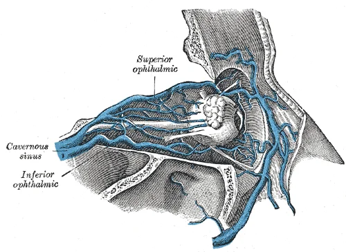
This fissure is an elongated, irregularly shaped opening located lateral to the optic canal. It also provides passage for sensory nerves and the nerves that supply the muscles involved in eye movements. The optic canal is located at the posterior apex of the orbit. It allows for the passage of the optic nerve from the retina to the brain. Venous drainage from the orbital region connects with the cavernous sinus, a critical anatomical relation for understanding orbital infections. [1]
Below the orbit, the infraorbital foramen serves as the point of emergence for a sensory nerve that supplies the anterior face below the orbit. These foramina are key landmarks for identifying the boundaries of the orbital region. [1]
The maxillary sinuses are located within the maxillary bones, just below the orbits. They are the largest of the paranasal sinuses. Because their connection to the nasal cavity is located high on their medial wall, they are difficult to drain. This anatomical feature makes them most commonly involved during sinus infections. Understanding these relationships is vital for diagnosing orbital complications from sinusitis. [1] [2]
Surface anatomy relies on identifying palpable landmarks that correspond to the bony boundaries. The superior orbital margin is formed by the frontal bone and can be palpated along the supraorbital ridge[1]. The lateral orbital margin is formed by the zygomatic bone, which is easily felt as the prominent cheekbone. The inferior orbital margin is formed primarily by the [2]
Figure 1Gray379
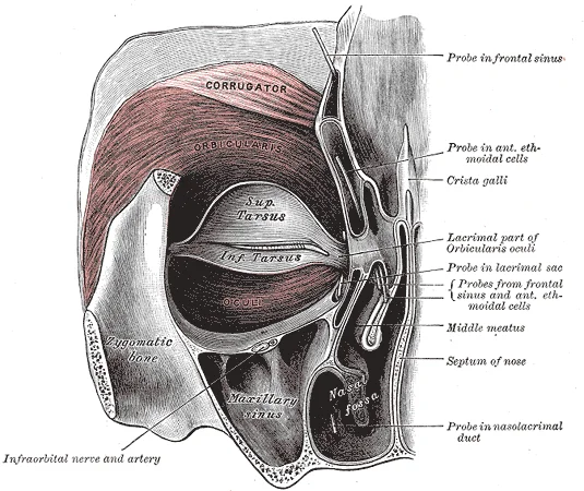
Long description
Left orbicularis oculi, seen from behind.

Figure 1: Left orbicularis oculi, seen from behind.
DefinitionOrbital Boundaries
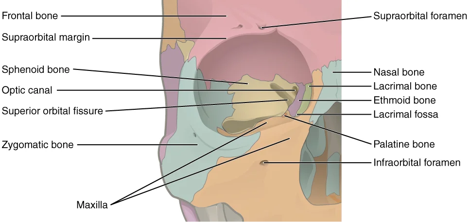
The orbit is bounded by seven skull bones: frontal, zygomatic, maxilla, palatine, ethmoid, lacrimal, and sphenoid. [1]
The walls of each orbit include contributions from seven skull bones. The frontal bone forms the roof and the zygomatic bone forms the lateral wall and lateral floor. The ethmoid bone and lacrimal bone make up much of the medial wall and the sphenoid bone forms the posterior orbit. The lateral portions of the ethmoid bone are located between the orbit and upper nasal cavity, and thus form the lateral nasal cavity wall and a portion of the medial orbit wall. Located inside this portion of the ethmoid bone are several small, air-filled spaces that are part of the paranasal sinus system of the skull. [1]
Figure 2710 Ethmoid Bone
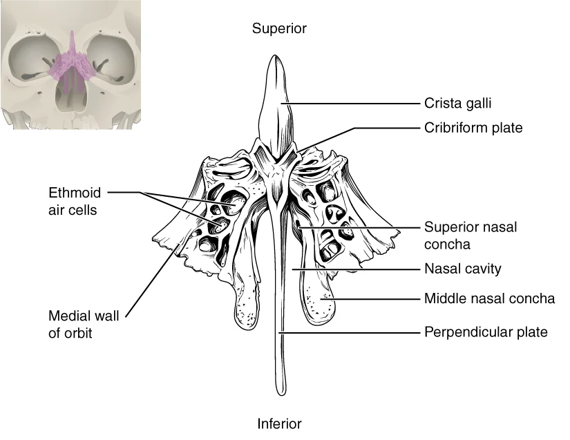
Long description
The unpaired ethmoid bone is located at the midline within the central skull. It has an upward projection, the crista galli, and a downward projection, the perpendicular plate, which forms the upper nasal septum. The cribriform plates form both the roof of the nasal cavity and a portion of the anter

Figure 2: The unpaired ethmoid bone is located at the midline within the central skull.
Figure 3Gray192
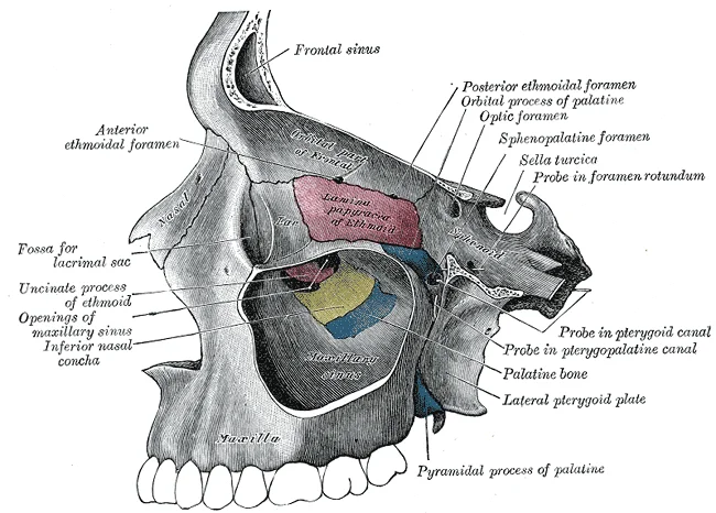
Long description
Medial wall of left orbit. Labelled illustration from Wikimedia Commons, used under its open licence.

Figure 3: Medial wall of left orbit.
The zygomatic bone forms the lateral wall. The maxilla forms the inferior wall. The ethmoid bone is medial, containing the ethmoidal air cells. The sphenoid bone is posterior. The extraocular muscles surround the globe.
Computed tomography (CT) scan images provide accurate anatomical data from different areas of the body that can be used to diagnose diseases. CT, however, excels at assessing cortical bone. Subtle increases in mucosal thickness can be appreciated on CT because the surrounding air and bone are of such different radiodensities. [2]
| Bone | Contribution to Orbit |
|---|---|
| Frontal | Roof |
| Zygomatic | Lateral wall and lateral floor |
| Maxilla | Medial floor |
| Palatine | Small contribution to medial floor |
| Ethmoid | Medial wall |
| Lacrimal | Medial wall |
| Sphenoid | Posterior orbit |
Figure 4Gray777
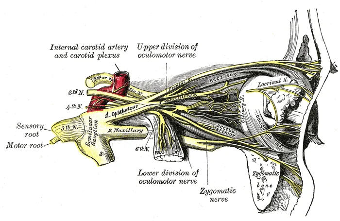
Long description
Nerves of the orbit, and the ciliary ganglion. Side view.

Figure 4: Nerves of the orbit, and the ciliary ganglion (side view).
The anterior skull is dominated by the facial bones, which provide structural support for the eyes and face. The orbit itself is formed by contributions from several cranial and facial bones. The frontal bone forms the superior boundary, while the zygomatic bone constitutes the lateral wall and the lateral-inferior margins of the anterior orbital opening. [1]
Figure 5713 Bones Forming Orbit
Long description
Seven skull bones contribute to the walls of the orbit. Opening into the posterior orbit from the cranial cavity are the optic canal and superior orbital fissure.

Figure 5: Seven skull bones contribute to the walls of the orbit.
Figure 6711 Maxilla
Long description
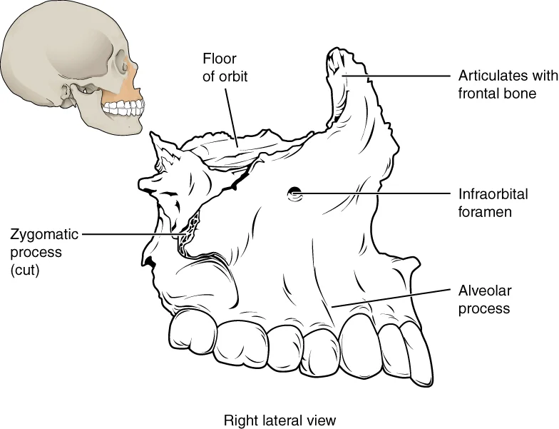
The maxillary bone forms the upper jaw and supports the upper teeth. Each maxilla also forms the lateral floor of each orbit and the majority of the hard palate.

Figure 6: The maxillary bone forms the upper jaw and supports the upper teeth.
The sutures between these bones are immobile joints filled with dense, fibrous connective tissue, which interlock tightly to add strength to the skull. [2] [1]
Orbital pathology often involves these bony structures. A blowout fracture typically occurs when increased intra-orbital pressure causes the thin bones of the orbital floor or medial wall to fracture, often involving the maxilla or ethmoid. Orbital cellulitis is a serious infection of the tissues within the orbit, which can spread from adjacent sinuses such as the ethmoid or maxillary sinuses. The proximity of the orbit to the nasal cavity and sinuses means that infections can easily extend into the orbital space. Understanding the precise bony landmarks is essential for diagnosing fractures and planning surgical interventions to repair orbital defects.
The orbit is not a single joint but a bony cavity formed by the articulation of seven distinct bones. These bones meet at sutures, which are immovable fibrous joints that provide structural stability while allowing for slight growth during development. The primary bones contributing to the orbital walls are the frontal, ethmoid, maxilla, lacrimal, sphenoid, zygomatic, and palatine bones. Each contributes specific segments to the roof, floor, medial wall, and lateral wall, creating a protective p [1]
The frontal bone forms the superior margin and roof. At the anterior midline, between the eyebrows, lies the glabella, a slight depression. The frontal bone also forms the supraorbital margin, which contains the supraorbital foramen near its middle. This foramen provides passage for a sensory nerve to the forehead. Just above the supraorbital margin, the frontal bone is thickened into rounded brow ridges, located just behind the eyebrows. These ridges vary in size among individuals but are generally larger in males. [1]
The medial wall is primarily formed by the ethmoid bone, which contributes to the medial wall of the orbit. Anterior to the ethmoid is the lacrimal bone, a small rectangular bone forming the anterior, medial wall of the orbit. The anterior portion of the lacrimal bone forms a shallow depression called the lacrimal fossa. Extending inferiorly from this fossa is the nasolacrimal canal. This canal allows lacrimal fluid to drain from the medial corner of the eye into the nasal cavity. [1]
On the anterior maxilla, just below the orbit, is the infraorbital foramen. This foramen is the point of exit for a sensory nerve that supplies the nose, upper lip, and anterior cheek. The posterior aspect of the orbit is formed by the sphenoid bone, which contains the optic canal and superior orbital fissure. These openings allow passage of nerves and vessels into the posterior orbit. The sutures between these bones are tight and interlocking, ensuring the integrity of the orbital cavity. [1]
The orbit is a pyramidal cavity whose walls are formed by seven bones: the frontal, sphenoid, zygomatic, maxilla, palatine, lacrimal, and ethmoid. Its apex lies at the optic canal, while its base is the orbital rim. The contents include the eyeball, extraocular muscles, nerves, vessels, and adipose tissue, all enclosed within a fibrous sheath known as the orbital fascia or periorbita. This periosteum lines the bony walls and reflects inward to form the suspensory ligament of Lockwood, which supp [1][3]
The superior rectus lies immediately above the optic nerve. The optic nerve is central, surrounded by the meningeal sheaths. The medial and lateral rectus muscles arise from the common tendinous ring (annulus of Zinn) at the orbital apex. This arrangement demonstrates how the muscles are organized around the optic nerve, with the fascial sheaths providing a pathway for neurovascular structures.
Understanding these compartments is crucial for interpreting imaging and performing surgical procedures.
DefinitionOrbital Fascia
The periosteum of the orbital bones that reflects to form the suspensory ligament of Lockwood and encloses the orbital contents.
The close relationship between the fascia and the bony walls allows for the transmission of infections or tumours from the orbit to the cranial cavity via the superior orbital fissure or optic canal. The fat compartments are divided into central and peripheral groups, separated by fascial septa. This organization helps localise pathology and guides surgical approaches.
| Structure | Relation |
|---|---|
| Optic Nerve | Central, surrounded by meningeal sheaths |
| Superior Rectus | Above optic nerve |
| Medial Rectus | Medial to optic nerve |
| Lateral Rectus | Lateral to optic nerve |
| Superior Oblique | Passes through trochlea |
Figure 7Gray893
Long description
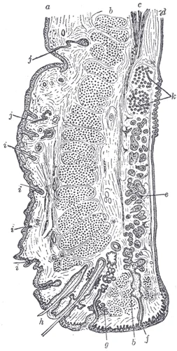
Sagittal section through the upper eyelid. (After Waldeyer.) a. Skin. b. Orbicularis oculi. b’. Marginal fasciculus of Orbicularis (ciliary bundle). c. Levator palpebræ. d. Conjunctiva. e. Tarsus. f. Tarsal gland. g. Sebaceous gland. h. Eyelashes. i. Small hairs of skin. Sweat glands. k. Posterior t

Figure 7: Sagittal section through the upper eyelid (After Waldeyer.) a.
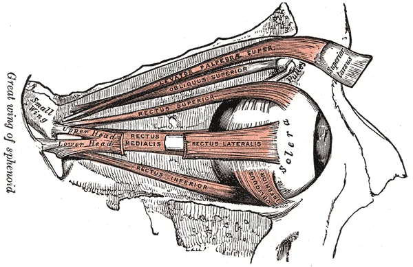
The orbit houses six primary extraocular muscles responsible for eye movement, plus the levator palpebrae superioris for eyelid elevation. These muscles are grouped into the superior, inferior, medial, and lateral compartments, with the oblique muscles occupying distinct spatial relationships. In this anterior view, the rectus muscles originate from the common tendinous ring (annulus of Zinn) at the apex of the orbit. The superior rectus arises from the su
Figure 8Gray889
Long description
Muscles of the right orbit. Labelled illustration from Wikimedia Commons, used under its open licence.

Figure 8: Muscles of the right orbit.
| Muscle | Origin | Insertion | Action | Nerve Supply |
|---|---|---|---|---|
| Superior Rectus | Common tendinous ring | Superior globe | Elevation, adduction, intorsion | Oculomotor (CN III) |
| Inferior Rectus | Common tendinous ring | Inferior globe | Depression, adduction, extorsion | Oculomotor (CN III) |
| Medial Rectus | Common tendinous ring | Medial globe | Adduction | Oculomotor (CN III) |
| Lateral Rectus | Common tendinous ring | Lateral globe | Abduction | Abducens (CN VI) |
| Superior Oblique | Sphenoid body (via trochlea) | Posterolateral globe | Intorsion, depression, abduction | Trochlear (CN IV) |
| Inferior Oblique | Orbital floor (anterior) | Posterolateral globe | Extorsion, elevation, abduction | Oculomotor (CN III) |
| Levator Palpebrae Superioris | Lesser wing of sphenoid | Superior tarsal plate | Eyelid elevation | Oculomotor (CN III) |
DefinitionCommon Tendinous Ring
A fibrous ring at the orbital apex from which four of the six rectus muscles originate.
Exam trapTrochlear Nerve
CN IV is the only cranial nerve to exit dorsally and innervate the superior oblique, which passes through the trochlea. [3]
The arterial supply to the orbit is derived primarily from the ophthalmic artery, a branch of the internal carotid artery. This vessel enters the orbit through the optic canal, accompanying the optic nerve. It initially lies inferior and lateral to the optic nerve before crossing over it to reach the medial wall of the orbit. The ophthalmic artery gives off several critical branches that supply the orbital contents, including the eye, extraocular muscles, and surrounding structures. [1]
The central structure is the optic nerve, surrounded by the ophthalmic artery.
DefinitionOphthalmic Artery
The primary arterial supply to the orbit, branching from the internal carotid artery and entering via the optic canal.
Occlusion of this artery leads to immediate and severe vision loss. The anterior and posterior ethmoidal arteries pierce the medial wall to supply the ethmoidal sinuses and the nasal cavity. The supraorbital artery exits through the supraorbital notch to supply the forehead and scalp.
| Branch | Origin | Course | Supply |
|---|---|---|---|
| Central Retinal Artery | Ophthalmic Artery | Enters optic nerve | Inner retina |
| Lacrimal Artery | Ophthalmic Artery | Lateral wall | Lacrimal gland, lateral rectus |
| Anterior Ethmoidal Artery | Ophthalmic Artery | Medial wall | Ethmoid sinuses, nasal cavity |
| Posterior Ethmoidal Artery | Ophthalmic Artery | Medial wall | Ethmoid sinuses |
| Supraorbital Artery | Ophthalmic Artery | Supraorbital notch | Forehead, scalp |
Collateral circulation is limited, making the orbit vulnerable to ischemic events. The anastomoses between the ophthalmic artery and external carotid branches, such as the facial and superficial temporal arteries, provide some compensatory flow but are often insufficient in acute occlusion. Understanding this vascular anatomy is crucial for surgical procedures and interpreting imaging studies.
Figure 9Gray572
Long description
Veins of orbit. (Poirier and Charpy.). Labelled illustration from Wikimedia Commons, used under its open licence.

Figure 9: Veins of orbit (Poirier and Charpy.).
This anatomical feature has significant clinical implications, particularly regarding the spread of infection from the face to the intracranial cavity.
The lack of valves in these veins means that pressure changes in the face can be transmitted directly to the intracranial venous system.
The cavernous sinus itself is a paired dural venous sinus situated on either side of the sella turcica. It receives blood from the ophthalmic veins, the superficial middle cerebral vein, and the sphenoparietal sinus. Blood leaves the cavernous sinus via the superior petrosal sinus and the inferior petrosal sinus. The superior petrosal sinus drains into the transverse sinus, while the inferior petrosal sinus drains into the internal jugular vein. This connection is critical because it links the orbital venous system to the systemic circulation.
Several cranial nerves from the brain exit the skull via this opening. It descends through the neck, lateral to the internal carotid artery and the vagus nerve. It eventually joins the subclavian vein to form the brachiocephalic vein. [1]
Clinical pearlClinical Pearl
The valveless nature of the ophthalmic veins allows infections from the 'danger triangle' of the face to spread to the cavernous sinus, causing cavernous sinus thrombosis.
| Structure | Origin | Termination | Key Relation |
|---|---|---|---|
| Superior Ophthalmic Vein | Medial angle of eye | Cavernous sinus | Superior to medial rectus |
| Inferior Ophthalmic Vein | Lower orbit | Cavernous sinus | Inferior to medial rectus |
| Cavernous Sinus | Superior orbital fissure | Petrosal sinuses | Lateral to sella turcica |
| Internal Jugular Vein | Jugular foramen | Brachiocephalic vein | Lateral to carotid artery |
Understanding this drainage pathway is essential for diagnosing conditions such as orbital cellulitis and cavernous sinus thrombosis. The direct communication between the facial veins and the intracranial sinuses underscores the importance of early treatment of facial infections.
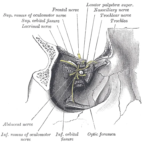
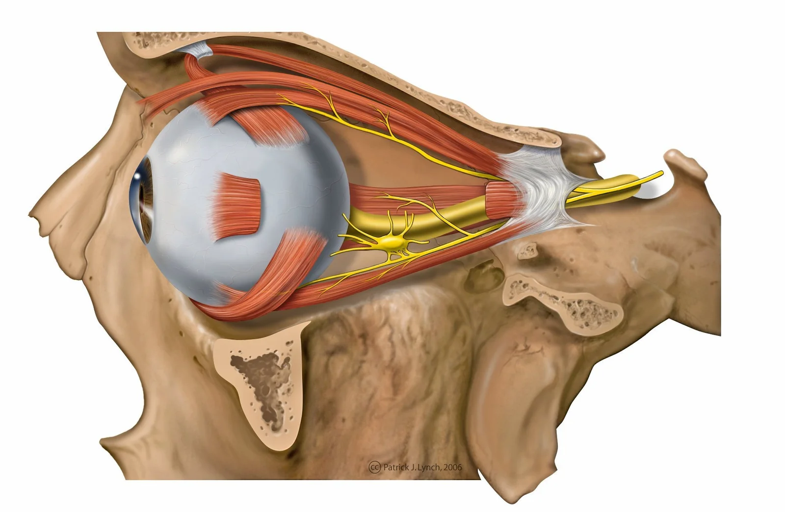
The orbit is a complex bony cavity that houses the eyeball and its associated structures. Understanding the neural supply requires distinguishing between motor innervation for movement and cutaneous sensation for the surrounding skin. The nerves entering the orbit do so through specific apertures, primarily the superior orbital fissure and the optic canal.
Figure 10Gray890
Long description
Dissection showing origins of right ocular muscles, and nerves entering by the superior orbital fissure.

Figure 10: Dissection showing origins of right ocular muscles, and nerves entering by the superior orbital fissure.
Figure 11Lateral orbit nerves chngd
Long description
Lateral eye and orbit anatomy with nerves

Figure 11: Lateral eye and orbit anatomy with nerves.
The central structure is the optic nerve (CN II), which passes through the optic canal. Lateral to the optic nerve lies the oculomotor nerve (CN III), which divides into superior and inferior divisions. The abducens nerve (CN VI) runs inferior to the oculomotor nerve, close to the lateral rectus muscle. The ophthalmic division of the trigeminal nerve (V1) enters via the superior orbital fissure, lateral to CN III.
| Nerve | Type | Muscles Supplied | Cutaneous Area Supplied |
|---|---|---|---|
| Oculomotor (CN III) | Motor | Superior, inferior, medial recti; inferior oblique; levator palpebrae superioris | None |
| Trochlear (CN IV) | Motor | Superior oblique | None |
| Abducens (CN VI) | Motor | Lateral rectus | None |
| Ophthalmic (V1) | Sensory | None | Forehead, scalp, upper eyelid, cornea, lacrimal gland |
| Optic (CN II) | Special Sensory | None | Retina (vision) |
Lesions of these nerves produce distinct clinical signs. Damage to CN III results in a "down and out" eye position due to unopposed action of the lateral rectus and superior oblique. Ptosis occurs due to levator palpebrae superioris paralysis. CN IV palsy causes vertical diplopia, worse on looking down and in. CN VI palsy leads to horizontal diplopia and inability to abduct the eye. Sensory loss in the V1 distribution indicates pathology affecting the ophthalmic nerve or its branches.
The lymphatic drainage of the orbit is a critical pathway for immune surveillance and potential spread of infection or malignancy. Understanding this system requires visualising the spatial relationships between the orbital contents and the surrounding facial structures.
The central structure is the optic nerve, surrounded by the extraocular muscles. The lymphatics from the lateral aspect of the orbit drain towards the preauricular nodes, located just anterior to the ear. Conversely, lymphatics from the medial aspect, including those from the caruncle and plica semilunaris, drain towards the submandibular nodes.
The conjunctiva, a mucous membrane lining the inner eyelids and covering the anterior sclera, possesses a rich lymphatic network. Lymphatics from the palpebral conjunctiva follow the course of the eyelid margins. Those from the lateral palpebral conjunctiva drain into the preauricular lymph nodes. These nodes are situated in front of the ear, superficial to the parotid gland. They receive lymph from the lateral aspects of the eyelids, the conjunctiva, and the lacrimal gland. Infection in these areas often presents with tender preauricular lymphadenopathy.
Figure 12Gray897
Long description
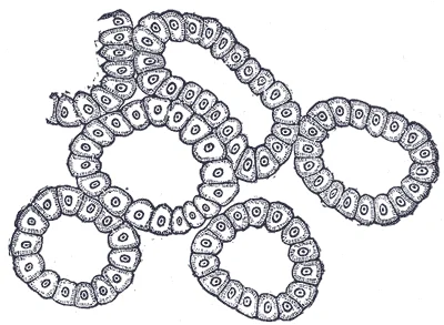
Alveoli of lacrimal gland. Labelled illustration from Wikimedia Commons, used under its open licence.

Figure 12: Alveoli of lacrimal gland.
Figure 13Gray895
Long description
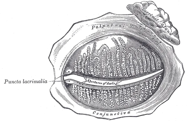
The tarsal glands, etc., seen from the inner surface of the eyelids.

Figure 13: The tarsal glands, etc., seen from the inner surface of the eyelids.
Lymphatics from the medial palpebral conjunctiva and the lacrimal sac drain into the submandibular lymph nodes. These nodes are located beneath the mandible, in the submandibular triangle. They receive lymph from the medial canthus, the lacrimal apparatus, and the nasal cavity. The caruncle, a small pink nodule at the medial canthus, is particularly rich in lymphatics. Inflammation here, such as in carunculitis, can lead to submandibular node enlargement.
Figure 14Gray896
Long description
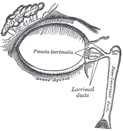
The lacrimal apparatus. Right side. Labelled illustration from Wikimedia Commons, used under its open licence.

Figure 14: The lacrimal apparatus (right side).
The skin of the eyelids also contributes to this drainage pattern. The upper eyelid skin drains primarily to the preauricular nodes. The lower eyelid skin drains to both the preauricular and submandibular nodes, depending on the lateral or medial position. This dual drainage explains why infections in the lower eyelid can present with bilateral lymphadenopathy.
The deep structures of the orbit, including the extraocular muscles and the optic nerve, have limited direct lymphatic drainage. Instead, they rely on the drainage of the surrounding connective tissue and the sclera. The sclera itself is avascular and has minimal lymphatic supply. However, the episcleral space contains lymphatic vessels that communicate with the conjunctival lymphatics. This connection allows for the spread of inflammatory mediators from the globe to the eyelid lymphatics.
Clinical correlation is essential. Orbital cellulitis, a serious infection of the orbital tissues, can spread via these lymphatic channels. Early recognition of lymph node involvement helps in diagnosing the source of infection. Preauricular lymphadenopathy suggests a lateral or superior orbital source, while submandibular lymphadenopathy points to a medial or inferior source. This anatomical knowledge guides clinical examination and management decisions.
The orbit is a pyramidal cavity that houses the eyeball and its associated structures. It is bounded by several bones. The orbital apex is formed by the sphenoid bone, which relates closely to the cavernous sinus and optic canal. The orbital floor is primarily formed by the maxilla, with contributions from other bones. The lateral wall is thick, formed mainly by the zygomatic and sphenoid bones. [1][2][3]
The frontal bone forms the superior roof[1]. The zygomatic bone forms the lateral wall[1]. The maxilla forms the inferior floor[1]. The ethmoid bone forms the medial wall[1]. The sphenoid bone forms the apex[1]. The palatine bone contributes to the posterior medial wall[1].
The superior orbital fissure transmits the oculomotor, trochlear, abducens, and ophthalmic nerves. The optic canal transmits the optic nerve and ophthalmic artery. The inferior orbital fissure transmits the infraorbital nerve and vessels. The nasolacrimal duct drains tears into the nasal cavity[1]. The extraocular muscles originate from the common tendinous ring at the orbital apex. The superior rectus, inferior rectus, medial rectus, and lateral rectus insert onto the sclera. The supe [3]
Figure 15Gray888
Long description
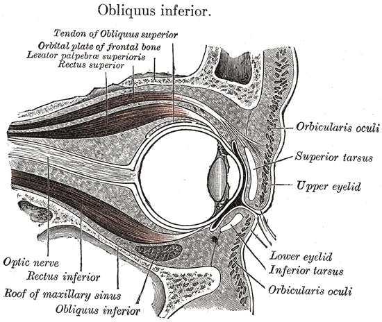
Sagittal section of right orbital cavity.

Figure 15: Sagittal section of right orbital cavity.
The central retinal artery branches from the ophthalmic artery. The superior and inferior divisions of the oculomotor nerve pass through the superior orbital fissure. The trochlear nerve passes through the superior orbital fissure. The abducens nerve passes through the superior orbital fissure. The ophthalmic division of the trigeminal nerve passes through the superior orbital fissure. [3]
It contains the internal carotid artery and cranial nerves III, IV, V1, V2, and VI. The optic nerve runs medial to the cavernous sinus. The pituitary gland lies inferior to the optic chiasm. The frontal sinus is located superior to the orbit[1]. The maxillary sinus is located inferior to the orbit[1]. The ethmoid sinuses are located between the orbit and upper nasal cavity[1]. The lacrimal sac is located in the lacrimal fossa[1]. The conjunctiva lines the [3]
| Muscle | Origin | Insertion | Action | Nerve Supply |
|---|---|---|---|---|
| Superior Rectus | Common tendinous ring | Sclera | Elevation | Oculomotor |
| Inferior Rectus | Common tendinous ring | Sclera | Depression | Oculomotor |
| Medial Rectus | Common tendinous ring | Sclera | Adduction | Oculomotor |
| Lateral Rectus | Common tendinous ring | Sclera | Abduction | Abducens |
| Superior Oblique | Sphenoid bone | Sclera | Intorsion | Trochlear |
| Inferior Oblique | Maxilla | Sclera | Extorsion | Oculomotor |
The orbit is a pyramidal cavity that houses the globe and its associated structures. Radiological assessment relies on multiple modalities to define its boundaries and contents. Plain X-ray provides limited detail but can screen for gross fractures or foreign bodies. Computed tomography (CT) offers high-resolution cross-sectional views in axial, coronal, and sagittal planes. Magnetic resonance imaging (MRI) excels at soft tissue contrast, visualising the [2]
The medial wall is thin and formed by the ethmoid bone. The lateral wall is thick and robust, composed mainly of the zygomatic bone and greater wing of the sphenoid. The floor is formed by the maxilla and zygomatic bone. The roof is formed by the frontal bone and lesser wing of the sphenoid. The superior orbital fissure lies between the greater and lesser wings of the sphenoid, transmitting cranial nerves III, IV, V1, and VI. The inferior orbital fissure connects the orbit to the pterygopalatine fossa.
DefinitionOrbital Walls
The orbit has four walls: roof, floor, medial, and lateral. Each wall has distinct radiological features and clinical significance.
CT is particularly valuable for identifying fractures. Blowout fractures typically involve the floor or medial wall. MRI is valuable for detecting optic neuritis or tumours. Recognising these variants is important for surgical planning. [3]
| Wall | Primary Bone | Key Structures |
|---|---|---|
| Roof | Frontal, Sphenoid | Lacrimal gland, trochlea |
| Floor | Maxilla, Zygomatic | Infraorbital nerve, fat |
| Medial | Ethmoid, Maxilla | Ethmoidal air cells, nasolacrimal duct |
| Lateral | Zygomatic, Sphenoid | Zygomaticofacial nerve |
Ultrasound can assess extraocular muscle thickness. It is operator-dependent but provides real-time imaging. The combination of modalities ensures comprehensive evaluation.
The orbit is a pyramidal cavity that houses the eyeball and its associated structures. Its boundaries are defined by seven bones: the frontal, sphenoid, zygomatic, maxilla, lacrimal, ethmoid, and palatine bones. Understanding these boundaries is essential for interpreting imaging and surgical approaches. The roof is formed primarily by the orbital plate of the frontal bone and the lesser wing of the sphenoid. The floor consists mainly of the orbital surface of the maxilla, with contributions from the zygomatic and palatine bones. The lateral wall is the thickest, formed by the zygomatic and greater wing of the sphenoid. [1]
DefinitionOrbital Apex
The posterior convergence of the orbit where the optic canal and superior orbital fissure are located.
Injuries to the orbit often result from blunt trauma. Fractures of the orbital floor, known as blowout fractures, typically involve the maxilla and ethmoid bones. These fractures can trap inferior rectus or inferior oblique muscles, leading to restricted eye movement. Nerve palsies are common complications. The optic nerve (CN II) passes through the optic canal. Injury here causes vision loss. The oculomotor nerve (CN III), trochlear nerve (CN IV), and abducens nerve (CN VI) pass through the superior orbital fissure. Palsy of CN III results in a "down and out" eye position, ptosis, and a dilated pupil. Palsy of CN IV causes vertical diplopia, worse on looking down. Palsy of CN VI leads to inability to abduct the eye. [1]
Surface landmarks are critical for clinical examination. The infraorbital foramen is found below the orbit, transmitting the infraorbital nerve and vessels. Palpation here assesses for tenderness or swelling.
| Muscle | Action | Nerve Supply |
|---|---|---|
| Levator palpebrae superioris | Elevates upper eyelid | Oculomotor (CN III) [3] |
| Superior rectus | Elevates, adducts, intorts | Oculomotor (CN III) |
| Inferior rectus | Depresses, adducts, extorts | Oculomotor (CN III) |
| Medial rectus | Adducts | Oculomotor (CN III) |
| Lateral rectus | Abducts | Abducens (CN VI) |
| Superior oblique | Depresses, abducts, intorts | Trochlear (CN IV) |
| Inferior oblique | Elevates, abducts, extorts | Oculomotor (CN III) |
Procedures such as orbital decompression require precise knowledge of bony boundaries. Surgeons remove parts of the medial wall and floor to relieve pressure in thyroid eye disease. The ethmoid air cells are key landmarks in this approach.
The superior orbital fissure is located between the greater and lesser wings of the sphenoid. The inferior orbital fissure lies between the maxilla and sphenoid.
Students must distinguish the spaces defined by this arch. The temporal fossa is the shallow space located on the lateral skull above the level of the zygomatic arch. In contrast, the infratemporal fossa is located below the zygomatic arch and deep to the ramus of the mandible. Confusion between these two fossae is a common trap in anatomy exams. [1]
The nasal cavity serves as the facial part of the respiratory tract, extending from the osseous nasal opening to the cribriform plate of the ethmoid bone. It is separated longitudinally into two symmetric halves by the median nasal septum. This septum continues caudally in the perpendicular plate of the ethmoid bone and rostrally in the cartilaginous, flexible part of the nasal septum. Each half of the nasal cavity contains the nasal conchae rostrally and the ethmoidal labyrinth caudally. [2]
The mandible comprises two parts, each divided into body and ramus. The body includes incisive teeth anteriorly and pre-molar and molar teeth posteriorly. In ruminants, a gap between the pre-molars and incisors is called the inter-alveolar space or diastema. The condylar and angular processes are located caudally. [2]
DefinitionDiastema
A gap between the pre-molars and incisors in ruminants.
NoteEthmoid Excavation
A small excavation in the perpendicular plate of the ethmoidal bone opens into the ethmoidal meatus. [2]
Clinical pearlMandibular Processes
The coronoid process participates in the TMJ dorsally.
Exam trapNasal Septum Composition
The septum includes both bone (perpendicular plate) and cartilage.
DefinitionZygomatic Arch Parts
Temporal process of zygomatic bone anteriorly.
NoteSinus Partition
Each sinus is subdivided by a small incomplete transverse septum. [2]
Exam trapFossa Depth
The infratemporal fossa is deep to the ramus.
DefinitionNasal Cavity Extent
From osseous nasal opening to cribriform plate.
NoteMandible Body
The anterior part is convex.
Students frequently confuse the bony contributions to the nasal septum, assuming the entire structure is cartilaginous or derived from a single bone. It is crucial to distinguish that the perpendicular plate of the ethmoid bone forms the upper portion of the nasal septum. The anterior and inferior aspects are supported by the septal cartilage, which is not found in the dry skull. Misidentifying these boundaries leads to errors in understanding the structural integrity of the nasal cavity. [1]
Another frequent error involves the lateral walls of the nasal cavity. Students often overlook the ethmoid bone’s role in forming the lateral walls of the upper nasal cavity. They may also fail to recognise that the superior and middle nasal conchae are projections of the ethmoid bone, whereas the inferior nasal concha is a separate, independent bone. The inferior nasal concha forms a curved bony plate that projects into the nasal cavity space from the lower lateral wall. It is the largest of the nasal conchae and can easily be seen when looking into the anterior opening of the nasal cavity. Confusing the origin of these conchae results in incorrect mapping of the meatuses and sinus drainage pathways. [1]
The relationship between the cranial fossae and the orbits is another area of confusion. The anterior cranial fossa is the most anterior and the shallowest of the three cranial fossae. It overlies the orbits and contains the frontal lobes of the brain. Students must remember that the anterior fossa is bounded anteriorly by the frontal bone, which also forms the majority of the floor for this space. The lesser wings of the sphenoid bone form the prominent ledge that marks the boundary between the anterior and middle cranial fossae. Mistaking this boundary can lead to errors in localising fractures or lesions. [1]
Finally, the distinction between the oral and nasal cavities is often blurred. The hard palate is the bony plate that forms the roof of the mouth and floor of the nasal cavity, separating the oral and nasal cavities. The oral cavity is divided into anterior and posterior parts by the hard and soft palate. The hard palate forms the roof of the oral cavity in the anterior part and is limited to four pairs of incisors from the front and the inter‐alveolar space is behind them. After inter‐alveolar, there are three pre‐molar teeth on each side and then three molar teeth. The roof of the oral cavity forms the soft palate at the end, which eventually ends in the larynx. Failing to appreciate this separation complicates the understanding of palatal defects and airway management. [1]
Exam trapSeptal Cartilage
The septal cartilage is not found in the dry skull. Do not look for it when examining bone specimens.
DefinitionInferior Concha
The inferior nasal concha forms a curved bony plate that projects into the nasal cavity space from the lower lateral wall.
This anatomical knowledge is fundamental at the bedside for managing nasal obstruction and epistaxis. Knowing that the inferior nasal concha is the largest of the nasal conchae helps clinicians predict areas of turbulent airflow and potential mucosal drying. When assessing a patient with a deviated septum, understanding that the perpendicular plate of the ethmoid bone forms the upper portion of the nasal septum guides surgical correction strategies. The presence of the septal cartilage allows for flexible reconstruction in the anterior septum.
In the operating theatre, precise identification of the cranial fossae boundaries is critical for neurosurgical and ENT procedures. The anterior cranial fossa overlies the orbits. Surgeons must respect the boundary marked by the lesser wings of the sphenoid bone to avoid entering the middle cranial fossa. The ethmoid bone’s role in forming the lateral walls of the upper nasal cavity is vital during endoscopic sinus surgery to prevent orbital or intracranial injury. The small superior nasal concha is well hidden above and behind the middle concha, requiring careful navigation to access the sphenoethmoidal recess. [1]
On imaging, radiologists use these landmarks to identify sinus pathology. The hard palate separates the oral and nasal cavities. Fractures involving the hard palate can disrupt the airway and feeding mechanisms. The relationship between the hard palate and the inter‐alveolar space is assessed in trauma to determine the extent of maxillofacial injury. The soft palate’s extension towards the larynx is evaluated in sleep apnoea studies to assess airway collapse potential.
In examinations, students must accurately describe the structures. The ear includes both auditory organs and balance. The external acoustic meatus has a distal cartilagenous part and a proximal osseous part. The tympanic membrane separates the middle ear from the external acoustic meatus. The middle ear is comprised of the tympanic cavity, auditory ossicles and auditory tube. The tympanic cavity is housed within the petrous temporal bone. The transmission of vibrations from the tympanic membrane across the tympanic cavity to the inner ear is mediated by the three auditory ossicles. Understanding these connections is essential for answering questions on hearing mechanisms and otological surgery.
Nasal Cavity Anatomy
- Identify SeptumUpper: Perpendicular plate of ethmoid; Anterior: Septal cartilage
- Question: Check Lateral Wall
Upper
- Ethmoid Bone
- Superior & Middle Conchae
Lower
- Inferior Concha
The perpendicular plate of the ethmoid bone forms the upper portion of the nasal septum. The ethmoid bone also forms the lateral walls of the upper nasal cavity. The inferior nasal concha forms a curved bony plate that projects into the nasal cavity space from the lower lateral wall. The inferior concha is the largest of the nasal conchae and can easily be seen when looking into the anterior opening of the nasal cavity. The small superior nasal concha is well hidden above and behind the middle concha. The anterior cranial fossa is the most anterior and the shallowest of the three cranial fossae. The hard palate is the bony plate that forms the roof of the mouth and floor of the nasal cavity, separating the oral and nasal cavities. The septal cartilage is not found in the dry skull.
Answer each one in your head first, then open the answer.
Which bone forms the upper portion of the nasal septum?
Show answerHide answer
Perpendicular plate of the ethmoid boneWhat is the largest nasal concha?
Show answerHide answer
Inferior nasal conchaWhich bone forms the lateral walls of the upper nasal cavity?
Show answerHide answer
Ethmoid boneWhat separates the oral and nasal cavities?
Show answerHide answer
Hard palateWhich part of the nasal septum is not found in the dry skull?
Show answerHide answer
Septal cartilage
- Betts JG, Young KA, Wise JA, et al. The Skull. OpenStax, Anatomy and Physiology 2e (CC BY-NC-SA 4.0). Accessed .
- Masoudifard M, Zehtabvar O, Modarres SH, et al. CT anatomy of the head in the Ile de France sheep. Veterinary medicine and science 2022;8(4):1694-1708. doi:10.1002/vms3.834. Accessed .
- Krahe AM, Ketterer MC, Offergeld C, Hildenbrand T. [Evaluation of a structured e-learning-based approach to CT anatomy of the paranasal sinuses for medical students : A pilot study]. HNO 2022;70(6):468-475. doi:10.1007/s00106-021-01141-x. Accessed .
Figure credits
- Figure 1: Henry Vandyke Carter, via Wikimedia Commons, Public domain, Public domain.
- Figure 2: OpenStax College, via Wikimedia Commons, CC BY 3.0, CC BY 3.0.
- Figure 3: Henry Vandyke Carter, via Wikimedia Commons, Public domain, Public domain.
- Figure 4: Henry Vandyke Carter, via Wikimedia Commons, Public domain, Public domain.
- Figure 5: OpenStax College, via Wikimedia Commons, CC BY 3.0, CC BY 3.0.
- Figure 6: OpenStax College, via Wikimedia Commons, CC BY 3.0, CC BY 3.0.
- Figure 7: Henry Vandyke Carter, via Wikimedia Commons, Public domain, Public domain.
- Figure 8: Henry Vandyke Carter, via Wikimedia Commons, Public domain, Public domain.
- Figure 9: Henry Vandyke Carter, via Wikimedia Commons, Public domain, Public domain.
- Figure 10: Henry Vandyke Carter, via Wikimedia Commons, Public domain, Public domain.
- Figure 11: Lateral_orbit_nerves.jpg: Patrick J. Lynch, medical illustrator derivative work: Anka Friedrich (talk), via Wikimedia Commons, CC BY 2.5, CC BY 2.5. resized.
- Figure 12: Henry Vandyke Carter, via Wikimedia Commons, Public domain, Public domain.
- Figure 13: Henry Vandyke Carter, via Wikimedia Commons, Public domain, Public domain.
- Figure 14: Henry Vandyke Carter, via Wikimedia Commons, Public domain, Public domain.
- Figure 15: Henry Vandyke Carter, via Wikimedia Commons, Public domain, Public domain.
Educational content for medical students and doctors. It is not a substitute for clinical judgement or local guidelines.