Eye anatomy
The eye is a hollow sphere of three tissue layers within the bony orbit, managed by six extraocular muscles and innervated by cranial nerves.
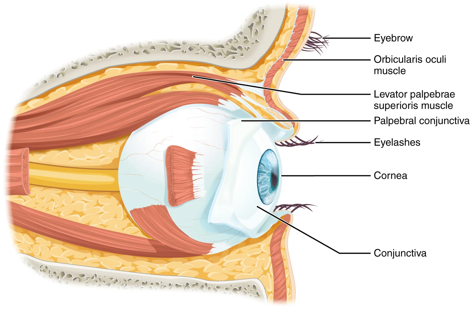
Vision is the special sense of sight that is based on the transduction of light stimuli received through the eyes. The eyes are located within either orbit in the skull. The bony orbits surround the eyeballs, protecting them and anchoring the soft tissues of the eye. The eyelids, with lashes at their leading edges, help to protect the eye from abrasions by blocking particles that may land on the surface of the eye. The inner surface of each lid is a thin membrane known as the palpebral conjuncti [1]
Figure 11411 Eye in The Orbit
Long description
The eye is located within the orbit and surrounded by soft tissues that protect and support its function. The orbit is surrounded by cranial bones of the skull.

Figure 1: The eye is located within the orbit and surrounded by soft tissues that protect and support its function.
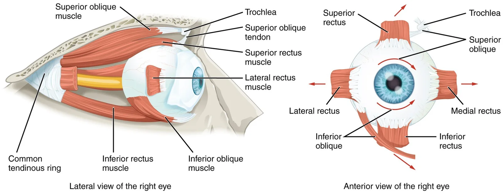
Movement of the eye within the orbit is accomplished by the contraction of six extraocular muscles that originate from the bones of the orbit and insert into the surface of the eyeball. Four of the muscles are arranged at the cardinal points around the eye and are named for those locations. They are the superior rectus, medial rectus, inferior rectus, and lateral rectus. When each of these muscles contract, the eye moves toward the contracting muscle. For example, when the superior rectus contra [1]
Figure 21412 Extraocular Muscles
Long description
The extraocular muscles move the eye within the orbit.

Figure 2: The extraocular muscles move the eye within the orbit.
The inferior oblique muscle originates from the floor of the orbit and inserts into the inferolateral surface of the eye. Rotation of the eye by the two oblique muscles is necessary because the eye is not perfectly aligned on the sagittal plane. When the eye looks up or down, the eye must also rotate slightly to compensate for the superior rectus pulling at approximately a 20-degree angle, rather than straight up. The same is true for the inferior rectus, which is compensated by contraction of the inferior oblique. [1]
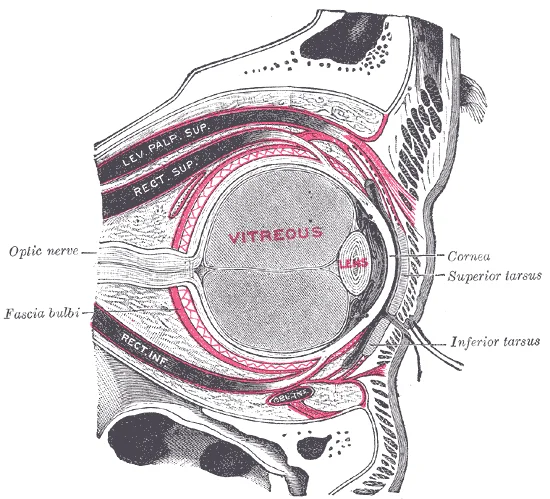
Figure 3Gray891
Long description
The right eye in sagittal section, showing the fascia bulbi (semidiagrammatic). (Testut.)

Figure 3: The right eye in sagittal section, showing the fascia bulbi (semidiagrammatic) (Testut.).
DefinitionExtraocular Muscles
Six muscles control eye movement within the bony orbit, originating from orbital bones and inserting onto the eyeball surface.
| Muscle | Nerve Supply |
|---|---|
| Lateral rectus | Abducens nerve |
| Superior oblique | Trochlear nerve |
| All other muscles | Oculomotor nerve |
The extraocular muscles are innervated by three cranial nerves. The lateral rectus, which causes abduction of the eye, is innervated by the abducens nerve. The superior oblique is innervated by the trochlear nerve. All of the other muscles are innervated by the oculomotor nerve, as is the levator palpebrae superioris. The motor nuclei of these cranial nerves connect to the brain stem, which coordinates eye movements. [1]
Eye Anatomy Outline
- StartIdentify the hollow sphere of the eyeball
- Question: Which layer?
Outer
- Fibrous tunic: Sclera and Cornea
Middle
- Vascular tunic: Choroid, Ciliary body, Iris
Inner
- Neural tunic: Retina
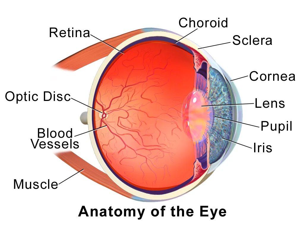
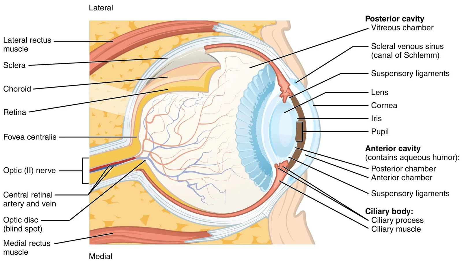
The eye itself is a hollow sphere composed of three layers of tissue. The outermost layer is the fibrous tunic, which includes the white sclera and clear cornea. The sclera accounts for five sixths of the surface of the eye, most of which is not visible, though humans are unique compared with many other species in having so much of the “white of the eye” visible. The transparent cornea covers the anterior part of the eye. It allows light to enter, and along with the lens, focuses light onto the [1]
These two structures bend the lens, allowing it to focus light on the back of the eye. Overlaying the ciliary body, and visible in the anterior eye, is the iris—the colored part of the eye. The iris is a smooth muscle that opens or closes the pupil, which is the hole at the center of the eye that allows light to enter. The iris constricts the pupil in response to bright light and dilates the pupil in response to dim light. The innermost layer of the eye is the neural tunic, or retina, which contains the nervous tissue responsible for photoreception. [1]
The eye is also divided into two cavities: the anterior cavity and the posterior cavity. The anterior cavity is the space between the cornea and lens, including the iris and ciliary body. It is filled with a watery fluid called the aqueous humor. The posterior cavity is the space behind the lens that extends to the posterior side of the interior eyeball, where the retina is located. The posterior cavity is filled with a more viscous fluid called the vitreous humor. [1]
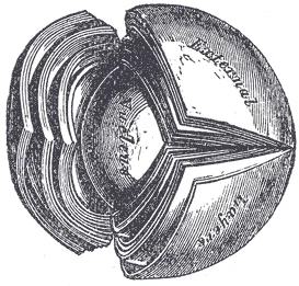
Figure 4Gray884
Long description
The crystalline lens, hardened and divided. (Enlarged.)

Figure 4: The crystalline lens, hardened and divided (Enlarged.).
Figure 5Gray875
Long description
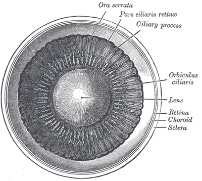
Interior of anterior half of bulb of eye.

Figure 5: Interior of anterior half of bulb of eye.
Figure 6Gray869
Long description
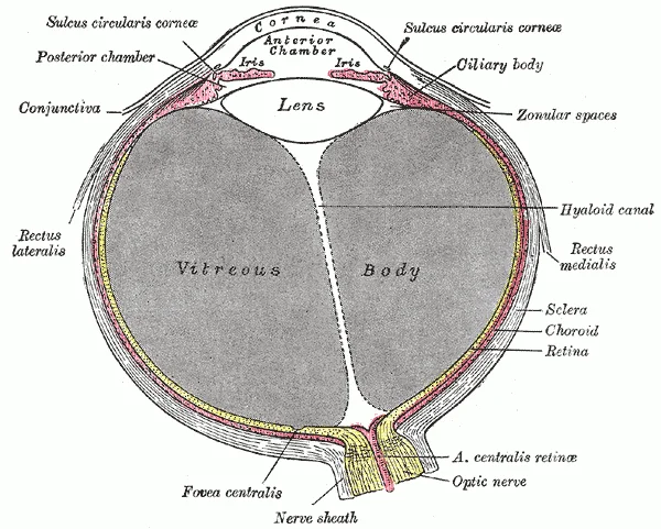
Horizontal section of the eyeball. Labelled illustration from Wikimedia Commons, used under its open licence.

Figure 6: Horizontal section of the eyeball.
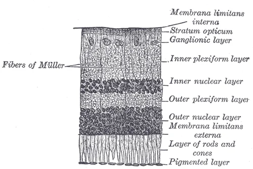
The retina is composed of several layers and contains specialized cells for the initial processing of visual stimuli. The photoreceptors (rods and cones) change their membrane potential when stimulated by light energy. The change in membrane potential alters the amount of neurotransmitter that the photoreceptor cells release onto bipolar cells in the outer synaptic layer. It is the bipolar cell in the retina that connects a photoreceptor to a retinal ganglion cell (RGC) in the inner synaptic layer. The axons of RGCs, which lie at the innermost layer of the retina, collect at the optic disc and leave the eye as the optic nerve. [1]
Figure 7Gray881
Long description
Section of retina. (Magnified.). Labelled illustration from Wikimedia Commons, used under its open licence.

Figure 7: Section of retina (Magnified.).
Figure 8Blausen 0388 EyeAnatomy 01
Long description
Anatomy of the Eye. See a related animation of this medical topic.

Figure 8: Anatomy of the Eye.
The viewer should identify the medial canthus, where the upper and lower eyelids meet near the nose, and the lateral canthus, where they meet towards the temple. The upper eyelid is elevated by the levator palpebrae superioris muscle, a movement that usually occurs in concert with elevation of the eye by the superior rectus. The lower eyelid provides inferior support. Between the eyelids lies the palpebral fissure. The skin of the eyelids is thin and continuous with the conjunctiva at the lid margins. [1]
Figure 9Gray894
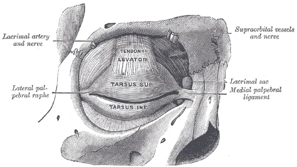
Long description
The tarsi and their ligaments. Right eye; front view.

Figure 9: The tarsi and their ligaments (right eye; front view).
The eyeball itself is protected by the bony orbit. Anteriorly, the cornea is the transparent, avascular dome that covers the iris and pupil. It is continuous with the sclera, the tough, white, fibrous outer coat of the eye that provides structural integrity. The sclera is visible as the 'white' of the eye. Posteriorly, the optic nerve exits the globe. The extraocular muscles originate from the common tendinous ring (annulus of Zinn) at the apex of the orbit and insert onto the sclera. These muscles control eye movement. The six primary muscles are the superior rectus, inferior rectus, medial rectus, lateral rectus, superior oblique, and inferior oblique. A seventh muscle, the levator palpebrae superioris, is responsible for elevating and retracting the upper eyelid.
The conjunctiva is a thin, transparent mucous membrane that lines the inner surface of the eyelids (palpebral conjunctiva) and reflects onto the anterior surface of the sclera (bulbar conjunctiva). It secretes mucus and tears to lubricate the eye. The lacrimal gland, located in the superolateral aspect of the orbit, produces tears that spread across the eye surface via blinking. The tear drainage system begins at the lacrimal puncta, small openings at the medial canthus of each eyelid. Tears flow into the lacrimal canaliculi, then into the lacrimal sac, and finally down the nasolacrimal duct into the inferior nasal meatus. [1]
Figure 10Gray892
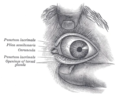
Long description
Front of left eye with eyelids separated to show medial canthus.

Figure 10: Front of left eye with eyelids separated to show medial canthus.
Vision involves the activation of photoreceptors. This special sense has a specific organ devoted to it, namely the eye. The external features described here facilitate the entry of light and protection of the delicate internal structures. The auricle, or pinna, is part of the external ear, not the eye, and directs sound waves toward the auditory canal. This distinction is crucial for anatomical accuracy. The eye relies on the cornea and lens for refraction, while the retina contains the photoreceptors. The extraocular muscles allow for precise alignment of the visual axis.
| Muscle | Origin | Insertion | Action | Nerve Supply |
|---|---|---|---|---|
| Superior Rectus | Annulus of Zinn | Superior sclera | Elevation, intorsion, adduction | Oculomotor (CN III) |
| Inferior Rectus | Annulus of Zinn | Inferior sclera | Depression, extorsion, adduction | Oculomotor (CN III) |
| Medial Rectus | Annulus of Zinn | Medial sclera | Adduction | Oculomotor (CN III) |
| Lateral Rectus | Annulus of Zinn | Lateral sclera | Abduction | Abducens (CN VI) |
| Superior Oblique | Annulus of Zinn | Posterolateral sclera | Intorsion, depression, abduction | Trochlear (CN IV) |
| Inferior Oblique | Maxillary bone | Posterolateral sclera | Extorsion, elevation, abduction | Oculomotor (CN III) |
| Levator Palpebrae Superioris | Sphenoid bone | Upper eyelid skin/tarsus | Elevates upper eyelid | Oculomotor (CN III) |
DefinitionAnatomical Landmarks
The medial canthus is the inner corner of the eye where the eyelids meet. The lateral canthus is the outer corner. The levator palpebrae superioris elevates the upper eyelid.
Exam trapCommon Confusion
Do not confuse the auricle (pinna) of the ear with eye structures. The auricle directs sound waves. The eye uses the cornea for light entry.
The eye is a complex sensory organ situated within the bony orbit, protected by the surrounding skeletal structure. Its position allows for optimal visual field coverage while maintaining mobility through extraocular muscle attachments. The globe rests within the orbital cavity, cushioned by retrobulbar fat which absorbs shock and permits smooth movement. Anteriorly, the eye is bounded by the eyelids and the conjunctiva, which provide a protective barrier against environmental debris and desicca The stimulus causes the sensory cell to produce an action potential that is relayed into the central nervous system (CNS). [1]
The ethmoid sinuses are adjacent to the medial wall.
DefinitionOrbital Relations
The eye is housed in the bony orbit, surrounded by fat and muscles, with critical neurovascular structures running alongside it.
The interior of the eye is a complex chambered system designed to transmit and process visual information. Understanding this anatomy requires viewing the eye not merely as a camera, but as an extension of the central nervous system. A major role of sensory receptors is to help us learn about the environment around us, or about the state of our internal environment. Stimuli from varying sources, and of different types, are received and changed into the electrochemical signals of the nervous system. [1]
The outermost layer, the fibrous tunic, consists of the sclera posteriorly and the transparent cornea anteriorly. The middle vascular tunic, or uvea, comprises the choroid, ciliary body, and iris. The innermost neural tunic is the retina. The lens sits behind the iris, suspended by zonular fibres from the ciliary body. The vitreous body fills the large posterior cavity between the lens and the retina. The optic nerve exits the posterior pole, carrying the axons of the retinal ganglion cells. [1]
Figure 11Gray873
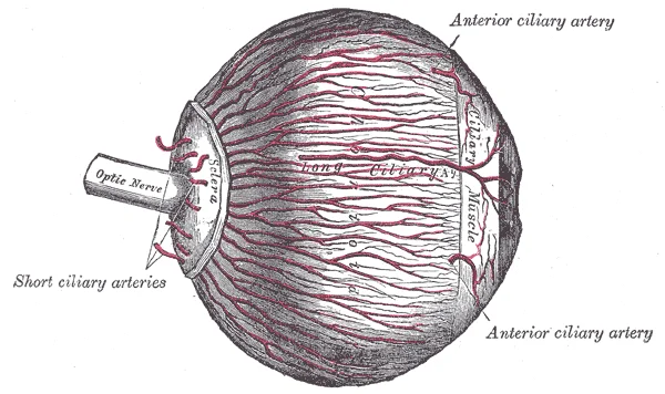
Long description
The arteries of the choroid and iris. The greater part of the sclera has been removed. (Enlarged.)

Figure 11: The arteries of the choroid and iris.
Figure 12Gray872
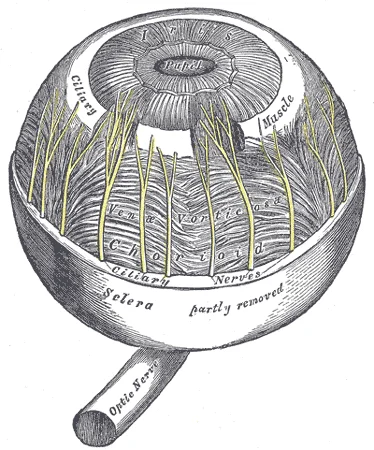
Long description
The choroid and iris. (Enlarged.). Labelled illustration from Wikimedia Commons, used under its open licence.

Figure 12: The choroid and iris (Enlarged.).
DefinitionRetinal Vascular Plexuses
The retina contains distinct vascular networks that can be visualized using advanced imaging techniques.
This technology demonstrates that PR-OCTA can visualize four unique vascular plexuses in the human retina with distinct vascular patterns, which vary based on depth and location from the nerve. Using PR-OCTA one can visualize the inter-plexus spaces and the interconnecting vascular segments in vivo. Furthermore, PR-OCTA preserves in situ flow signal in deeper vessels, resulting in visualization of continuous capillary networks in the ICP and DCP. [2]
| Plexus | Location | Characteristics |
|---|---|---|
| Superficial | Inner retina | Part of the four unique vascular plexuses |
| Intermediate | Inner nuclear layer | Distinct vascular patterns |
| Deep | Outer plexiform layer | Continuous capillary networks |
| Choriocapillaris | Choroid | High flow, supports outer retina |
These boundaries were defined in terms of retinal anatomic layers that could be easily segmented from structural OCT images. [2]
Clinical pearlClinical Relevance
Retinal vascular anatomy is crucial for diagnosing neurodegenerative diseases like glaucoma.
Atrophy and thinning of these structures are objective and precisely quantifiable disease indicators. However, retinal edema can confound thickness measurements, and in advanced glaucoma these structures are dominated by residual glial tissue and no longer serve as reliable disease indicators. Optical coherence tomography angiography of the vascular supplies to these structure is a promising alternate approach to diagnose and monitor these diseases. [2]
This system forms the foundational blood supply for the inner retinal layers. The SVP acts as a central network of large and small vessels, connected directly to the retinal arteries and veins. It supplies all other vascular plexuses within the retina. In the macula, the SVP is arranged in a centripetal pattern that converges on the parafoveal capillary ring. In the peripheral retina, the superior and inferior circulations converge in an interleaved comb pattern, similar to the retinal v [2]
Figure 13Gray879
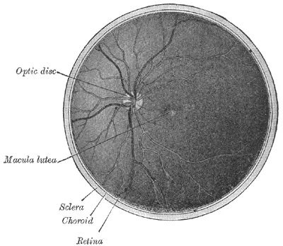
Long description
Interior of posterior half of bulb of left eye. The veins are darker in appearance than the arteries.

Figure 13: Interior of posterior half of bulb of left eye.
Primate histology demonstrates that the SVP supplies all other vascular plexuses through vertical pre-capillary arterial segments. These segments typically ascend to the nerve fibre layer (NFL) in the case of the RPCP, and descend to the deeper layers, including the inner nuclear layer (ICP) and deep capillary plexus (DCP). These vertical segments originate off the first-order branches from the main arcades and terminal extensions of the SVP arterial segments. The post-capillary segments often drain directly into the main venous arcades. [2]
It can be uniquely visualised within the superficial vascular complex by segmenting on the NFL/ganglion cell layer (GCL) interface. They are fed by pre-capillary arterial segments from the SVP and drained by post-capillary venous segments into the SVP. [2]
In the periphery, the interleaved comb pattern of the superior and inferior circulations is apparent. The asymmetric butterfly distribution around the optic nerve and vascular arcades is clearly demonstrated. The post-capillary segments draining into the main venous arcades are also indicated.
DefinitionSuperficial Vascular Complex
The SVP and RPCP together form the superficial vascular complex, supplying the inner retinal layers through a network of arteries, capillaries, and veins.
| Feature | Description |
|---|---|
| Location | Peripapillary region, posterior pole only |
| Path | Longer and straighter than other plexuses |
| Anastomoses | Fewer anastomoses within the plane |
| Supply | Fed by pre-capillary arterial segments from SVP |
| Drainage | Drained by post-capillary venous segments into SVP |
| Distribution | Asymmetric butterfly pattern around optic nerve and arcades |
The venous drainage of the eye is a complex system that mirrors the arterial supply and ensures the removal of deoxygenated blood and metabolic waste from the highly active ocular tissues. Understanding this drainage requires an appreciation of the layered structure of the eyeball and the specific anatomical pathways for each component.
The eyeball consists of three main layers: the fibrous tunic, the vascular tunic, and the neural tunic. The fibrous tunic comprises the sclera and cornea. The vascular tunic, or uvea, includes the choroid, ciliary body, and iris. The neural tunic is the retina. Venous drainage begins within these layers. The choroid is a layer of highly vascularized connective tissue that provides a blood supply to the eyeball. The retina has its own vascular supply, with blood draining into the central retinal vein. This vein emerges from [1]
Figure 14Gray883
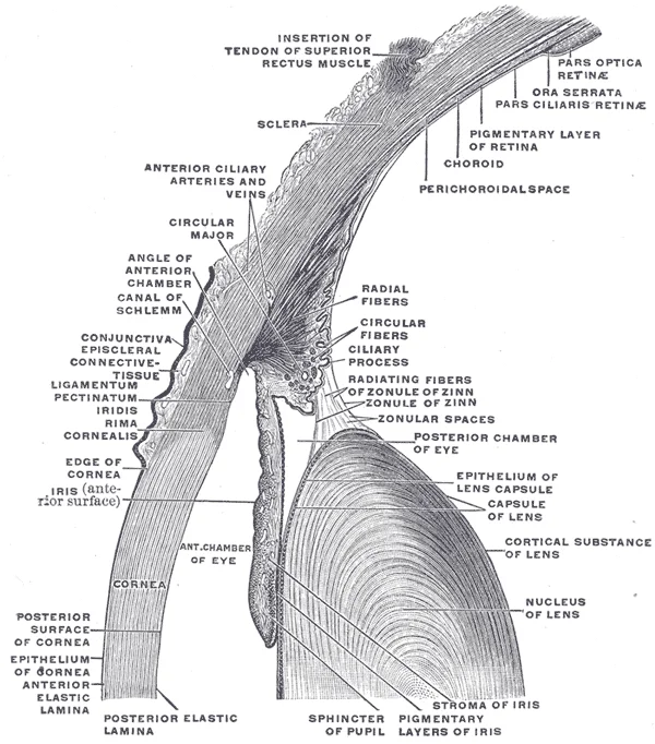
Long description
The upper half of a sagittal section through the front of the eyeball.

Figure 14: The upper half of a sagittal section through the front of the eyeball.
Figure 15Gray877
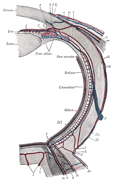
Long description
Diagram of the blood vessels of the eye, as seen in a horizontal section. (Leber, after Stöhr.). Course of vasa centralia retinæ: a. Arteria. a. 147 Vena centralis retinæ. B. Anastomosis with vessels of outer coats. C. Anastomosis with branches of short posterior ciliary arteries. D. Anastomosis wit

Figure 15: Diagram of the blood vessels of the eye, as seen in a horizontal section (Leber, after Stöhr.).
Figure 16Gray874
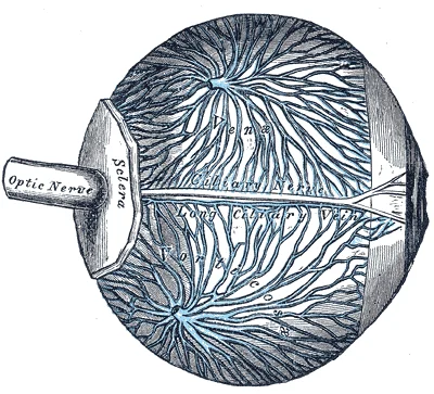
Long description
The veins of the choroid. (Enlarged.). Labelled illustration from Wikimedia Commons, used under its open licence.

Figure 16: The veins of the choroid (Enlarged.).
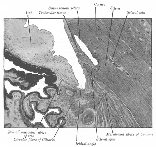
The anterior and posterior chambers are filled with aqueous humour, which is produced by the ciliary body. This fluid flows from the posterior chamber, through the pupil, into the anterior chamber, and then drains via the drainage angle. The drainage angle is formed by the cornea and the iris. It contains the trabecular meshwork and Schlemm’s canal. Aqueous humour passes through the trabecular meshwork into Schlemm’s canal, which then drains into the episcleral veins. The lens is avascular and relies on diffusion from the aqueous humour and vitreous humour for nutrients. It does not have direct venous drainage. [1]
Figure 17Gray870
Long description
Enlarged general view of the iridial angle. (Arthur Thomson.)

Figure 17: Enlarged general view of the iridial angle (Arthur Thomson.).
The eyeball consists of three main layers: the fibrous tunic, the vascular tunic, and the neural tunic. The fibrous tunic comprises the sclera and cornea. The vascular tunic, or uvea, includes the choroid, ciliary body, and iris. The neural tunic is the retina. Venous drainage begins within these layers. The choroid contains a rich network of large vessels known as the vortex veins. The retina has its own vascular supply, with blood draining into the central retinal vein. The axons of RGCs, which lie at the innermost layer of the retina, collect at the optic disc and leave the eye as the optic nerve (see ). [1]
The eyelids have a rich venous supply that drains into the facial vein. The lacrimal apparatus includes the lacrimal gland, which produces tears, and the drainage system consisting of the puncta, canaliculi, lacrimal sac, and nasolacrimal duct. Tears drain from the eye surface into the puncta, then through the canaliculi to the lacrimal sac, and finally into the nasolacrimal duct, which empties into the nasal cavity. The venous drainage of the lacrimal gland follows the arterial supply, draining into the ophthalmic veins.
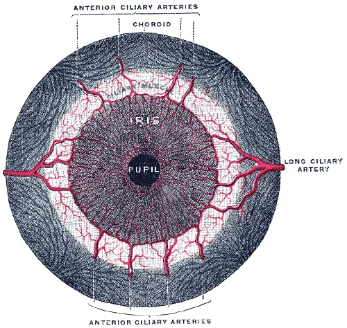
The pupillary muscles, including the sphincter pupillae and dilator pupillae, are smooth muscles within the iris. They do not have a distinct venous drainage system separate from the iris vasculature.
Figure 18Gray878
Long description
Iris, front view. Labelled illustration from Wikimedia Commons, used under its open licence.

Figure 18: Iris, front view.
Figure 191413 Structure of the Eye
Long description
The sphere of the eye can be divided into anterior and posterior chambers. The wall of the eye is composed of three layers: the fibrous tunic, vascular tunic, and neural tunic. Within the neural tunic is the retina, with three layers of cells and two synaptic layers in between. The center of the ret

Figure 19: The sphere of the eye can be divided into anterior and posterior chambers.
The pupillary muscles are indicated within the iris.
The lymphatic drainage of the eye is a complex network that follows specific anatomical pathways, ensuring fluid balance and immune surveillance within the orbital and facial regions. Understanding these pathways is crucial for diagnosing infections and metastatic spread.
The retrobulbar lymph nodes are indicated in the posterior orbit, near the apex. This visual guide helps students correlate the anatomical position of the drainage pathways with the clinical presentation of lymphadenopathy.
The conjunctiva has a rich lymphatic supply. The bulbar conjunctiva drains primarily to the preauricular lymph nodes. This pathway is clinically significant in conditions such as trachoma or viral conjunctivitis, where preauricular lymphadenopathy is a common finding. This distribution explains why infections in different parts of the conjunctiva may present with different patterns of lymph node enlargement.
These vessels can become prominent during inflammation, such as in keratitis. The drainage from the limbus follows the same pathways as the conjunctiva.
These nodes are located in the posterior orbit. Drainage from these structures is less direct and may involve the pterygoid venous plexus as an alternative route. This anatomical arrangement is important in understanding the spread of intraocular tumours.
DefinitionPreauricular Lymph Nodes
Lymph nodes located anterior to the tragus of the ear, receiving drainage from the lateral eyelids and conjunctiva.
DefinitionParotid Lymph Nodes
Lymph nodes embedded within the parotid gland, receiving drainage from the lateral aspects of the eye and face.
| Structure | Primary Drainage Pathway | Key Lymph Nodes |
|---|---|---|
| Bulbar Conjunctiva | Medial and Lateral Canthi | Preauricular, Parotid |
| Palpebral Conjunctiva | Medial and Lateral Canthi | Preauricular, Parotid |
| Cornea | Limbal Network | Preauricular, Parotid |
| Sclera | Episcleral Vessels | Preauricular, Parotid |
| Ciliary Body | Deep Orbital Vessels | Retrobulbar |
| Choroid | Deep Orbital Vessels | Retrobulbar |
Clinical pearlClinical Pearl
Preauricular lymphadenopathy is a hallmark sign of viral conjunctivitis and trachoma, aiding in differential diagnosis.
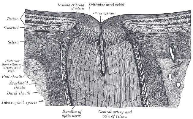
Figure 20Gray880
Long description
The terminal portion of the optic nerve and its entrance into the eyeball, in horizontal section. (Toldt.)

Figure 20: The terminal portion of the optic nerve and its entrance into the eyeball, in horizontal section (Toldt.).
The eye receives a complex innervation involving both somatic and autonomic components, alongside a dedicated vascular supply. Understanding this requires distinguishing between sensory perception, motor control, and vascular regulation.
Somatic sensation from the eye and its surrounding structures is primarily mediated by the trigeminal nerve (CN V). This nerve conveys general senses, including pain, temperature, and touch, from the face and head. The ophthalmic division (V1) provides sensory innervation to the upper eyelid, cornea, and conjunctiva. These sensations are transmitted via free nerve endings, which act as nociceptors and thermoreceptors. Nociceptors respond to potentially damaging stimuli, such as mechanical, chemical, or thermal inputs exceeding a specific threshold. For instance, the cornea is densely innervated, making it highly sensitive to pain, which serves as a protective mechanism.
Autonomic control regulates the pupil and lens, ensuring optimal focus and light adaptation. The sympathetic nervous system dilates the pupil (mydriasis) and elevates the eyelid. Parasympathetic input, carried by the oculomotor nerve (CN III), constricts the pupil (miosis) and facilitates accommodation by contracting the ciliary muscle. This balance allows the eye to adjust to varying light conditions and focal distances.
Pain from the eye is often perceived in specific regions of the head due to shared neural pathways. For example, irritation of the cornea may be felt as deep ocular pain or referred to the forehead. This phenomenon occurs because sensory fibers from the eye converge with those from the scalp and face in the trigeminal nucleus.
The eye’s function depends on a precise arrangement of nerves and vessels. This vascular network is critical for maintaining retinal health. Disruption of this supply can lead to rapid vision loss.
DefinitionNociceptor
A sensory receptor that responds to damaging or potentially damaging stimuli by sending signals to the spinal cord and brain.
| Nerve | Type | Function |
|---|---|---|
| Optic (CN II) | Sensory | Vision |
| Oculomotor (CN III) | Motor/Autonomic | Pupil constriction, accommodation |
| Trochlear (CN IV) | Motor | Superior oblique muscle |
| Abducens (CN VI) | Motor | Lateral rectus muscle |
| Trigeminal (CN V1) | Sensory | Corneal sensation, pain |
Link to the histology page.
The eye comprises distinct layers that facilitate sensory transduction. The outer fibrous layer includes the sclera and cornea. The middle vascular layer, or uvea, contains the choroid, ciliary body, and iris. The inner neural layer is the retina, which processes visual information.
DefinitionRetinal Layers
The retina consists of ten layers, including the photoreceptor layer, outer nuclear layer, inner nuclear layer, and ganglion cell layer.
The cornea is the transparent anterior portion of the fibrous tunic. It provides most of the eye's refractive power. The sclera is the opaque, white posterior portion. It maintains the shape of the globe and provides attachment for extraocular muscles.
The uvea is highly vascular and pigmented. The choroid lies between the sclera and retina, supplying nutrients to the outer retina. The ciliary body produces aqueous humour and controls lens shape. The iris regulates the amount of light entering the eye via the pupil.
The retina contains photoreceptors (rods and cones) that convert light into neural signals. Rods are sensitive to low light, while cones mediate colour vision. The macula is the central region responsible for high-acuity vision. The optic disc is the point where ganglion cell axons exit the eye to form the optic nerve. [1]
| Structure | Location | Function |
|---|---|---|
| Photoreceptors | Outer retina | Light detection |
| Bipolar cells | Inner nuclear layer | Signal relay |
| Ganglion cells | Ganglion cell layer | Axon output |
| Macula | Central retina | High acuity |
Visual stimuli cause changes in the photopigment molecule that lead to changes in membrane potential of the photoreceptor cell. A single unit of light is called a photon. Light with a wavelength of 380 nm is blue whereas light with a wavelength of 720 nm is dark red. [1]
NoteClinical Note
OCTA allows layer-by-layer visualization of vascular networks, distinguishing superficial and deep plexuses based on depth.
The eye develops from three primary embryonic sources: the neuroectoderm, surface ectoderm, and mesenchyme. Understanding these origins clarifies the adult anatomy and clinical presentations of congenital anomalies.
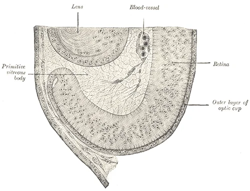
Figure 21Gray868
Long description
Section of developing eye of trout. (Szily.)

Figure 21: Section of developing eye of trout (Szily.).
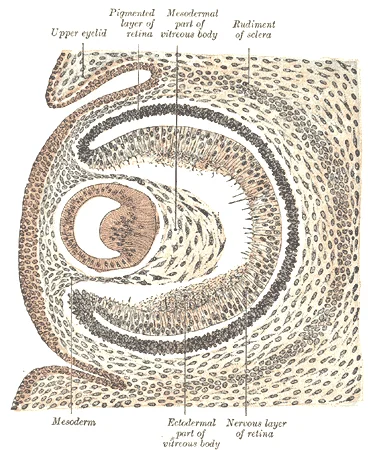
Figure 22Gray867
Long description
Sagittal section of eye of human embryo of six weeks. (Kollmann.)

Figure 22: Sagittal section of eye of human embryo of six weeks (Kollmann.).
The optic vesicles evaginate from the diencephalon of the forebrain. These vesicles contact the overlying surface ectoderm, inducing the formation of the lens placode. The optic vesicle then invaginates to form the optic cup, which consists of an inner and an outer layer. The inner layer becomes the neural retina, containing photoreceptors and neurons. The outer layer becomes the retinal pigment epithelium. The space between these layers is the potential space that may lead to retinal detachment. The optic stalk connects the optic cup to the brain, forming the optic nerve.
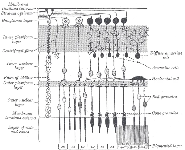
Figure 23Gray882
Long description
Plan of retinal neurons. (After Cajal.). Labelled illustration from Wikimedia Commons, used under its open licence.

Figure 23: Plan of retinal neurons (After Cajal.).
The lens fibres elongate and fill the lens vesicle, creating the mature lens. The corneal epithelium develops from the surface ectoderm directly above the developing eye. The underlying mesenchyme condenses to form the corneal stroma and endothelium.
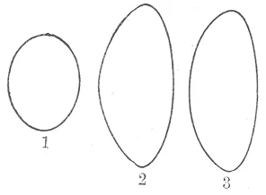
Figure 24Gray886
Long description
Profile views of the lens at different periods of life. 1. In the fetus. 2. In adult life. 3. In old age.

Figure 24: Profile views of the lens at different periods of life (1).
Figure 25Gray885
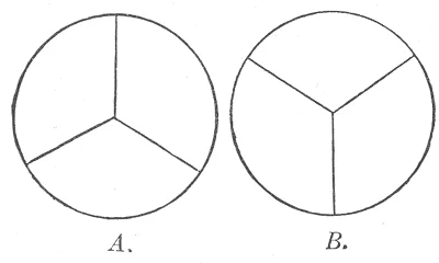
Long description
Diagram to show the direction and arrangement of the radiating lines on the front and back of the fetal lens. A. From the front. B. From the back.

Figure 25: Diagram to show the direction and arrangement of the radiating lines on the front and back of the fetal lens (a).
DefinitionMesenchyme
The mesenchyme, derived from neural crest cells and mesoderm, forms the sclera, choroid, ciliary muscle, and most of the vascular structures of the eye.
The mesenchyme surrounding the optic cup differentiates into the vascular tunic (uvea) and the fibrous tunic (sclera and cornea). The ciliary body and iris stroma arise from this mesenchyme.
The vitreous body develops from the primary and secondary vitreous. The primary vitreous forms from the hyaloid artery and surrounding mesenchyme. It is largely resorbed, leaving the secondary vitreous, which constitutes the bulk of the adult vitreous humour. The hyaloid canal, a remnant of the hyaloid artery, runs through the vitreous to the optic disc.
Exam trapCommon Error
Do not confuse the origin of the lens epithelium with the neural retina. Both arise from the optic cup, but the lens is induced by surface ectoderm, while the retina is an outpouching of the brain.
Congenital defects often result from failures in these developmental processes. Coloboma occurs when the optic fissure fails to close, typically affecting the inferior aspect of the iris or retina. Microphthalmia results from incomplete development of the optic vesicle. Aniridia arises from mutations affecting the development of the iris from the neuroectoderm.
Figure 26Gray865
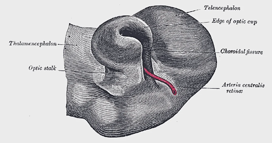
Long description
Optic cup and choroidal fissure seen from below, from a human embryo of about four weeks. (Kollmann.)

Figure 26: Optic cup and choroidal fissure seen from below, from a human embryo of about four weeks (Kollmann.).
NoteClinical Relevance
Knowledge of embryological origins helps predict associated anomalies. For example, defects in neural crest cell migration can affect both the eye and facial structures, as seen in Treacher Collins syndrome.
The blood supply to the developing eye is established by the hyaloid artery, which regresses before birth. The central retinal artery, a branch of the ophthalmic artery, supplies the inner retina. The choroidal circulation, derived from mesenchyme, supplies the outer retina. Understanding this vascular development is crucial for interpreting retinal findings in premature infants, such as retinopathy of prematurity, where abnormal angiogenesis occurs due to oxygen-induced vaso-obliteration.
| Muscle | Origin | Insertion | Action | Nerve Supply |
|---|---|---|---|---|
| Medial Rectus | Annulus of Zinn | Sclera | Adduction | Oculomotor (CN III) |
| Lateral Rectus | Annulus of Zinn | Sclera | Abduction | Abducens (CN VI) |
| Superior Rectus | Annulus of Zinn | Sclera | Elevation | Oculomotor (CN III) |
| Inferior Rectus | Annulus of Zinn | Sclera | Depression | Oculomotor (CN III) |
| Superior Oblique | Sphenoid Bone | Sclera | Intorsion | Trochlear (CN IV) |
| Inferior Oblique | Maxillary Bone | Sclera | Extorsion | Oculomotor (CN III) |
The eye is a complex sensory organ situated within the bony orbit, protected by the eyelids and extraocular muscles. Understanding its anatomy requires a systematic approach to layers, vascular supply, and neural pathways. The globe consists of three concentric coats: the fibrous tunic, the vascular tunic, and the neural tunic.
The inner retina is supplied by the central retinal artery, a branch of the ophthalmic artery. This vessel enters the optic nerve and divides into superior and inferior temporal and nasal branches. These arteries form a dense capillary network within the retinal layers. To assess the health of this network, clinicians often use Optical Coherence Tomography Angiography (OCTA). This non-invasive imaging technique allows for the visualization of blood flow without the need for dye injection.
The depth of the retinal vascular plexuses can be visualised using OCTA volumes. In clinical practice, these volumes are aligned at the level of the retinal pigment epithelium (RPE) to ensure accurate layer segmentation. This alignment is crucial for distinguishing between the superficial and deep capillary plexuses. [2]
Quantifying blood flow in these plexuses involves calculating capillary density. This metric is expressed as the percentage of flow per area. The count of flow pixels is then divided by the total number of pixels in that sample area. [2]
However, large vessels can distort this measurement. Arteries and veins carry high flow signals that may be misinterpreted as capillary density. Therefore, these areas are excluded from the analysis. This exclusion is achieved by filtering the signal using a thresholding algorithm. A large vessel mask is created from the en face angiogram of the superficial vascular plexus (SVP). [2]
This threshold helps to identify pixels with significant flow. Only connected regions larger than 50 voxels are kept in the mask. This procedure ensures that only the finer capillary network is analysed. The same procedure is applied to all groups in comparative studies to maintain consistency. [2]
Lesions in the retinal vascular plexuses can lead to vision loss. Conditions such as diabetic retinopathy and retinal vein occlusion affect these microvascular networks. By excluding large vessels, clinicians can more accurately assess capillary dropout. This information is vital for staging disease and determining treatment strategies. The ability to visualise these structures in three dimensions provides a comprehensive view of retinal health.
In the eye, light falling on the retina causes chemical changes to pigment molecules in the photoreceptors, ultimately leading to a change in the activity of the RGCs. Photoreceptor cells have two parts, the inner segment and the outer segment (). The inner segment contains the nucleus and other common organelles of a cell, whereas the outer segment is a specialized region in which photoreception takes place. There are two types of photoreceptors—rods and cones—which differ in the shape of their ou [1]
Exam trapPhotoreceptor Structure
Rods have stacked discs; cones have membrane infoldings. Do not confuse the location of rhodopsin with cone opsins.
Clinical pearlVascular Plexuses
In the parafoveal and perifoveal regions, there are three distinct peaks corresponding to the three vascular plexuses (SVP, ICP, and DCP). [2]
Exam trapFAZ Boundary
The 3 plexuses merge into one at the edge of the foveal avascular zone (FAZ). [2]
DefinitionCapillary Density
In the peripapillary region, the RPCP and the SVP demonstrate two peaks of capillary density, but with no distinct trough separating them. [2]
Students frequently confuse the fundamental definitions of sensory processing, leading to errors in clinical reasoning. It is crucial to distinguish between sensation and perception. Sensation is the activation of sensory receptor cells at the level of the stimulus. Perception is the central processing of sensory stimuli into a meaningful pattern. Perception is dependent on sensation, but not all sensations are perceived. A receptor cell is changed directly by a stimulus. Confusing these stages can lead to misinterpreting neurological deficits as purely sensory when they may be central processing errors. [1]
Another significant error involves the classification of receptors. A transmembrane protein receptor is a protein in the cell membrane that mediates a physiological change in a neuron, most often through the opening of ion channels or changes in the cell signaling processes. Transmembrane receptors are activated by chemicals called ligands. Students often assume all receptors work via ligand binding, ignoring physical stimuli. Stimuli are of three general types. Some stimuli are ions and macromo This occurs when a stimulus changes the cell membrane potential of a sensory neuron. [1]
In taste physiology, a common mistake is ignoring the established submodalities. Only a few recognized submodalities exist within the sense of taste, or gustation. Until recently, only four tastes were recognized: sweet, salty, sour, and bitter. Research at the turn of the 20th century led to recognition of the fifth taste, umami, during the mid-1980s. Umami is a Japanese word that means “delicious taste,” and is often translated to mean savory. While recent research suggests a sixth taste for fats, the core five remain the standard for examination.
Regarding sour taste, students often misunderstand the ionic mechanism. Sour taste is the perception of H+ concentration. Just as with sodium ions in salty flavors, these hydrogen ions enter the cell and trigger depolarization. Sour flavors are, essentially, the perception of acids in our food. Increasing hydrogen ion concentrations in the saliva (lowering saliva pH) triggers progressively stronger graded potentials in the gustatory cells. For example, orange juice—which contains citric acid—will taste sour because it has a pH value of approximately 3. [1]
In the eye, a critical anatomical error is misidentifying the photopigment sensitivity. The opsins are sensitive to limited wavelengths of light. Rhodopsin, the photopigment in rods, is most sensitive to light at a wavelength of 498 nm. The three color opsins have peak sensitivities of 564 nm, 534 nm, and 420 nm corresponding roughly to the primary colors of red, green, and blue. The absorbance of rhodopsin in the rods is much more sensitive than in the cone opsins; specifically, rods are sensitive to vision in low light conditions, and cones are sensitive to brighter conditions. In normal sunlight, rhodopsin will be constantly bleached while the cones are active. In a darkened room, there is not enough light to activate cone opsins, and vision is entirely dependent on rods. Rods are so sensitive to light that a single photon can result in an action potential from a rod’s corresponding RGC. [1]
Finally, students may overestimate the role of memory in dark adaptation. If you think that you can see colors in the dark, it is most likely because your brain knows what color something is and is relying on that memory. True scotopic vision is monochromatic.
Exam trapSensation vs Perception
Sensation is the activation of sensory receptor cells at the level of the stimulus. Perception is the central processing of sensory stimuli into a meaningful pattern.
This knowledge is applied at the bedside to differentiate peripheral nerve lesions from central cortical deficits. Somatosensation is considered a general sense, as opposed to the special senses discussed in this section. Somatosensation is the group of sensory modalities that are associated with touch, proprioception, and interoception. These modalities include pressure, vibration, light touch, tickle, itch, temperature, pain, proprioception, and kinesthesia. This means that its receptors are not associated with a specialized organ, but are instead spread throughout the body in a variety of organs. Many of the somatosensory receptors are located in the skin, but receptors are also found in muscles, tendons, joint capsules, ligaments, and in the walls of visceral organs.
In the operating theatre, understanding the middle ear anatomy is vital for otological procedures. The three ossicles are the malleus, incus, and stapes, which are Latin names that roughly translate to hammer, anvil, and stirrup. The malleus is attached to the tympanic membrane and articulates with the incus. The incus, in turn, articulates with the stapes. The stapes is then attached to the inner ear, where the sound waves will be transduced into a neural signal. The middle ear is connected to the pharynx through the Eustachian tube. [1]
On imaging, particularly SD-OCT, segmentation errors can mimic pathology. Additionally, due to flow projection artifact, the true plexus boundaries between the superficial and deeper vasculature could not be well characterized, which led to segmentation at the INL/IPL border on SD-OCT. Unfortunately, this anatomic boundary yields unpredictable segmentation of the ICP between the superficial and deep slabs. Clinicians must recognize that apparent defects may be artifacts of the imaging plane rather than true anatomical loss.
In examinations, understanding retinal vascular density is key. As a whole, the SVC presents an extremely dense vascular network in the peripapillary region that decreases in density with distance from disc along the maculopapillary axis. This agrees with previous histologic evidence – Snodderly et al. reported the percentage of retinal area covered by capillaries and found that it was highest in the peripapillary region, often exceeding 60%. Prior work has suggested that this layer nourishes the NFL, and therefore is densest in the peripapillary region and reduces in thickness as the NFL decreases.
Visual Adaptation
- StartAssess lighting condition
- Question: Is light bright?
Yes
- Cones activeColor vision, high acuity
- Rhodopsin bleachedLow rod sensitivity
No
- Rods activeMonochromatic, high sensitivity
- Single photon detection1 photon = action potential
Sensation is the activation of sensory receptor cells at the level of the stimulus. Perception is the central processing of sensory stimuli into a meaningful pattern. A receptor cell is changed directly by a stimulus. Sour taste is the perception of H+ concentration. The three ossicles are the malleus, incus, and stapes. Rhodopsin, the photopigment in rods, is most sensitive to light at a wavelength of 498 nm. The three color opsins have peak sensitivities of 564 nm, 534 nm, and 420 nm. Rods are sensitive to vision in low light conditions, and cones are sensitive to brighter conditions.
Answer each one in your head first, then open the answer.
What is the definition of sensation?
Show answerHide answer
Sensation is the activation of sensory receptor cells at the level of the stimulus.What are the three ossicles?
Show answerHide answer
The malleus, incus, and stapes.What wavelength is rhodopsin most sensitive to?
Show answerHide answer
498 nm.What does sour taste perceive?
Show answerHide answer
H+ concentration.Where are somatosensory receptors located?
Show answerHide answer
Spread throughout the body in a variety of organs, including skin, muscles, tendons, joint capsules, ligaments, and visceral organs.
- Betts JG, Young KA, Wise JA, et al. Sensory Perception. OpenStax, Anatomy and Physiology 2e (CC BY-NC-SA 4.0). Accessed .
- Campbell JP, Zhang M, Hwang TS, et al. Detailed Vascular Anatomy of the Human Retina by Projection-Resolved Optical Coherence Tomography Angiography. Scientific reports 2017;7:42201. doi:10.1038/srep42201. Accessed .
Figure credits
- Figure 1: OpenStax College, via Wikimedia Commons, CC BY 3.0, CC BY 3.0. resized.
- Figure 2: OpenStax College, via Wikimedia Commons, CC BY 3.0, CC BY 3.0. resized.
- Figure 3: Henry Vandyke Carter, via Wikimedia Commons, Public domain, Public domain.
- Figure 4: Henry Vandyke Carter, via Wikimedia Commons, Public domain, Public domain.
- Figure 5: Henry Vandyke Carter, via Wikimedia Commons, Public domain, Public domain.
- Figure 6: Henry Vandyke Carter, via Wikimedia Commons, Public domain, Public domain.
- Figure 7: Henry Vandyke Carter, via Wikimedia Commons, Public domain, Public domain.
- Figure 8: BruceBlaus. When using this image in external sources it can be cited as: Blausen.com staff (2014). "Medical gallery of Blausen Medical 2014". WikiJournal of Medicine 1 (2). DOI:10.15347/wjm/2014.010. ISSN 2002-4436., via Wikimedia Commons, CC BY 3.0, CC BY 3.0.
- Figure 9: Henry Vandyke Carter, via Wikimedia Commons, Public domain, Public domain.
- Figure 10: Henry Vandyke Carter, via Wikimedia Commons, Public domain, Public domain.
- Figure 11: Henry Vandyke Carter, via Wikimedia Commons, Public domain, Public domain.
- Figure 12: Henry Vandyke Carter, via Wikimedia Commons, Public domain, Public domain.
- Figure 13: Henry Vandyke Carter, via Wikimedia Commons, Public domain, Public domain.
- Figure 14: Henry Vandyke Carter, via Wikimedia Commons, Public domain, Public domain.
- Figure 15: Henry Vandyke Carter, via Wikimedia Commons, Public domain, Public domain.
- Figure 16: Henry Vandyke Carter, via Wikimedia Commons, Public domain, Public domain.
- Figure 17: Henry Vandyke Carter, via Wikimedia Commons, Public domain, Public domain.
- Figure 18: Henry Vandyke Carter, via Wikimedia Commons, Public domain, Public domain.
- Figure 19: OpenStax College, via Wikimedia Commons, CC BY 3.0, CC BY 3.0. resized.
- Figure 20: Henry Vandyke Carter, via Wikimedia Commons, Public domain, Public domain.
- Figure 21: Henry Vandyke Carter, via Wikimedia Commons, Public domain, Public domain.
- Figure 22: Henry Vandyke Carter, via Wikimedia Commons, Public domain, Public domain.
- Figure 23: Henry Vandyke Carter, via Wikimedia Commons, Public domain, Public domain.
- Figure 24: Henry Vandyke Carter, via Wikimedia Commons, Public domain, Public domain.
- Figure 25: Henry Vandyke Carter, via Wikimedia Commons, Public domain, Public domain.
- Figure 26: Henry Vandyke Carter, via Wikimedia Commons, Public domain, Public domain.
Educational content for medical students and doctors. It is not a substitute for clinical judgement or local guidelines.