Nasal cavity and paranasal sinuses
Anatomical structures of the nasal cavity and paranasal sinuses including septum, conchae, and clinical correlations.
Figure 1Gray153
Long description
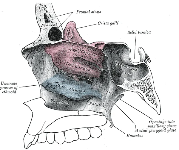
Lateral wall of nasal cavity, showing ethmoid bone in position.

Figure 1: Lateral wall of nasal cavity, showing ethmoid bone in position.
Figure 2710 Ethmoid Bone
Long description
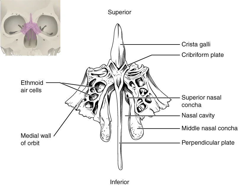
The unpaired ethmoid bone is located at the midline within the central skull. It has an upward projection, the crista galli, and a downward projection, the perpendicular plate, which forms the upper nasal septum. The cribriform plates form both the roof of the nasal cavity and a portion of the anter

Figure 2: The unpaired ethmoid bone is located at the midline within the central skull.
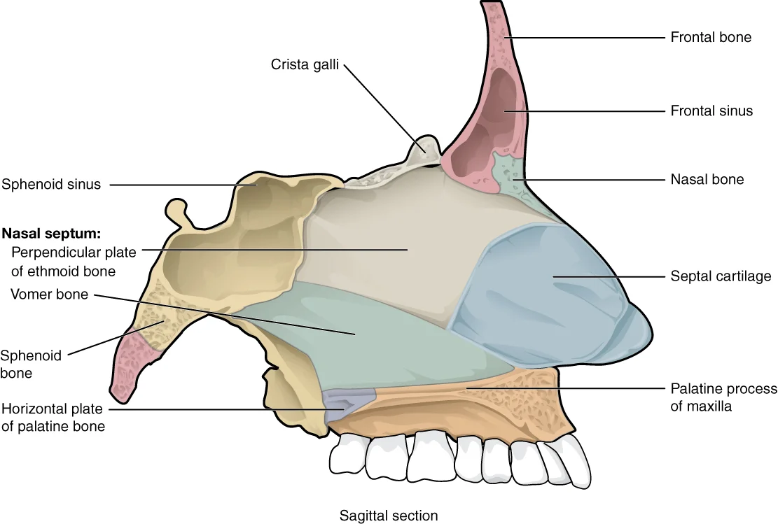
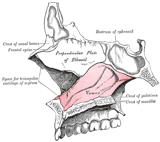
The nasal cavity is the central airway of the upper respiratory tract, situated within the facial skeleton. It is divided into left and right halves by the nasal septum. The septum comprises both bony and cartilaginous elements. The upper portion is formed by the perpendicular plate of the ethmoid bone. The lower and posterior parts are formed by the triangular-shaped vomer bone. The anterior nasal septum is formed by the septal cartilage, a flexible plate that fills the gap between the ethmoid and vomer bones. This cartilage is the portion you can touch with your fingers. [1] [2]
Figure 3714 Bone of Nasal Cavity
Long description
The nasal septum is formed by the perpendicular plate of the ethmoid bone and the vomer bone. The septal cartilage fills the gap between these bones and extends into the nose.

Figure 3: The nasal septum is formed by the perpendicular plate of the ethmoid bone and the vomer bone.
Figure 4704 Skull-01
Long description
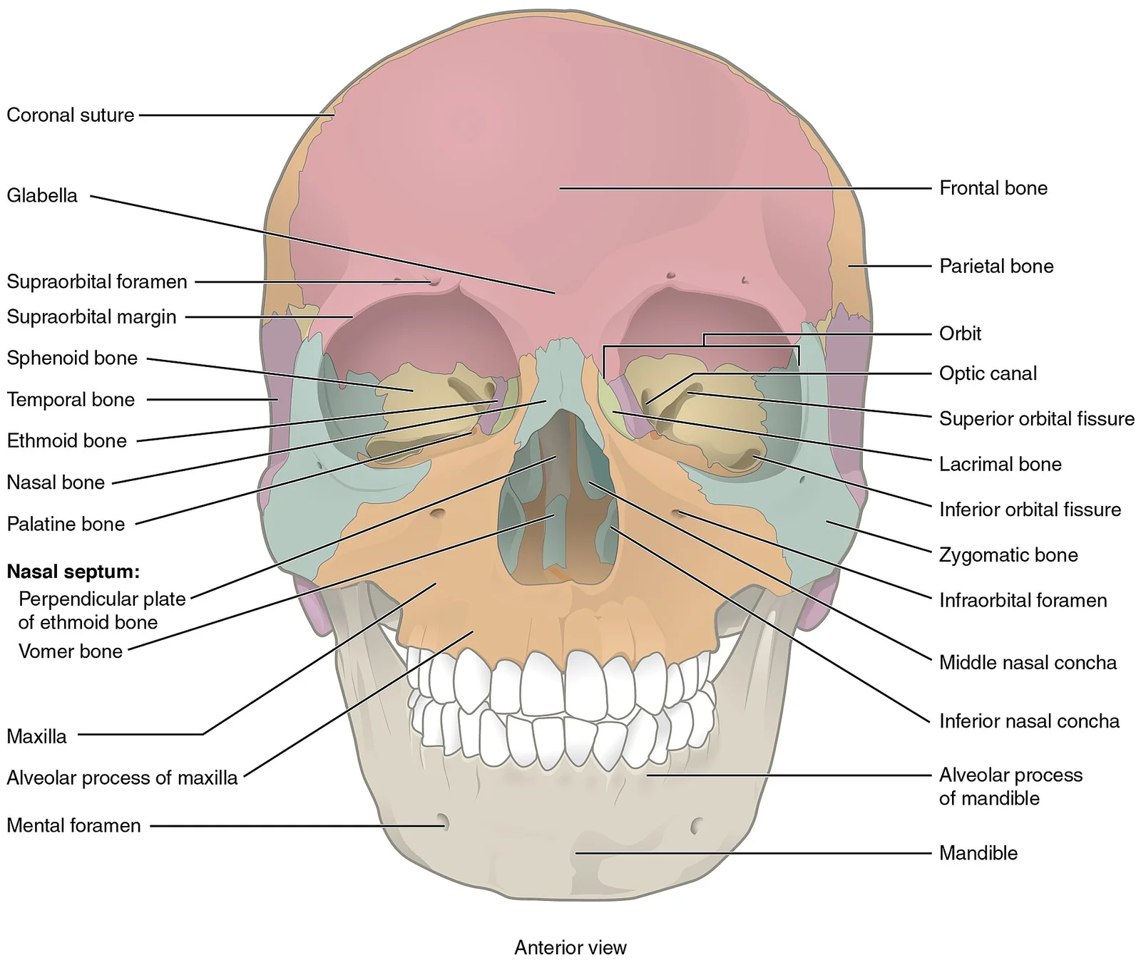
An anterior view of the skull shows the bones that form the forehead, orbits (eye sockets), nasal cavity, nasal septum, and upper and lower jaws.

Figure 4: An anterior view of the skull shows the bones that form the forehead, orbits (eye sockets), nasal cavity, nasal septum, and upper and lower jaws.
Figure 5Gray173
Long description
Median wall of left nasal cavity showing vomer in situ.

Figure 5: Median wall of left nasal cavity showing vomer in situ.
Each lateral wall of the nasal cavity is triangular in shape, with a broad inferior space that narrows superiorly. Projecting from each lateral wall are three bony plates known as the nasal conchae. These structures curve downward into the nasal cavity space. They serve to swirl the incoming air, which helps to warm and moisturize it before the air moves into the delicate air sacs of the lungs. This turbulence also allows mucus, secreted by the tissue lining the nasal cavity, to trap incoming dust, pollen, bacteria, and viruses.
Figure 6Gray171
Long description
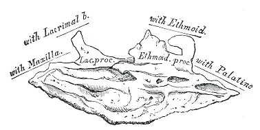
Right inferior nasal concha. Medial surface.

Figure 6: Right inferior nasal concha (medial surface).
Figure 7Gray856
Long description
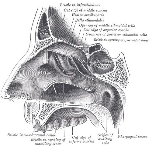
Lateral wall of nasal cavity; the three nasal conchæ have been removed.

Figure 7: Lateral wall of nasal cavity; the three nasal conchæ have been removed.
Figure 8Gray855
Long description
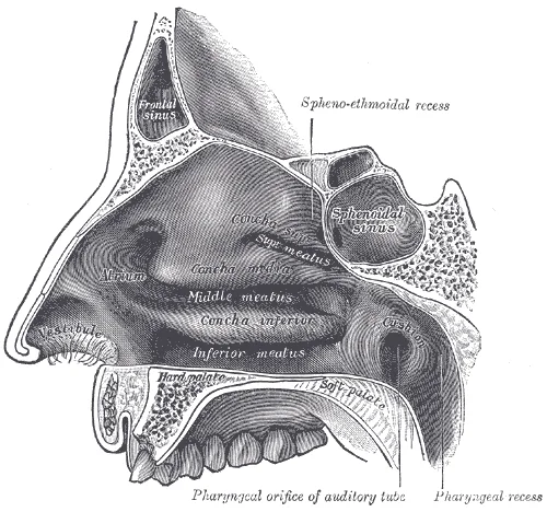
Lateral wall of nasal cavity. Labelled illustration from Wikimedia Commons, used under its open licence.

Figure 8: Lateral wall of nasal cavity.
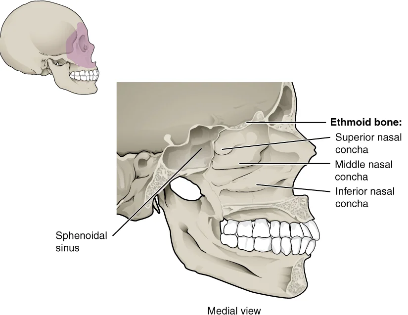
The inferior nasal concha is the largest of the nasal conchae and is an independent bone of the skull. It can easily be seen when looking into the anterior opening of the nasal cavity. Located just above the inferior concha is the middle nasal concha, which is part of the ethmoid bone. A third bony plate, the superior nasal concha, is also part of the ethmoid bone. It is much smaller and out of sight, located just lateral to the perpendicular plate in the upper nasal cavity. When looking into the anterior nasal opening of the skull, only the inferior and middle conchae can be seen. [1]
DefinitionNasal Cavity
The air-filled space within the skull divided by the septum and lined with mucosa, containing the conchae to condition inhaled air.
The paranasal sinuses are hollow, air-filled spaces located within certain bones of the skull. All of the sinuses communicate with the nasal cavity. They are lined with nasal mucosa. The sinuses serve to reduce bone mass and thus lighten the skull. They also add resonance to the voice. This second feature is most obvious when you have a cold or sinus congestion. These produce swelling of the mucosa and excess mucus production, which can obstruct the narrow passageways between the sinuses and the nasal cavity. This blockage can also allow the sinuses to fill with fluid, with the resulting pressure producing pain and discomfort. [1]
NoteClinical Pearl
Sinusitis pain results from mucosal swelling obstructing sinus drainage, leading to fluid accumulation and pressure within the confined bony spaces.
The facial bones include 14 bones, with six paired bones and two unpaired bones. The paired bones are the maxilla, palatine, zygomatic, nasal, lacrimal, and inferior nasal conchae bones. The unpaired bones are the vomer and mandible bones. Although classified with the brain-case bones, the ethmoid bone also contributes to the nasal septum and the walls of the nasal cavity and orbit. The ethmoid bone also forms the lateral walls of the upper nasal cavity. Extending from each lateral wall are the superior nasal concha and middle nasal concha, which are thin, curved projections that extend into the nasal cavity. [1]
Figure 9731 Nasal Cavity
Long description
The three nasal conchae are curved bones that project from the lateral walls of the nasal cavity. The superior nasal concha and middle nasal concha are parts of the ethmoid bone. The inferior nasal concha is an independent bone of the skull.

Figure 9: The three nasal conchae are curved bones that project from the lateral walls of the nasal cavity.
Figure 10Gray172
Long description
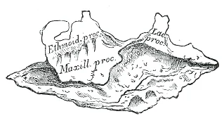
Right inferior nasal concha. Lateral surface.

Figure 10: Right inferior nasal concha (lateral surface).
| Structure | Bone of Origin | Position/Relation |
|---|---|---|
| Perpendicular Plate | Ethmoid | Upper portion of septum |
| Vomer | Unpaired | Lower and posterior septum |
| Septal Cartilage | Cartilage | Anterior septum |
| Inferior Concha | Independent Bone | Lower lateral wall |
| Middle Concha | Ethmoid | Middle lateral wall |
| Superior Concha | Ethmoid | Upper lateral wall |
Figure 11724 Paranasal Sinuses
Long description
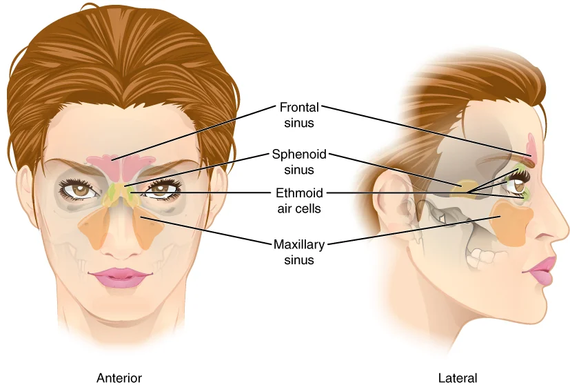
The paranasal sinuses are hollow, air-filled spaces named for the skull bone that each occupies. The most anterior is the frontal sinus, located in the frontal bone above the eyebrows. The largest are the maxillary sinuses, located in the right and left maxillary bones below the orbits. The most pos

Figure 11: The paranasal sinuses are hollow, air-filled spaces named for the skull bone that each occupies.
The nose serves as the primary entrance and exit for the respiratory system. For anatomical study, it is divided into two main sections: the external nose and the internal nasal cavity. [2]
Figure 12Gray859
Long description
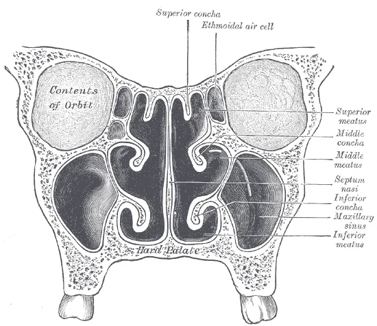
Coronal section of nasal cavities. Labelled illustration from Wikimedia Commons, used under its open licence.

Figure 12: Coronal section of nasal cavities.
The external nose consists of the surface and skeletal structures that determine its outward appearance. The most superior region, situated between the eyebrows, is the root. This connects to the bridge, which is the part of the nose linking the root to the rest of the structure. The length of the nose is termed the dorsum nasi, which terminates at the tip, known as the apex. [2]
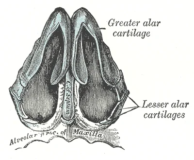
The nostrils, or nares, are the openings of the nasal cavity. Each naris is bordered laterally by an ala (plural: alae), which is a cartilaginous structure. Finally, the philtrum is the concave surface that connects the apex of the nose to the upper lip. [2]
Figure 13Gray853
Long description
Cartilages of the nose, seen from below.

Figure 13: Cartilages of the nose, seen from below.
Figure 14Gray852
Long description
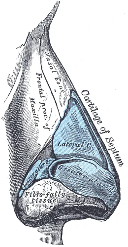
Cartilages of the nose. Side view. Labelled illustration from Wikimedia Commons, used under its open licence.

Figure 14: Cartilages of the nose (side view).
DefinitionNares
The nostrils or openings that serve as the entrance to the nasal cavity.
Moving inferiorly, the bridge connects the root to the dorsum nasi, which represents the main length of the nose. The apex is the most inferior point of the midline. On either side of the apex are the alae, which form the lateral boundaries of the nares. The philtrum is the depression between the apex and the upper lip.
The framework of the external nose is composed of a bony upper portion and a cartilaginous lower portion.
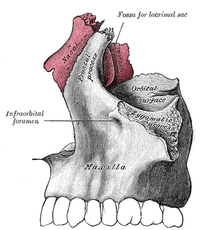
The bony base, or bridge, is formed by a pair of nasal bones. These bones articulate superiorly with the frontal bone and laterally with the maxillary bones. They provide the necessary support for the cartilages that form the lateral walls of the nose. [1]
Figure 15Gray154
Long description
Articulation of nasal and lacrimal bones with maxilla.

Figure 15: Articulation of nasal and lacrimal bones with maxilla.
Figure 16Gray158
Long description
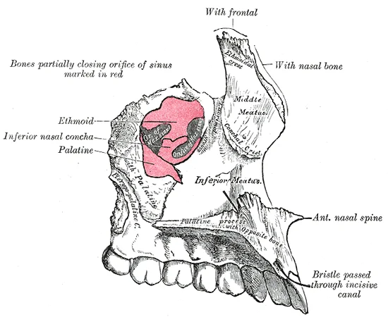
Left maxilla. Nasal surface. Labelled illustration from Wikimedia Commons, used under its open licence.

Figure 16: Left maxilla (nasal surface).
Figure 17Gray157
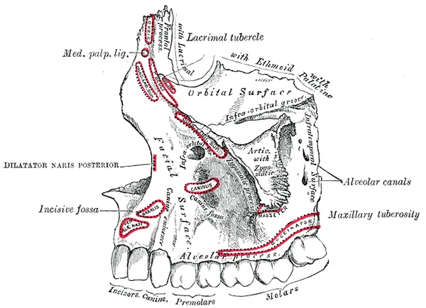
Long description
Left maxilla. Outer surface. Labelled illustration from Wikimedia Commons, used under its open licence.

Figure 17: Left maxilla (outer surface).
Figure 18Gray854
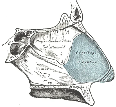
Long description
Bones and cartilages of septum of nose. Right side.

Figure 18: Bones and cartilages of septum of nose (right side).
The protruding portion of the nose consists of cartilage. The dorsum nasi is formed by the septal cartilage, a flexible hyaline cartilage that connects to the nasal bone. The apex of the nose and the area surrounding the naris are formed by the alar cartilage. [2]
NoteClinical Correlation
The nasal bones are the specific structures damaged when the nose is broken. [1]
| Structure | Composition | Location | Relation or Function |
|---|---|---|---|
| Nasal bone | Bone | Root and bridge | Articulates with frontal and maxillary bones |
| Septal cartilage | Hyaline cartilage | Dorsum nasi | Connects to the nasal bone |
| Alar cartilage | Cartilage | Apex and naris | Surrounds the nostril opening |
The nasal cavity is a central opening located within the anterior skull, which is composed of the facial bones. It serves as the primary entry point of the conducting zone of the respiratory system[1].
The nasal cavity is the central opening of the anterior skull. Surrounding this are the paranasal sinuses, which are air-filled spaces located within the frontal, maxillary, sphenoid, and ethmoid bones. Posteriorly, the nasal cavity is continuous with the nasopharynx[1], which is flanked by the conchae[1]. [2]
The lateral and posterior boundaries of the nasal cavity are defined by the sphenoid bone. Specifically, the medial pterygoid plates are thin, vertically oriented bony plates that form the posterior, lateral walls of the nasal cavity. [1]
The cavity is associated with the paranasal sinuses, which are air-filled spaces distributed across four specific bones.
| Sinus | Bone Location |
|---|---|
| Frontal | Frontal bone |
| Maxillary | Maxillary bone |
| Sphenoid | Sphenoid bone |
| Ethmoid | Ethmoid bone |
The nasal cavity is continuous posteriorly with the nasopharynx[1]. The nasopharynx is flanked by the conchae of the nasal cavity[1]. At the superior portion of the nasopharynx lie the pharyngeal tonsils, also known as adenoids, which are aggregates of lymphoid reticular tissue[1]. The nasopharynx is bordered inferiorly by the oropharynx[1].
The internal surface of the nasal cavity is modified by conchae and meatuses, which expand the surface area to facilitate the warming and humidifying of incoming air[1].
DefinitionRespiratory Epithelium
Pseudostratified ciliated columnar epithelium containing goblet cells that produce mucus to trap debris[1].
This epithelium lines the conchae, meatuses, and paranasal sinuses[1]. Capillaries located just beneath this epithelium warm the air by convection[1].
Figure 192301 Major Respiratory Organs
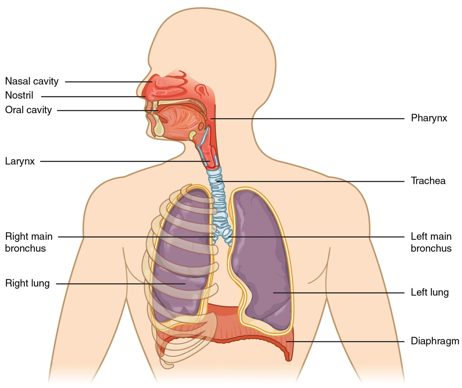
Long description
The major respiratory structures span the nasal cavity to the diaphragm.

Figure 19: The major respiratory structures span the nasal cavity to the diaphragm.
The nasal cavity serves as the primary entry point of the conducting zone. The fundamental purpose of this zone is to provide a route for incoming and outgoing air, remove pathogens and debris, and warm and humidify the air. The internal lining is specialised, as the epithelium of the nasal passages is essential for sensing odours. [2]
DefinitionConducting Zone
The portion of the respiratory system that transports, cleans, and conditions air before it reaches the gas-exchange surfaces.
The nasal cavity is formed by the facial bones. There are 14 facial bones in total: the paired nasal, lacrimal, zygomatic, maxillary, palatine, and inferior conchae bones, alongside the unpaired vomer and mandible. Additionally, the ethmoid bone contributes to the formation of these facial structures. [1]
The floor of the cavity is the hard palate. This is a composite structure where the maxilla forms the larger anterior portion. The posterior quarter of the hard palate is formed by the horizontal plates of the paired palatine bones, which join together at the midline. [1]
Figure 20Gray160
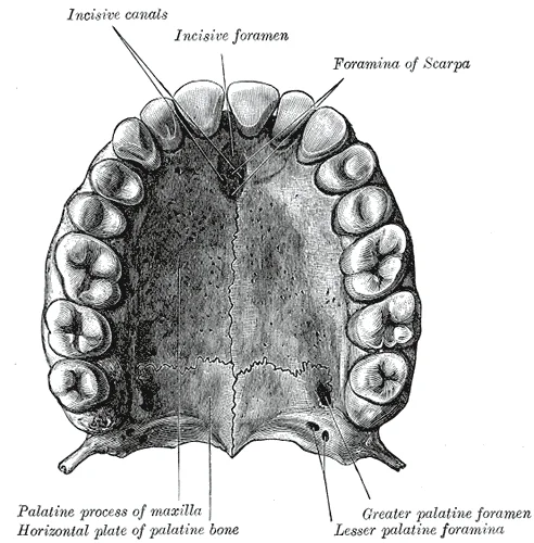
Long description
The bony palate and alveolar arch. Labelled illustration from Wikimedia Commons, used under its open licence.

Figure 20: The bony palate and alveolar arch.
| Bone | Type | Contribution |
|---|---|---|
| Maxilla | Paired | Anterior portion of the hard palate |
| Palatine | Paired | Posterior quarter of the hard palate and lateral walls |
| Inferior Conchae | Paired | Lateral wall projections |
| Ethmoid | Unpaired | General facial structure contribution |
| Vomer | Unpaired | General facial structure contribution |
The lateral walls of the nasal cavity are partially formed by the palatine bones. These walls contain the nasal conchae, which increase the surface area for air conditioning. The inferior conchae are distinct paired bones. Above these lie the middle conchae. The superior nasal concha is the smallest and is well hidden, situated above and behind the middle concha. [1]
The nasal cavities terminate posteriorly where they are continuous with the pharynx. The pharynx is a muscular tube lined by mucous membrane. The most superior region of the pharynx, which directly follows the nasal cavity, is the nasopharynx. [2]
The floor is the hard palate, consisting of the maxilla anteriorly and the palatine bones posteriorly. On the lateral walls, the inferior conchae are the lowest bony projections. Above these are the middle conchae, and the superior nasal concha is the highest projection, tucked behind the middle concha. The cavity opens posteriorly into the nasopharynx.
The arterial supply to the nasal cavity and paranasal sinuses is derived from both the internal and external carotid systems. This dual supply ensures robust perfusion but also creates a rich vascular network that is prone to bleeding. The anterior and posterior ethmoidal arteries, branches of the ophthalmic artery from the internal carotid system, supply the superior aspects of the nasal cavity. The sphenopalatine artery, a terminal branch of the maxillary artery from the external carotid system, is the primary blood supply to the lateral nasal wall and the posterior septum. The greater palatine artery also contributes to the inferior nasal cavity via the incisive canal. [1]
The supply lines trace the sphenopalatine artery entering through the sphenopalatine foramen and branching into the lateral nasal and septal branches. The blood supply network is dense in the anterior septum, correlating with the common site of nosebleeds.
DefinitionLittle Area
A vascular area on the anterior inferior part of the nasal septum where multiple arteries anastomose. It is the most common site of epistaxis.
The Little area, or Kiesselbach’s plexus, is located on the anterior inferior nasal septum. It is formed by the anastomosis of four arteries: the anterior ethmoidal artery, the sphenopalatine artery, the greater palatine artery, and the superior labial artery. This convergence creates a dense capillary bed that is superficial and vulnerable to trauma and dry air. Consequently, this area is the origin of the majority of anterior nosebleeds. Understanding the vascular anatomy here is crucial for managing epistaxis. [1]
| Artery | Origin | Territory |
|---|---|---|
| Anterior Ethmoidal | Ophthalmic Artery | Superior and Anterior Septum |
| Posterior Ethmoidal | Ophthalmic Artery | Superior Lateral Wall |
| Sphenopalatine | Maxillary Artery | Posterior Septum and Lateral Wall |
| Greater Palatine | Maxillary Artery | Inferior Septum and Floor |
| Superior Labial | Facial Artery | Anterior Septum (Little Area) |
The paranasal sinuses receive their blood supply from branches that also supply the nasal cavity. The frontal sinus is supplied by the anterior ethmoidal artery and supraorbital artery. The maxillary sinus receives branches from the posterior superior alveolar artery and the infraorbital artery. The ethmoidal sinuses are supplied by the anterior and posterior ethmoidal arteries. The sphenoid sinus is supplied by the posterior ethmoidal artery and sphenopalatine artery. This rich vascularisation supports the mucosal lining and facilitates immune responses within the sinuses.
Figure 21Gray857
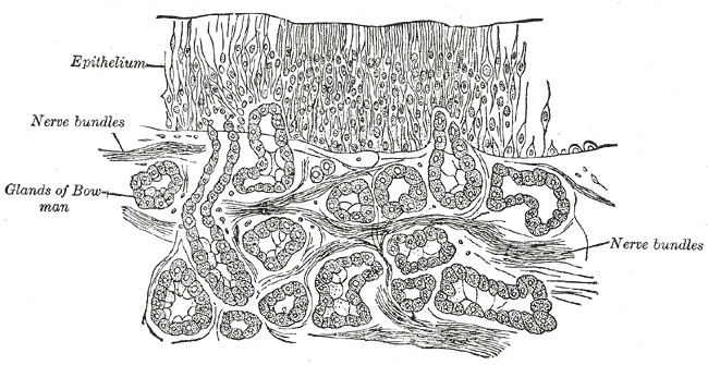
Long description
Section of the olfactory mucous membrane. (Cadiat.)

Figure 21: Section of the olfactory mucous membrane (Cadiat.).
The venous drainage of the nasal cavity and paranasal sinuses is a critical anatomical pathway that connects the facial region with the intracranial cavity. Understanding this system requires tracing the flow from the superficial facial veins to the deep dural sinuses. The primary route involves the angular vein, the ophthalmic veins, and the cavernous sinus.
The maxillary sinus is located inferiorly, with its venous drainage contributing to the pterygoid plexus. The sphenoid sinus is positioned centrally, posterior to the ethmoid sinuses. The cavernous sinus is a paired dural venous sinus located on either side of the sella turcica.
The angular vein begins at the medial angle of the eye. It is formed by the union of the supratrochlear and supraorbital veins. It runs downwards and laterally along the side of the nose. It continues as the facial vein. The facial vein drains the anterior part of the scalp, the forehead, the nose, and the upper lip. It passes deep to the platysma muscle. It joins the posterior facial vein to form the common facial vein. The common facial vein usually empties into the internal jugular vein. However, it also communicates with the cavernous sinus via the ophthalmic veins.
The ophthalmic veins are divided into superior and inferior divisions. The superior ophthalmic vein is the larger of the two. It receives blood from the angular vein and the supraorbital vein. It passes through the superior orbital fissure. It enters the cavernous sinus. The inferior ophthalmic vein is smaller. It drains the lower eyelid and the lower part of the orbit. It may join the superior ophthalmic vein or drain directly into the cavernous sinus. It also communicates with the pterygoid plexus.
The pterygoid plexus is a network of veins located in the infratemporal fossa. It surrounds the lateral pterygoid muscle. It receives blood from the deep structures of the face, including the maxillary sinus. It communicates with the facial vein via the deep facial vein. It also communicates with the cavernous sinus via emissary veins. These connections provide a route for infection to spread from the face to the cavernous sinus.
The cavernous sinus is a paired dural venous sinus. It is located on either side of the sella turcica. It receives blood from the ophthalmic veins, the superficial middle cerebral vein, and the sphenoparietal sinus. It drains into the superior and inferior petrosal sinuses. The superior petrosal sinus drains into the transverse sinus. The inferior petrosal sinus drains into the internal jugular vein. The cavernous sinus contains the internal carotid artery and several cranial nerves. The abducens nerve runs through the sinus. The oculomotor, trochlear, and ophthalmic nerves run in the lateral wall.
The sphenoid bone is a single, complex bone of the central skull. It serves as a “keystone” bone, because it joins with almost every other bone of the skull. The sphenoid forms much of the base of the central skull and also extends laterally to contribute to the sides of the skull. Inside the cranial cavity, the right and left lesser wings of the sphenoid bone, which resemble the wings of a flying bird, form the lip of a prominent ridge that marks the boundary between the anterior and middle cranial fossae. The sella turcica (“Turkish saddle”) is located at the midline of the middle cranial fossa. This bony region of the sphenoid bone is named for its resemblance to the horse saddles used by the Ottoman Turks, with a high back and a tall front. [1]
Clinical pearlClinical Significance
Infections in the 'danger triangle' of the face can spread to the cavernous sinus via valveless veins, causing cavernous sinus thrombosis.
Lymph from the anterior part of the nasal septum and the lateral wall drains primarily towards the submandibular nodes. Lymph from the posterior nasal cavity and the sphenoid sinus drains posteriorly towards the retropharyngeal nodes. The maxillary sinus lymphatics drain inferiorly and laterally towards the upper deep cervical nodes. The ethmoid sinuses drain both anteriorly to the submandibular nodes and posteriorly to the retropharyngeal nodes. The frontal sinus lymphatics drain anteriorly towards the preauricular and upper deep cervical nodes.
These nodes are located in the submandibular triangle, beneath the body of the mandible. They receive lymph from the face, nose, and upper lip. These nodes lie behind the pharynx, between the prevertebral fascia and the visceral fascia. They are small and often difficult to palpate clinically. The maxillary sinus drains into the upper deep cervical lymph nodes. These nodes are located along the internal jugular vein, from the base of the skull to the omohyoid muscle. The preauricular nodes are located in front of the ear.
DefinitionClinical Pearl
Malignancies of the nasal cavity and paranasal sinuses typically metastasize to the regional lymph nodes corresponding to their specific anatomical drainage pathways.
The upper deep cervical nodes are the final common pathway for most of the head and neck lymphatics. They drain into the jugular trunk, which then empties into the thoracic duct on the left or the right lymphatic duct on the right. This convergence explains why distant metastases can occur if the disease progresses beyond the regional nodes.
| Sinus | Primary Drainage Nodes | Secondary Drainage Nodes |
|---|---|---|
| Maxillary | Upper deep cervical | Submandibular |
| Ethmoid (Anterior) | Submandibular | Upper deep cervical |
| Ethmoid (Posterior) | Retropharyngeal | Upper deep cervical |
| Sphenoid | Retropharyngeal | Upper deep cervical |
| Frontal | Preauricular | Upper deep cervical |
Figure 22Lateral wall of the nasal cavity: cross-section showing the
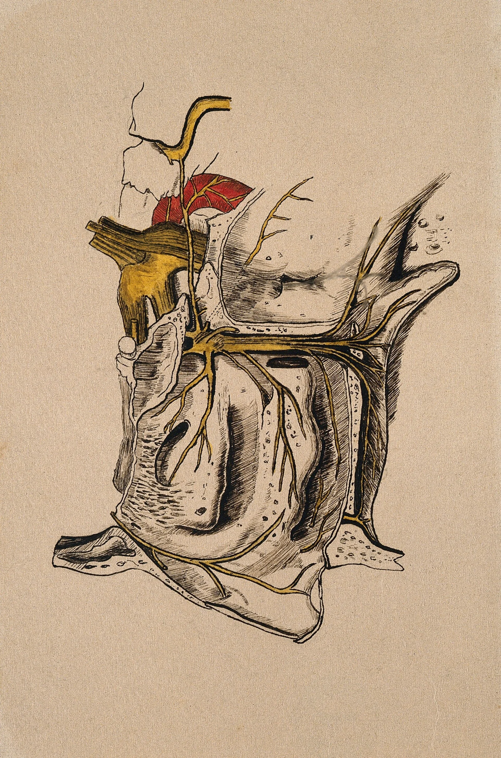
Long description
Lateral wall of the nasal cavity: cross-section showing the trigeminal nerve and sphenopalatine ganglion. Pen and watercolour drawing after H. Gray, ca. 1890. Iconographic Collections Keywords: Henry Gray

Figure 22: Lateral wall of the nasal cavity: cross-section showing the trigeminal nerve and sphenopalatine ganglion.
The sensory innervation of the nasal cavity and paranasal sinuses is derived from the ophthalmic (V1) and maxillary (V2) divisions of the trigeminal nerve (CN V). This somatic supply provides general sensation, including pain, temperature, and touch, to the mucosal lining and bony structures. Understanding this distribution is critical for localising pathology and understanding referred pain patterns.
It exits the nasal cavity through the anterior ethmoidal foramen. It supplies the skin of the lower eyelid, side of the nose, and upper lip, as well as the mucosa of the maxillary sinus.
Figure 23Gray858
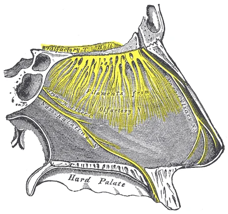
Long description
Nerves of septum of nose. Right side. Labelled illustration from Wikimedia Commons, used under its open licence.

Figure 23: Nerves of septum of nose (right side).
Autonomic innervation is provided by the parasympathetic and sympathetic fibres. Parasympathetic fibres originate from the facial nerve (CN VII) via the greater petrosal nerve and the nerve of the pterygoid canal, synapsing in the pterygopalatine ganglion. Postganglionic fibres then join the maxillary nerve to supply the nasal mucosa, promoting secretion and vasodilation.
Referred pain from the sinuses often follows the distribution of these nerves. For instance, maxillary sinusitis may present as pain in the upper teeth or cheek, supplied by the infraorbital nerve. Ethmoid sinusitis may cause pain between the eyes, supplied by the anterior ethmoidal nerve.
DefinitionOlfaction
The sense of smell, mediated by the olfactory nerve (CN I), which passes through the cribriform plate to reach the olfactory epithelium.
| Structure | Nerve Supply | Origin |
|---|---|---|
| Anterior Superior Septum | Anterior Ethmoidal Nerve | Ophthalmic Nerve (V1) |
| Lateral Nasal Wall (Anterior) | Anterior Ethmoidal Nerve | Ophthalmic Nerve (V1) |
| Maxillary Sinus | Posterior Superior Alveolar Nerves | Maxillary Nerve (V2) |
| Lower Eyelid, Side of Nose, Upper Lip | Infraorbital Nerve | Maxillary Nerve (V2) |
| Olfactory Epithelium | Olfactory Nerve (CN I) | Special Sensory |
Epistaxis (nosebleed) is common in the anterior inferior part of the nasal septum, known as Kiesselbach's plexus. The rich vascular supply and superficial location make this area prone to bleeding.
The nasal cavity and paranasal sinuses are lined by respiratory epithelium, which is pseudostratified ciliated columnar epithelium with goblet cells. This histological arrangement facilitates the mucociliary escalator, a critical defence mechanism that traps particulate matter and pathogens in mucus and propels them towards the pharynx for swallowing or expectoration. The underlying lamina propria contains seromucous glands that contribute to the mucus layer, providing hydration and antimicrobial properties. [2]
In the anterior part of the nasal septum, specifically in Kiesselbach’s plexus, the epithelium is thinner and the underlying vascular network is dense. This area is highly susceptible to epistaxis due to the superficial location of the anastomosing arteries, including branches of the sphenopalatine, greater palatine, anterior ethmoidal, and superior labial arteries. Understanding this vascular supply is essential for managing anterior nosebleeds, where local pressure or chemical cautery is often effective.
The paranasal sinuses, including the maxillary, frontal, ethmoid, and sphenoid sinuses, are also lined by respiratory epithelium. However, the thickness and cellularity of the epithelium can vary depending on the sinus and physiological state. The ethmoid sinuses, being the most complex, consist of multiple air cells that vary in size and shape. These cells are separated by thin bony septa and are closely related to the orbit and cranial cavity, making them prone to complications such as orbital cellulitis or intracranial extension of infection.
The olfactory epithelium, located in the roof of the nasal cavity, is distinct from the respiratory epithelium. It contains olfactory receptor neurons, supporting cells, and basal cells. The cilia of the olfactory neurons project into the mucus layer, where they bind odorant molecules. This specialized histology is crucial for the sense of smell, which plays a significant role in taste perception and environmental awareness.
For further details on the cellular composition and structural variations, please refer to the dedicated histology page. This resource provides comprehensive diagrams and descriptions of the epithelial types, glandular structures, and vascular networks within the nasal cavity and sinuses.
The nasal placodes invaginate to form nasal pits, which deepen into nasal sacs. These sacs communicate with the stomodeum via the primitive nasal apertures. The medial and lateral nasal prominences grow around these pits, eventually fusing to form the philtrum of the upper lip and the primary palate. The secondary palate forms from the palatine shelves, which elevate and fuse to separate the nasal and oral cavities. This fusion is critical for normal facial development; failure results in cleft [1]
The paranasal sinuses develop as outgrowths of the nasal cavity. The maxillary sinus appears first, as an evagination from the middle meatus around the sixth week. It remains small at birth and expands significantly during childhood. The ethmoidal sinuses develop from the ethmoidal bulla, appearing as small air cells at birth. They continue to grow and pneumatize throughout childhood. It is not fully developed until puberty. [1]
Figure 24Gray862
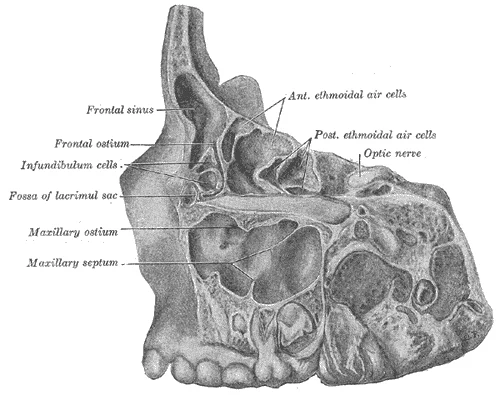
Long description
Specimen from a child eight years, eight months, and one day old. Lateral view of frontal, ethmoidal and maxillary sinus areas, the lateral portion of each having been removed by sagittal cuts. Note that the sinus frontalis developed directly from the infundibulum ethmoidale. Note also the incomplet

Figure 24: Specimen from a child eight years, eight months, and one day old.
Figure 25Gray861
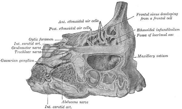
Long description
Specimen from a child one year, four months, and seven days old. Lateral view of frontal, ethmoidal, and maxillary sinus areas. (Davis.)

Figure 25: Specimen from a child one year, four months, and seven days old.
Figure 26Gray860
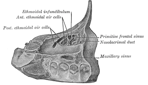
Long description
—Specimen from a child eight days old. By sagittal sections removing the lateral portion of frontal bone, lamina papyracea of ethmoid, and lateral portion of maxilla—the sinus maxillaris, cellulæ ethmoidales, anterior and posterior, infundibulum ethmoidale, and the primitive sinus frontalis are brou

Figure 26: —Specimen from a child eight days old.
This epithelium is derived from the endoderm of the nasal sacs. It contains olfactory receptor neurons, supporting cells, and basal cells. The olfactory nerve fibres pass through the cribriform plate to synapse in the olfactory bulb. [1]
The sphenopalatine artery, a branch of the maxillary artery, supplies the posterior and inferior parts. Kiesselbach’s plexus, located in the anterior inferior septum, is a common site for epistaxis. [1]
The olfactory sensation is mediated by the olfactory nerve (CN I). [1]
DefinitionKiesselbach’s plexus
A vascular network on the anterior inferior nasal septum, commonly involved in epistaxis.
| Artery | Source | Area Supplied |
|---|---|---|
| Sphenopalatine | Maxillary | Posterior and inferior septum and lateral wall |
| Anterior Ethmoidal | Ophthalmic | Anterior superior septum and lateral wall |
| Greater Palatine | Maxillary | Inferior nasal cavity and septum |
| Superior Labial | Facial | Anterior inferior septum |
Figure 27Gray170
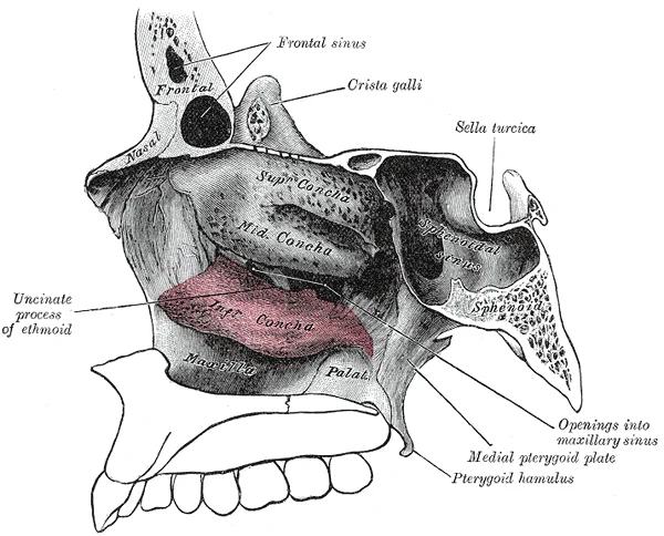
Long description
Lateral wall of right nasal cavity showing inferior concha in situ.

Figure 27: Lateral wall of right nasal cavity showing inferior concha in situ.
The nasal cavity is divided into two halves by the nasal septum. On the radiological image, the septum appears as a vertical midline structure, typically composed of the perpendicular plate of the ethmoid bone superiorly and the vomer inferiorly. Lateral to the septum are the nasal conchae, which project medially. The superior, middle, and inferior conchae are visible as curved bony shelves. The spaces beneath each concha are termed the superior, middle, and inferior meatuses, respectively. These meatuses serve as drainage pathways for the paranasal sinuses and the nasolacrimal duct. The inferior meatus receives the opening of the nasolacrimal duct, which drains tears from the eye. The middle meatus is particularly important as it receives drainage from the frontal, maxillary, and anterior ethmoid sinuses. The sphenoethmoidal recess, located above the superior concha, receives the sphenoid sinus.
Figure ior nasal concha (medial surface).
Figure ved.
The paranasal sinuses are air-filled cavities within the surrounding bones. The frontal sinuses are located within the frontal bone, above the orbits. On the coronal view, they appear as paired, variable-sized cavities superior to the nasal cavity. The ethmoid sinuses, or ethmoidal air cells, are located between the eyes, lateral to the superior nasal cavity. They are divided into anterior, middle, and posterior groups. The anterior and middle ethmoid cells drain into the middle meatus, while the posterior cells drain into the sphenoethmoidal recess. The maxillary sinuses are the largest of the paranasal sinuses, located within the body of the maxilla. They are situated lateral to the nasal cavity and inferior to the orbits. Their floor forms the roof of the oral cavity and is often close to the roots of the upper molars. The sphenoid sinus is located within the body of the sphenoid bone, posterior to the ethmoid sinuses. It lies inferior to the sella turcica and is separated from the optic nerves and internal carotid arteries by thin bone.
Understanding the relationships between these structures is vital for interpreting imaging. The lamina papyracea, a thin plate of the ethmoid bone, separates the ethmoid sinuses from the orbit. Fractures here can lead to orbital complications. The skull base, formed by the cribriform plate of the ethmoid bone, separates the nasal cavity from the anterior cranial fossa. This area is a potential route for cerebrospinal fluid leaks. The maxillary sinus is bounded inferiorly by the alveolar process of the maxilla. Radiological assessment of sinus opacification, mucosal thickening, or air-fluid levels is essential for diagnosing sinusitis. The patency of the osteomeatal complex, a functional unit in the middle meatus, is crucial for sinus drainage. Obstruction here can lead to recurrent sinus infections.
These conchae increase the surface area for warming and humidifying inspired air.
Figure 28Nose and nasal cavities
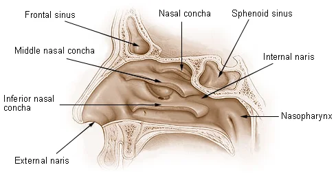
Long description
Diagram of the nose and the nasal cavities.

Figure 28: Diagram of the nose and the nasal cavities.
Below it, the middle nasal concha projects laterally, creating the middle meatus where the frontal, maxillary, and anterior ethmoid sinuses drain. The inferior nasal concha is a separate bone situated at the bottom, forming the inferior meatus which receives the nasolacrimal duct. The nasal septum lies medially, separating the two cavities.
DefinitionKey Anatomical Landmark
The osteomeatal complex is the functional unit of the maxillary, frontal, and anterior ethmoid sinuses, and its obstruction is a primary cause of sinusitis.
| Sinus | Drainage Site | Key Relation |
|---|---|---|
| Maxillary | Middle meatus | Alveolar process of maxilla |
| Frontal | Middle meatus | Orbit and anterior cranial fossa |
| Ethmoid (Anterior) | Middle meatus | Orbit and nasal cavity |
| Sphenoid | Superior meatus | Pituitary gland and optic nerve |
Sutures are immobile joints where dense fibrous connective tissue unites adjacent skull bones. The coronal suture joins the frontal bone to the right and left parietal bones. The sagittal [1]
DefinitionZygomatic Arch Composition
Remember the anterior component is the zygomatic bone, and the posterior is the temporal bone.
| Suture | Bones Joined |
|---|---|
| Coronal | Frontal and Parietal |
| Sagittal | Right and Left Parietal |
| Lambdoid | Occipital and Parietal/Temporal |
| Squamous | Squamous Temporal and Parietal |
NoteVocal Cord Anatomy
True vocal cords are attached to thyroid and arytenoid cartilages; false vocal cords are vestibular folds.
DefinitionRespiratory Membrane
This barrier consists of alveolar epithelium and capillary endothelium, facilitating gas exchange by simple diffusion. [2]
Clinical pearlSuture Strength
Irregular, twisting suture paths interlock bones, adding strength to the skull for brain protection. [1]
NoteMandibular Notch
The mandibular notch is the broad U-shaped curve between the coronoid and condylar processes. [1]
DefinitionConducting Zone Lining
Pseudostratified ciliated columnar epithelium with goblet cells lines the conducting zone, trapping debris in mucus. [2]
Students frequently confuse the structural composition of the skull with the functional units of the respiratory system. A primary error is assuming the hyoid bone is part of the skull. The hyoid bone is an independent bone that does not contact any other bone and thus is not part of the skull[1]. It is located in the upper neck near the level of the inferior mandible, with the tips of the “U” pointing posteriorly[1]. It serves as the base for the tongue above, and is attached to the larynx below and the pharynx posteriorly.
Another frequent mistake involves the boundaries of the cranial fossae. Students often misidentify the boundary between the anterior and middle cranial fossae. The lesser wings of the sphenoid bone form the prominent ledge that marks the boundary between the anterior and middle cranial fossae[1]. The anterior cranial fossa is the most anterior and the shallowest of the three cranial fossae[1]. It overlies the orbits and contains the frontal lobes of the brain[1]. Anteriorly, the anterior fossa is bounded by the frontal bone, which also forms the majority of the floor for this space[1].
Confusion also arises regarding the zygomatic bone. The zygomatic bone is also known as the cheekbone[1]. Each of the paired zygomatic bones forms much of the lateral wall of the orbit and the lateral-inferior margins of the anterior orbital opening[1]. The short temporal process of the zygomatic bone projects posteriorly, where it forms the anterior portion of the zygomatic arch[1]. This arch is a key landmark for distinguishing the temporal fossa from the infratemporal fossa.
The temporal fossa and infratemporal fossa are often conflated. On the lateral side of the brain case, above the level of the zygomatic arch, is a shallow space called the temporal fossa[1]. Below the level of the zygomatic arch and deep to the vertical portion of the mandible is another space called the infratemporal fossa[1]. Both the temporal fossa and infratemporal fossa contain muscles that act on the mandible during chewing[1].
Regarding the mandible, students may forget its developmental origin. The mandible forms the lower jaw and is the only moveable bone of the skull[1]. At the time of birth, the mandible consists of paired right and left bones, but these fuse together during the first year to form the single U-shaped mandible of the adult skull[1]. Each side of the mandible consists of a horizontal body and posteriorly, a vertically oriented ramus of the mandible[1]. The outside margin of the mandible, where the body and ramus come together is called the angle of the mandible[1].
Finally, the cartilaginous structures of the nose are often overlooked in dry skull anatomy. This cartilage also extends outward into the nose where it separates the right and left nostrils[1]. The septal cartilage is not found in the dry skull[1]. This distinction is vital when identifying nasal structures on radiological imaging versus physical examination.
This anatomical knowledge is applied at the bedside during airway management and facial trauma assessment. The mandible forms the lower jaw and is the only moveable bone of the skull[1]. Clinicians assess the angle of the mandible and the ramus for fractures. The hyoid bone is located in the upper neck and does not join with any other bone[1]. Its position is critical in strangulation cases or hyoid fractures during intubation.
In the operating theatre, understanding the temporal and infratemporal fossae is essential for accessing the mandibular nerve and muscles of mastication. Both the temporal fossa and infratemporal fossa contain muscles that act on the mandible during chewing[1]. Surgeons navigate these spaces carefully to avoid damaging the neurovascular bundles. The zygomatic bone is also known as the cheekbone[1]. Repair of zygomaticomaxillary complex fractures requires precise restoration of the lateral orbital wall and the zygomatic arch[1].
On imaging, the cranial fossae boundaries help localise intracranial pathology. The anterior cranial fossa is the most anterior and the shallowest of the three cranial fossae[1]. It overlies the orbits and contains the frontal lobes of the brain[1]. The lesser wings of the sphenoid bone form the prominent ledge that marks the boundary between the anterior and middle cranial fossae[1]. Radiologists use these landmarks to identify fractures or tumours.
In examinations, students must distinguish the bone composition of the skull. In the the adult, the skull consists of 22 individual bones, 21 of which are immobile and united into a single unit[1]. The 22nd bone is the mandible (lower jaw), which is the only moveable bone of the skull[1]. The brain case consists of eight bones[1]. These include the paired parietal and temporal bones, plus the unpaired frontal, occipital, sphenoid, and ethmoid bones[1].
The respiratory system's anatomical context is also tested. The major organs of the respiratory system function primarily to provide oxygen to body tissues for cellular respiration, remove the waste product carbon dioxide, and help to maintain acid-base balance. Portions of the respiratory system are also used for non-vital functions, such as sensing odors, speech production, and for straining, such as during childbirth or coughing. [2]
Airway Anatomy Assessment
- Assess Airway PatencyCheck for obstruction
- Question: Is Hyoid Fracture Suspected?
Yes
- Palpate Upper NeckCheck for tenderness near inferior mandible
No
- Evaluate MandibleCheck angle and ramus for fractures
NoteMandibular Development
At the time of birth, the mandible consists of paired right and left bones, but these fuse together during the first year to form the single U-shaped mandible of the adult skull[1].
The skull consists of 22 individual bones, 21 of which are immobile and united into a single unit[1]. The 22nd bone is the mandible (lower jaw), which is the only moveable bone of the skull[1]. The brain case consists of eight bones[1]. These include the paired parietal and temporal bones, plus the unpaired frontal, occipital, sphenoid, and ethmoid bones[1]. The anterior cranial fossa is the most anterior and the shallowest of the three cranial fossae[1]. It overlies the orbits and contains the frontal lobes of the brain[1]. The hyoid bone is an independent bone that does not contact any other bone and thus is not part of the skull[1]. It is a small U-shaped bone located in the upper neck near the level of the inferior mandible, with the tips of the “U” pointing posteriorly[1].
Answer each one in your head first, then open the answer.
How many bones are in the adult skull and which is the only moveable one?
Show answerHide answer
22 bones; the mandible is the only moveable bone[1].What forms the boundary between the anterior and middle cranial fossae?
Show answerHide answer
The lesser wings of the sphenoid bone[1].Is the hyoid bone part of the skull?
Show answerHide answer
No, it is an independent bone that does not contact any other bone[1].Where is the temporal fossa located relative to the zygomatic arch?
Show answerHide answer
Above the level of the zygomatic arch[1].What is the primary function of the respiratory system organs?
Show answerHide answer
To provide oxygen to body tissues for cellular respiration, remove carbon dioxide, and maintain acid-base balance.
- Betts JG, Young KA, Wise JA, et al. The Skull. OpenStax, Anatomy and Physiology 2e (CC BY-NC-SA 4.0). Accessed .
- Betts JG, Young KA, Wise JA, et al. Organs and Structures of the Respiratory System. OpenStax, Anatomy and Physiology 2e (CC BY-NC-SA 4.0). Accessed .
Figure credits
- Figure 1: Henry Vandyke Carter, via Wikimedia Commons, Public domain, Public domain.
- Figure 2: OpenStax College, via Wikimedia Commons, CC BY 3.0, CC BY 3.0.
- Figure 3: OpenStax College, via Wikimedia Commons, CC BY 3.0, CC BY 3.0.
- Figure 4: OpenStax College, via Wikimedia Commons, CC BY 3.0, CC BY 3.0. resized.
- Figure 5: Henry Vandyke Carter, via Wikimedia Commons, Public domain, Public domain.
- Figure 6: Henry Vandyke Carter, via Wikimedia Commons, Public domain, Public domain.
- Figure 7: Henry Vandyke Carter, via Wikimedia Commons, Public domain, Public domain.
- Figure 8: Henry Vandyke Carter, via Wikimedia Commons, Public domain, Public domain.
- Figure 9: OpenStax College, via Wikimedia Commons, CC BY 3.0, CC BY 3.0.
- Figure 10: Henry Vandyke Carter, via Wikimedia Commons, Public domain, Public domain.
- Figure 11: OpenStax College, via Wikimedia Commons, CC BY 3.0, CC BY 3.0.
- Figure 12: Henry Vandyke Carter, via Wikimedia Commons, Public domain, Public domain.
- Figure 13: Henry Vandyke Carter, via Wikimedia Commons, Public domain, Public domain.
- Figure 14: Henry Vandyke Carter, via Wikimedia Commons, Public domain, Public domain.
- Figure 15: Henry Vandyke Carter, via Wikimedia Commons, Public domain, Public domain.
- Figure 16: Henry Vandyke Carter, via Wikimedia Commons, Public domain, Public domain.
- Figure 17: Henry Vandyke Carter, via Wikimedia Commons, Public domain, Public domain.
- Figure 18: Henry Vandyke Carter, via Wikimedia Commons, Public domain, Public domain.
- Figure 19: OpenStax College, via Wikimedia Commons, CC BY 3.0, CC BY 3.0. resized.
- Figure 20: Henry Vandyke Carter, via Wikimedia Commons, Public domain, Public domain.
- Figure 21: Henry Vandyke Carter, via Wikimedia Commons, Public domain, Public domain.
- Figure 22: https://wellcomeimages.org/indexplus/obf_images/ce/49/005a8fdacde657da77e30f58fea1.jpg Gallery: https://wellcomeimages.org/indexplus/image/V0008038.html Wellcome Collection gallery (2018-03-30): https://wellcomecollection.org/works/y8afgr6y CC-BY-4.0, via Wikimedia Commons, CC BY 4.0, CC BY 4.0. resized.
- Figure 23: Henry Vandyke Carter, via Wikimedia Commons, Public domain, Public domain.
- Figure 24: Henry Vandyke Carter, via Wikimedia Commons, Public domain, Public domain.
- Figure 25: Henry Vandyke Carter, via Wikimedia Commons, Public domain, Public domain.
- Figure 26: Henry Vandyke Carter, via Wikimedia Commons, Public domain, Public domain.
- Figure 27: Henry Vandyke Carter, via Wikimedia Commons, Public domain, Public domain.
- Figure 28: This file is lacking author information., via Wikimedia Commons, Public domain, Public domain.
Educational content for medical students and doctors. It is not a substitute for clinical judgement or local guidelines.