Oral cavity
Anatomical structures of the oral cavity including palate, tongue, and floor of mouth.
The oral cavity, also known as the buccal cavity, is framed by the cheeks, tongue, and palate[1]. At the entrance lie the lips, or labia, which transition from skin to mucous membrane[1]. The cheeks form the sidewalls, containing the buccinator muscle between skin and non-keratinized stratified squamous epithelium[1].
The oral vestibule is the pocket-like space framed internally by gums and teeth, and externally by cheeks and lips[1]. The oral cavity proper extends from the gums to the fauces, the opening into the oropharynx[1]. The palate forms the arched roof, separating the oral and nasal cavities[1]. The anterior hard palate consists of maxillary and palatine bones, providing a rigid shelf for the tongue[1]. The posterior soft palate is composed mainly of skeletal muscle, allowing manipulation for yawning or swallowing[1].
DefinitionSoft Palate
Composed of skeletal muscle, it elevates during swallowing to prevent food entry into the nasal cavity.
The uvula hangs from the posterior soft palate, aiding in preventing nasal regurgitation[1]. The palatoglossal and palatopharyngeal arches flank the uvula, with palatine tonsils located between them[1]. The tongue rests on the floor of the mouth, attached to the mandible, styloid processes, and hyoid bone[1]. A medial septum divides the tongue into symmetrical halves[1].
| Papilla Type | Shape | Function |
|---|---|---|
| Fungiform | Mushroom-shaped | Contains taste buds |
| Filiform | Long and thin | Touch receptors, abrasive surface |
The lingual frenulum tethers the tongue to the floor of the mouth[1]. Minor salivary glands in the lips, cheeks, palate, and tongue secrete saliva, averaging 1 to 1.5 liters daily[1]. Saliva moistens food and initiates carbohydrate breakdown[1].
During deglutition, the tongue pushes the bolus backward against the palate into the oropharynx[1]. Clinical inspection of the fauces assesses for infection and tests cranial nerve function[1]. [2]
The vestibule lies between the cheeks and lips externally and the teeth and gums internally. The oral cavity proper is bounded by the teeth and gums on the sides and the hard and soft palates superiorly. The floor of the mouth forms the inferior boundary, supporting the tongue. The tongue itself occupies the central space, resting on the floor of the mouth. The teeth are embedded in the alveolar processes of the maxilla and mandible. The gingivae, or gums, are the soft tissues that line these alveolar processes and surround the necks of the teeth. The palate consists of the hard palate anteriorly, formed by the palatine processes of the maxilla and horizontal plates of the palatine bones, and the soft palate posteriorly, which is muscular. The supply to these structures involves specific nerves and blood vessels, while lymphatic drainage follows defined pathways to regional nodes. [1]
DefinitionOral Cavity
The mouth is the first organ of the upper alimentary canal, receiving food for mechanical and chemical digestion.
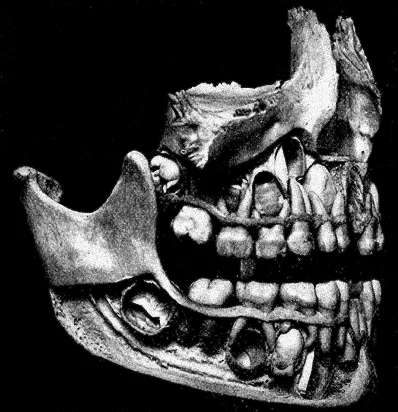
The teeth, or dentes, are organs similar to bones used to tear, grind, and mechanically break down food. Humans have two sets of teeth in their lifetime. The first set comprises 20 deciduous teeth, or baby teeth, which first begin to appear at about 6 months of age. Between approximately age 6 and 12, these are replaced by 32 permanent teeth. The teeth are secured in the alveolar processes, or sockets, of the maxilla and the mandible. The gingivae are soft tissues that line the alveolar processes and surround the necks of the teeth. Teeth are also held in their sockets by a connective tissue called the periodontal ligament. [1]
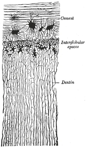
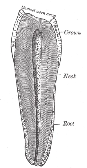
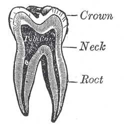
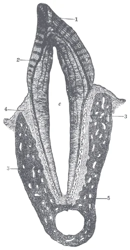
Each tooth has two main parts: the crown, which projects above the gum line, and the root, which is embedded within the maxilla and mandible. Both parts contain an inner pulp cavity with loose connective tissue, nerves, and blood vessels. The region of the pulp cavity running through the root is called the root canal. Surrounding the pulp cavity is dentin, a bone-like tissue. In the root, the dentin is covered by cementum, a layer of modified bone. In the crown, the dentin is covered by enamel, the hardest substance in the body. [1]
Figure 1Gray1008
Long description
Transverse section of a portion of the root of a canine tooth. X 300.

Figure 1: Transverse section of a portion of the root of a canine tooth (x 300).
Figure 2Gray1007
Long description
Vertical section of a premolar tooth. (Magnified.)

Figure 2: Vertical section of a premolar tooth (Magnified.).
Figure 3Gray1006
Long description
Vertical section of a molar tooth. Labelled illustration from Wikimedia Commons, used under its open licence.

Figure 3: Vertical section of a molar tooth.
Figure 4Gray1005
Long description
Vertical section of a tooth in situ. X 15. c is placed in the pulp cavity, opposite the neck of the tooth; the part above it is the crown, that below is the root. 1. Enamel with radial and concentric markings. 2. Dentin with tubules and incremental lines. 3. Cement or crusta petrosa, with bone corpu

Figure 4: Vertical section of a tooth in situ (x 15).
Figure 5Gray998
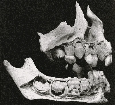
Long description
Maxillæ at about one year. (Noyes.). Labelled illustration from Wikimedia Commons, used under its open licence.

Figure 5: Maxillæ at about one year (Noyes.).
The palate forms the roof of the mouth. The hard palate provides a rigid structure for the tongue to press against food during swallowing. The soft palate is muscular and rises during swallowing to close off the nasopharynx. The floor of the mouth supports the tongue and contains the openings of the submandibular and sublingual salivary glands. The tongue begins mechanical digestion in the mouth. Saliva begins chemical digestion here. Saliva is essentially 99.4 percent water. The remaining 0.6 percent includes ions, glycoproteins, enzymes, growth factors, and waste products. Salivary amylase initiates the breakdown of carbohydrates. Bicarbonate and phosphate ions maintain saliva at a pH between 6.35 and 6.85. Salivary mucus lubricates food, facilitating movement and bolus formation. Saliva contains immunoglobulin A to prevent microbial penetration and lysozyme for antimicrobial action. [1]
| Gland | Nerve Supply | Ganglion |
|---|---|---|
| Parotid | Glossopharyngeal | Otic |
| Submandibular | Facial | Pterygopalatine |
| Sublingual | Facial | Pterygopalatine |
Neurons in the salivary nuclei project as preganglionic fibers. The parasympathetic fibers of the glossopharyngeal nerve synapse in the otic ganglion, projecting to the parotid gland. Salivation is based on a visceral reflex arc. [2]
Clinical pearlDigestion Start
Mechanical digestion starts with teeth; chemical digestion starts with salivary amylase.
The oral cavity is a complex anatomical space defined by its boundaries and the structures that traverse or border it. Understanding these relations is critical for localising pathology and planning surgical interventions. The cavity is bounded anteriorly by the lips, laterally by the cheeks, superiorly by the hard and soft palates, inferiorly by the floor of the mouth, and posteriorly by the oropharyngeal isthmus. The roof is formed by the palate, which separates the oral cavity from the nasal cavity above. The floor is formed by the mylohyoid muscle, the genioglossus, and the geniohyoid muscles, supporting the tongue.
The lingual nerve runs superior and lateral to the hypoglossal nerve for much of its course, providing sensory innervation to the anterior two-thirds of the tongue. The chorda tympani, a branch of the facial nerve, joins the lingual nerve and provides parasympathetic fibres to the submandibular and sublingual glands. The genioglossus muscle, responsible for protruding the tongue, is innervated by the hypoglossal nerve. If the hypoglossal nerve is damaged on one side, the tongue will deviate towards the side of the lesion upon protrusion due to the unopposed action of the healthy genioglossus muscle. This clinical sign is a key localising feature for hypoglossal nerve lesions. The close proximity of the lingual and hypoglossal nerves means that lesions in this region, such as those in the medulla or along the nerve's course, can affect both sensory and motor functions of the tongue. The trigeminal nerve controls the muscles of chewing, which are tested for stretch reflexes, and its branches are closely related to the oral cavity structures. The facial nerve's motor functions are usually obvious if facial expressions are compromised, but its parasympathetic fibres via the chorda tympani are crucial for salivary gland function. The cranial nerves serve the head and neck, and their examination allows directed tests of forebrain and brain stem structures. The special senses are served through the cranial nerves, as well as the general senses of the head and neck. The movement of the eyes, face, tongue, throat, and neck are all under the control of cranial nerves. Preganglionic parasympathetic nerve fibers that control pupillary size, salivary glands, and the thoracic and upper abdominal viscera are found in four of the nerves. Tests of these functions can provide insight into damage to specific regions of the brain stem and may uncover deficits in adjacent regions. If the nerve is not completely severed, the anosmia may be temporary as new neurons can eventually reconnect. Failure to discriminate these stimuli can localize problems within the brain stem. If a patient cannot recognize a painful stimulus, that might indicate damage to the spinal trigeminal nucleus in the medulla. The medulla also contains important regions that regulate the cardiovascular, respiratory, and digestive systems, as well as being the pathway for ascending and descending tracts between the brain and spinal cord. Damage, such as a stroke, that results in changes in sensory discrimination may indicate these unrelated regions are affected as well. As the name suggests, the abducens nerve is responsible for abducting the eye, which it controls through contraction of the lateral rectus muscle. The oculomotor nerve controls all the other extraocular muscles, as well as a muscle of the upper eyelid. Movements of the two eyes need to be coordinated to locate and track visual stimuli accurately. Testing eye movement is simply a matter of having the patient track the tip of a pen as it is passed through the visual field. This may appear similar to testing visual field deficits related to the optic nerve, but the difference is that the patient is asked to not move the eyes while the examiner moves a stimulus into the peripheral visual field. Here, the extent of movement is the point of the test. The examiner is watching for conjugate movements representing proper function of the related nuclei and the MLF. Failure of one eye to abduct while the other adducts in a horizontal movement is referred to as internuclear ophthalmoplegia. When this occurs, the patient will experience diplopia, or double vision, as the two eyes are temporarily pointed at different stimuli. The final aspect of testing eye movements is to move the tip of the pen in toward the patient’s face. As visual stimuli move closer to the face, the two medial recti muscles cause the eyes to move in the one nonconjugate movement that is part of gaze control. When the two eyes move to look at something closer to the face, they both adduct, which is referred to as convergence. To keep the stimulus in focus, the eye also needs to change the shape of the lens, which is controlled through the parasympathetic fibers of the oculomotor nerve. The change in focal power of the eye is referred to as accommodation. Accommodation ability changes with age; focusing on nearer objects, such as the written text of a book or on a computer screen, may require corrective lenses later in life. These two superficial muscles are important for changing the position of the head. Both muscles also receive input from cervical spinal nerves. Along with the spinal accessory nerve, these nerves contribute to elevating the scapula and clavicle through the trapezius, which is tested by asking the patient to shrug both shoulders, and watching for asymmetry. Movement of the head and neck using the sternocleidomastoid and trapezius muscles is controlled by the accessory nerve. Flexing of the neck and strength testing of those muscles reviews the function of that nerve. The trigeminal nerve controls the muscles of chewing, which are tested for stretch reflexes. Motor functions of the facial nerve are usually obvious if facial expressions are compromised, but can be tested by having the patient raise their eyebrows, smile, and frown. Movements of the tongue, soft palate, or superior pharynx can be observed directly while the patient swallows, while the gag reflex is elicited, or while the patient says repetitive consonant sounds. [2]
Figure 6Gray1013
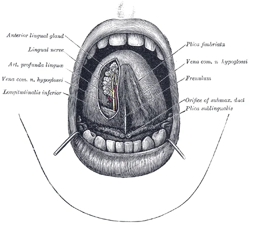
Long description
The mouth cavity. The apex of the tongue is turned upward, and on the right side a superficial dissection of its under surface has been made.

Figure 6: The mouth cavity.
Clinical pearlClinical Pearl
Deviation of the tongue to one side on protrusion indicates a lesion of the hypoglossal nerve on that same side.
| Muscle Group | Muscles | Nerve Supply | Action |
|---|---|---|---|
| Extrinsic | Genioglossus, Hyoglossus, Styloglossus, Palatoglossus | Hypoglossal (CN XII) except Palatoglossus | Protrusion, depression, retraction, elevation |
| Intrinsic | Superior longitudinal, Inferior longitudinal, Transverse, Vertical | Hypoglossal (CN XII) | Change shape of tongue |
DefinitionHypoglossal Nerve
The hypoglossal nerve is the motor nerve that controls the muscles of the tongue, except for the palatoglossus muscle, which is controlled by the vagus nerve. [2]
Exam trapCommon Trap
Do not confuse the hypoglossal nerve with the lingual nerve. The hypoglossal is motor to the tongue muscles, while the lingual is sensory to the anterior two-thirds of the tongue.
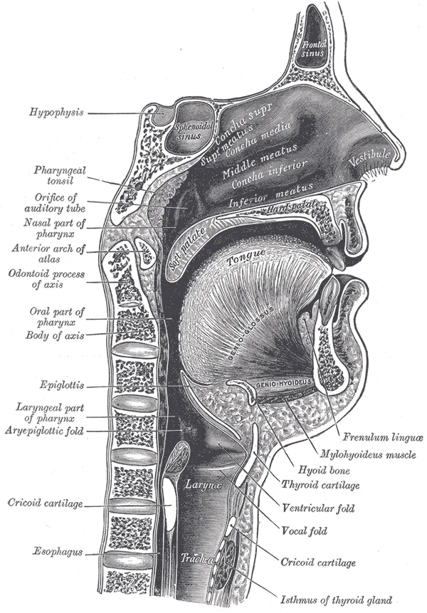
The oral cavity's internal structure includes several important components that facilitate essential functions such as breathing, speaking, swallowing, and digestion. The pharynx is a short tube of skeletal muscle lined with a mucous membrane that extends from the posterior oral and nasal cavities to the openings of the esophagus and larynx. This structure is divided into three distinct subdivisions: the nasopharynx, oropharynx, and laryngopharynx. [1]
The nasopharynx is the most superior subdivision and is exclusively involved in breathing and speech functions. Positioned inferior to the nasopharynx, the oropharynx serves both breathing and digestion purposes and continues below with the laryngopharynx. The laryngopharynx connects inferiorly to the esophagus while its anterior portion connects to the larynx, allowing air to flow into the bronchial tree. [1]
During swallowing, important protective reflexes are activated. The soft palate and uvula rise reflexively to close off the entrance to the nasopharynx, preventing food from entering the nasal cavity. Simultaneously, the larynx is pulled superiorly and the cartilaginous epiglottis folds inferiorly, covering the glottis (the opening to the larynx). This process effectively blocks access to the trachea and bronchi, ensuring food enters the esophagus rather than the respiratory tract. When food accidentally enters the trachea, the body responds with a cough reflex that typically forces the food back into the pharynx. [1]
The vascular architecture of the oral cavity is derived primarily from the external carotid artery, ensuring a rich blood supply necessary for thermoregulation and tissue repair. The principal vessel is the facial artery, which arises from the external carotid artery in the neck. It courses anteriorly across the mandible, where it can be palpated against the bone, before entering the face. Within the oral cavity, its submental and inferior labial branches supply the lower lip and chin region. The superior labial branch ascends to supply the upper lip and the nasal septum. This arterial network anastomoses extensively with branches from the contralateral side and with the internal maxillary artery, providing collateral circulation.
DefinitionFacial Artery
The facial artery is the main blood supply to the lower face and oral structures, branching from the external carotid artery.
Lymphatic drainage from the oral cavity follows a predictable pattern that mirrors the arterial supply. Lymph from the central part of the lower lip and the floor of the mouth drains into the submental lymph nodes. These nodes are located in the submental triangle, beneath the anterior belly of the digastric muscle. From here, lymph flows into the submandibular lymph nodes, situated beneath the mandible. The submandibular nodes receive drainage from the lateral aspects of the lower lip, the cheeks, and the anterior part of the tongue. Efferent vessels from the submandibular nodes pass to the deep cervical lymph nodes, specifically the jugulodigastric node. This node is located at the angle of the mandible, beneath the sternocleidomastoid muscle. [1]
Figure 7Gray1014
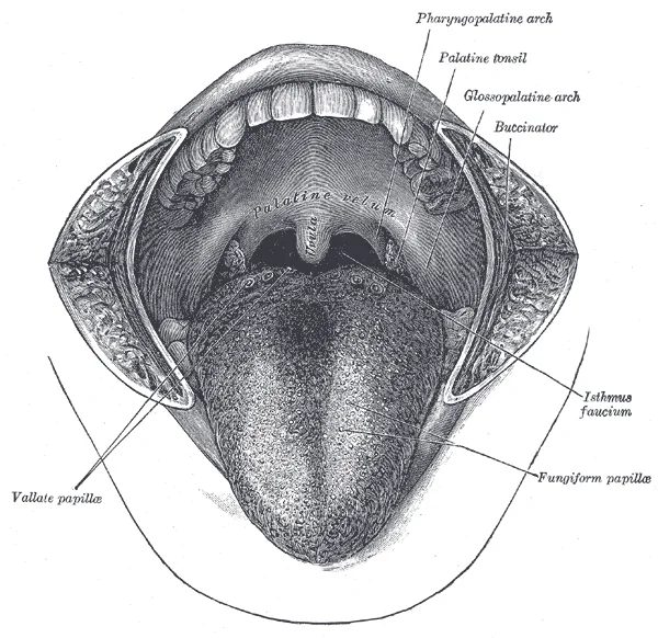
Long description
The mouth cavity. The cheeks have been slit transversely and the tongue pulled forward.

Figure 7: The mouth cavity.
| Structure | Primary Drainage Node | Secondary Drainage Node |
|---|---|---|
| Central Lower Lip | Submental | Submandibular |
| Lateral Lower Lip | Submandibular | Deep Cervical |
| Anterior Tongue | Submandibular | Deep Cervical |
| Floor of Mouth | Submandibular | Deep Cervical |
The clinical significance of this drainage pattern is profound. Infections or malignancies in the oral cavity often spread to these nodal basins. The submandibular nodes are key for lesions of the anterior tongue and floor of the mouth. The deep cervical nodes, especially the jugulodigastric node, are the final common pathway for most oral lymphatic drainage. Enlargement of these nodes can indicate pathology in the oral cavity. Therefore, examination of these lymph node groups is essential in any clinical assessment of oral disease. The rich vascular and lymphatic networks ensure rapid dissemination of pathogens and malignant cells, underscoring the importance of early diagnosis and treatment. Understanding these anatomical relationships allows clinicians to predict the spread of disease and plan appropriate surgical or medical interventions.
The venous drainage of the oral cavity is complex, reflecting its rich vascular supply and critical role in head and neck physiology. Understanding this system requires tracing blood from the mucosal capillaries through increasingly larger veins until it reaches the systemic circulation. The primary vessel responsible for this task is the facial vein, which serves as the main collector for the anterior structures of the face and mouth.
The vein then crosses the inferior border of the mandible, where it receives tributaries from the submental and submandibular regions. As it continues inferiorly, it joins the anterior division of the retromandibular vein to form the common facial vein. This common trunk then empties into the internal jugular vein.
DefinitionFacial Vein
The facial vein is a major vessel that drains blood from the face and anterior oral cavity, communicating with the cavernous sinus via the ophthalmic veins and deep facial vein.
The pterygoid plexus is another crucial component of oral venous drainage. This network of veins is located in the infratemporal fossa, surrounding the lateral pterygoid muscle. It receives blood from the deep structures of the face, including the teeth and gums, via the maxillary and middle temporal veins. The plexus communicates with the facial vein through the deep facial vein and with the cavernous sinus through the emissary veins passing through the foramen ovale and foramen lacerum. This anatomical arrangement explains why infections in the 'danger triangle' of the face can lead to serious intracranial complications.
| Structure | Origin | Termination | Clinical Significance |
|---|---|---|---|
| Facial Vein | Medial angle of eye | Internal jugular vein | Route for septic emboli to cavernous sinus |
| Pterygoid Plexus | Deep facial veins | Maxillary vein | Communication with cavernous sinus via emissary veins |
| Lingual Vein | Tongue muscles | Internal jugular vein | Drains anterior two-thirds of tongue |
| Submental Vein | Submental region | Facial vein | Drains chin and lower lip |
The lingual vein drains the tongue, particularly the anterior two-thirds. It arises from the deep lingual veins and runs alongside the lingual artery. It typically empties directly into the internal jugular vein, although variations exist where it may join the facial vein. The submental vein drains the chin and lower lip, ascending to join the facial vein. These vessels ensure efficient removal of deoxygenated blood from the highly active tissues of the oral cavity.
Exam trapInfection Spread
Do not assume all facial infections drain directly to the heart; the connection to the cavernous sinus via valveless veins allows retrograde spread of infection.
The internal jugular vein serves as the final common pathway for most of these tributaries. It receives the common facial vein, the lingual vein, and the superior thyroid vein. This convergence ensures that blood from the entire oral and facial region is efficiently returned to the right atrium of the heart via the brachiocephalic vein and superior vena cava.
The lymphatic drainage of the oral cavity is complex and follows specific anatomical rules that are critical for understanding the spread of pathology. The pattern of drainage is largely determined by the embryological origin and the specific region of the oral cavity involved.
The anterior two-thirds of the tongue drains primarily to the submandibular lymph nodes. These nodes are located within the submandibular triangle, beneath the mylohyoid muscle. From the submandibular nodes, efferent vessels pass to the deep cervical lymph nodes. This pathway is significant because malignancies in this region often metastasize to the submandibular nodes before reaching the deep cervical chain. The submental lymph nodes also receive drainage from the central part of the tongue, specifically the tip and the anterior midline. These nodes lie between the anterior bellies of the digastric muscles. Drainage from the submental nodes proceeds directly to the submandibular nodes or to the deep cervical nodes. [1]
The posterior one-third of the tongue has a different drainage pattern. It drains directly into the deep cervical lymph nodes. This direct drainage bypasses the submandibular nodes. The deep cervical nodes are located along the internal jugular vein. This anatomical difference is crucial because it affects the staging and management of tongue cancers. The base of the tongue is part of the posterior third and shares this drainage pattern. [1]
The floor of the mouth drains to the submandibular and submental lymph nodes. The anterior part of the floor of the mouth drains to the submental nodes. The posterior part drains to the submandibular nodes. From these nodes, lymph flows into the deep cervical lymph nodes. The rich lymphatic supply in this area makes it prone to early metastasis.
The buccal mucosa drains to the buccal lymph nodes, which are located near the masseter muscle. These nodes then drain into the parotid lymph nodes or the deep cervical nodes. The upper and lower gums drain to the submandibular lymph nodes. The upper gums may also drain to the submental nodes. [1]
Clinical pearlClinical Pearl
Always consider the primary drainage site when evaluating neck masses in patients with oral lesions.
The oral cavity receives a complex innervation involving both somatic and autonomic components, facilitating sensation, movement, and secretion. Understanding this requires distinguishing between general sensation, taste, and motor control of the muscles involved in mastication and deglutition.
General somatic afferent sensation from the anterior two-thirds of the tongue is provided by the lingual nerve, a branch of the mandibular division of the trigeminal nerve. The trigeminal nerve is a mixed nerve that carries the general somatic senses from the head, similar to those coming through spinal nerves from the rest of the body. This includes touch, pain, and temperature. In contrast, taste sensation is relayed to the brain stem through fibers of the facial and glossopharyngeal nerves. The facial nerve carries taste from the anterior two-thirds via the chorda tympani, while the glossopharyngeal nerve handles the posterior third. The facial and glossopharyngeal nerves convey gustatory stimulation to the brain. [2]
Autonomic innervation controls salivary secretion. Parasympathetic fibres travel with the facial nerve to the submandibular and sublingual glands, and with the glossopharyngeal nerve to the parotid gland. Sympathetic fibres modulate blood flow and viscosity.
The tongue is a workhorse, facilitating ingestion, mechanical digestion, chemical digestion (lingual lipase), sensation (of taste, texture, and temperature of food), swallowing, and vocalization. Beneath its mucous membrane covering, each half of the tongue is composed of the same number and type of intrinsic and extrinsic skeletal muscles. [1]
The intrinsic muscles (those within the tongue) are the longitudinalis inferior, longitudinalis superior, transversus linguae, and verticalis linguae muscles. These allow you to change the size and shape of your tongue, as well as to stick it out, if you wish. Having such a flexible tongue facilitates both swallowing and speech. [1]
The extrinsic muscles of the tongue are the palatoglossus, hyoglossus, styloglossus, and genioglossus muscles. These muscles originate outside the tongue and insert into connective tissues within the tongue. The mylohyoid is responsible for raising the tongue, the hyoglossus pulls it down and back, the styloglossus pulls it up and back, and the genioglossus pulls it forward. Working in concert, these muscles perform three important digestive functions in the mouth: (1) position food for optimal chewing, (2) gather food into a bolus (rounded mass), and (3) position food so it can be swallowed. [1]
Figure 8Gray1028
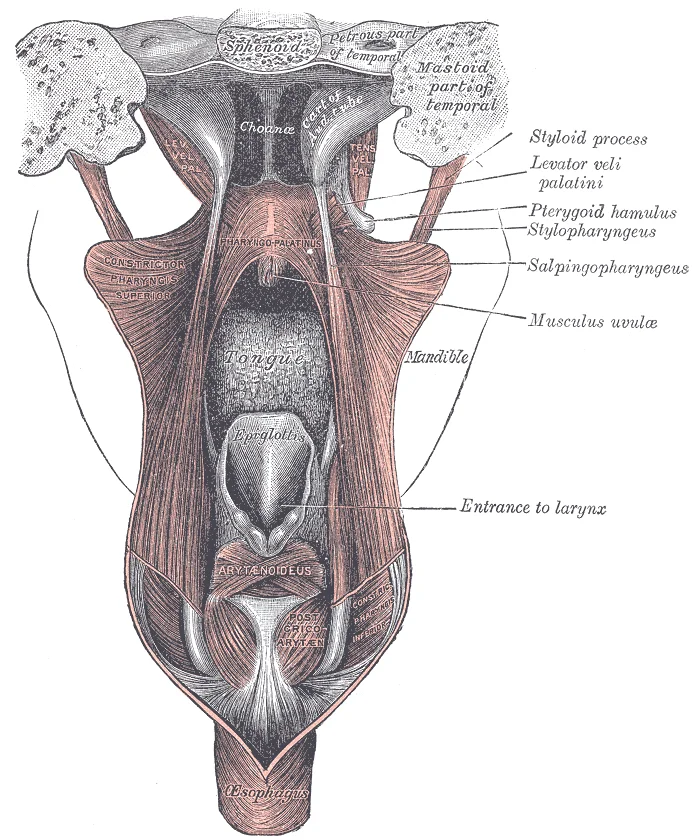
Long description
Dissection of the muscles of the palate from behind.

Figure 8: Dissection of the muscles of the palate from behind.
All intrinsic and extrinsic muscles of the tongue are innervated by the hypoglossal nerve (CN XII), except the palatoglossus, which is innervated by the vagus nerve (CN X) via the pharyngeal plexus. Loss of function of the tongue can interfere with speech and swallowing. [2]
Pain from the oral cavity can be referred due to shared dermatomal or segmental innervation. For instance, pathology in the tongue may present as ear pain because the glossopharyngeal nerve (CN IX) and vagus nerve (CN X) share pathways with cervical nerves that also supply the ear. This phenomenon underscores the importance of considering distant structures when evaluating oral pain.
Clinical pearlClinical Pearl
Taste and general sensation are carried by different nerves; testing both is essential for localising lesions.
| Muscle | Origin | Insertion | Action | Nerve Supply |
|---|---|---|---|---|
| Genioglossus | Mandible | Tongue | Protrudes tongue | Hypoglossal (CN XII) |
| Hyoglossus | Hyoid bone | Tongue | Depresses and retracts tongue | Hypoglossal (CN XII) |
| Styloglossus | Styloid process | Tongue | Elevates and retracts tongue | Hypoglossal (CN XII) |
| Palatoglossus | Palatine aponeurosis | Tongue | Elevates posterior tongue | Vagus (CN X) |
The oral cavity is lined by mucosa, which varies significantly depending on location and function. This variation is primarily determined by the type of epithelium present. Areas subject to abrasion, such as the hard palate and gingiva, are lined by non-keratinised or keratinised stratified squamous epithelium. In contrast, the soft palate, floor of the mouth, and ventral surface of the tongue often feature non-keratinised epithelium to allow for flexibility and moisture retention. The underlying lamina propria provides structural support and houses blood vessels and nerves.
DefinitionMucosa
The lining of the oral cavity, consisting of epithelium and lamina propria, adapted for protection, sensation, and secretion.
For a detailed exploration of cellular structures, staining characteristics, and specific histological features of the oral tissues, please refer to the dedicated histology page. This resource expands on the basic principles outlined here, providing microscopic views essential for understanding pathological changes.
The oral cavity serves as the initial segment of the digestive tract. It is bounded anteriorly by the lips, laterally by the cheeks, superiorly by the hard and soft palates, and inferiorly by the floor of the mouth. The tongue occupies the central space, playing a crucial role in mechanical digestion and speech. The vestibule is the potential space between the lips/cheeks and the teeth/gums. The oral cavity proper lies within the dental arches.
| Boundary | Structure |
|---|---|
| Anterior | Lips (Labia) |
| Lateral | Cheeks (Buccae) |
| Superior | Hard and Soft Palate |
| Inferior | Floor of the Mouth |
| Posterior | Oropharyngeal Isthmus |
The mucosa of the oral cavity is continuous with the skin at the lips and with the pharyngeal mucosa at the oropharyngeal isthmus. This continuity ensures a seamless transition for food and air. The submucosa beneath the mucosa contains minor salivary glands, which secrete mucus to lubricate the oral contents. These glands are particularly dense in the lips, cheeks, and soft palate.
NoteClinical Relevance
Minor salivary gland cysts, known as mucoceles, often result from trauma to these ducts, particularly in the lower lip.
Understanding the histological basis of the oral mucosa is fundamental for diagnosing lesions and understanding physiological functions. The presence of keratin provides protection against mechanical stress, while non-keratinised areas allow for greater permeability and sensation. The vascular supply is rich, facilitating rapid healing and immune response. Nerve endings are abundant, providing tactile, thermal, and nociceptive sensations.
The transition from oral mucosa to pharyngeal mucosa marks the beginning of the digestive process's next phase. As food is manipulated by the tongue and mixed with saliva, it forms a bolus ready for swallowing. The histological features of the oral cavity support this preparatory role, ensuring durability and sensitivity.
The oral cavity arises from the stomodeum, an ectodermal depression that communicates with the foregut. Initially separated by the buccopharyngeal membrane, this barrier ruptures to establish continuity between the primitive mouth and the pharynx. The roof of the stomodeum forms the primary palate, while the lateral palatine shelves grow medially to form the secondary palate. Fusion of these shelves occurs posterior to the incisive foramen, creating the hard palate. The tongue develops from multiple prominences around the first and second pharyngeal arches. The anterior two-thirds derive from the first arch, specifically the lateral lingual swellings and the tuberculum impar. The posterior one-third arises from the hypobranchial eminence, derived from the third and fourth arches. The line of demarcation is the sulcus terminalis, a V-shaped groove. At the apex of this V lies the foramen cecum, the remnant of the thyroglossal duct. The muscles of the tongue are derived from occipital myotomes, except for the palatoglossus, which originates from the fourth arch. This distinction is clinically significant as it dictates the nerve supply. The anterior two-thirds receive general sensation via the lingual nerve, a branch of the mandibular division of the trigeminal nerve. Taste sensation in this region is carried by the chorda tympani, a branch of the facial nerve. The posterior one-third receives both general and special sensory innervation from the glossopharyngeal nerve. The development of the floor of the mouth involves the submandibular and sublingual glands, which bud from the oral epithelium. The sublingual gland develops from multiple small ducts, whereas the submandibular gland has a single duct. The palatine tonsils develop from the second pharyngeal pouch, lined by endoderm. The crypts of the tonsils are invaginations of the surface epithelium. The dental lamina forms from the ectoderm of the stomodeum and gives rise to the tooth buds. Each tooth develops from the enamel organ, dental papilla, and dental follicle. The enamel is derived from ectoderm, while the dentin and pulp are derived from neural crest cells. The eruption of teeth follows a predictable sequence, with primary teeth appearing first. The permanent teeth replace the primary teeth in a specific order. The maxillary and mandibular processes fuse to form the upper and lower jaws. Failure of fusion can result in cleft lip or palate. The cleft lip results from failure of the medial nasal prominence to fuse with the maxillary prominence. Cleft palate results from failure of the palatine shelves to fuse. These defects can affect feeding, speech, and hearing. The oral mucosa is lined by stratified squamous epithelium, which is keratinized in some areas. The gingiva is keratinized to withstand masticatory forces. The floor of the mouth and ventral tongue are non-keratinized. The salivary glands develop as outgrowths of the oral epithelium. The parotid gland is the largest and develops last. The submandibular and sublingual glands develop earlier. The innervation of the oral cavity is complex. The trigeminal nerve provides general sensation. The facial, glossopharyngeal, and vagus nerves provide taste and visceral sensation. The motor supply to the muscles of mastication is from the mandibular nerve. The muscles of facial expression are supplied by the facial nerve. The development of the oral cavity is a complex process involving multiple embryological structures. Understanding this development is essential for diagnosing congenital anomalies. The stomodeum is the precursor to the oral cavity proper. The pharyngeal arches contribute to the formation of the tongue and pharynx. The neural crest cells migrate to the facial region to form the skeletal structures. The ectoderm and endoderm interact to form the oral mucosa. The mesoderm contributes to the muscles and connective tissue. The vascular supply develops from the aortic arches. The venous drainage follows the arterial supply. The lymphatic drainage is to the cervical lymph nodes. The development of the oral cavity is complete by the end of the embryonic period. The fetal period involves growth and maturation. The teeth continue to develop throughout childhood. The eruption of permanent teeth continues into adolescence. The oral cavity is a dynamic structure that changes throughout life. The understanding of its development provides a foundation for clinical practice.
Figure 9Gray1027
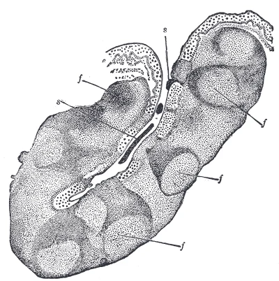
Long description
Section through one of the crypts of the tonsil. (Stöhr.) Magnified. e. Stratified epithelium of general surface, continued into crypt. f, f. Nodules of lymphoid tissue—opposite each nodule numbers of lymph cells are passing into or through the epithelium. s, s. Cells which have thus escaped to mix

Figure 9: Section through one of the crypts of the tonsil (Stöhr.) Magnified.
Figure 10Gray1015
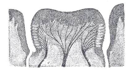
Long description
Circumvallate papilla in vertical section, showing arrangement of the taste-buds and nerves.

Figure 10: Circumvallate papilla in vertical section, showing arrangement of the taste-buds and nerves.
Figure 11Gray1012
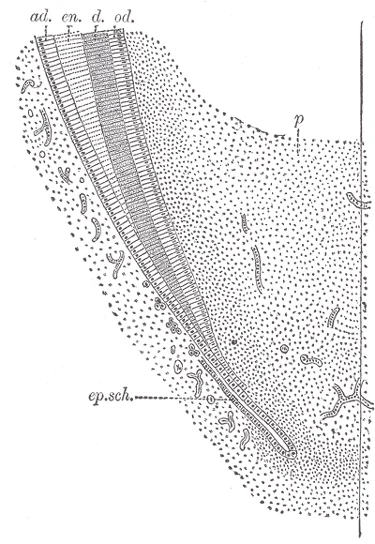
Long description
Longitudinal section of the lower part of a growing tooth, showing the extension of the layer of adamantoblasts beyond the crown to mark off the limit of formation of the dentin of the root. (Röse.) ad. Adamantoblasts, continuous below with ep.sch., the epithelial sheath of Hertwig. d. Dentin. en. E

Figure 11: Longitudinal section of the lower part of a growing tooth, showing the extension of the layer of adamantoblasts beyond the crown to mark off the limit of formation of the dentin of the root (Röse.) ad.
Figure 12Gray1017
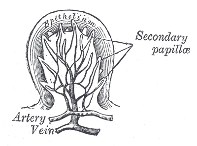
Long description
Section of a fungiform papilla. Magnified.

Figure 12: Section of a fungiform papilla (magnified).
Figure 13Gray1000
Long description
The complete temporary dentition and the first permanent molar. Note the relation of the bicuspids to the temporary molars. (In the seventh year.) (Noyes.)

Figure 13: The complete temporary dentition and the first permanent molar.
Figure 14Gray994
Long description
Sagittal section of nose mouth, pharynx, and larynx.

Figure 14: Sagittal section of nose mouth, pharynx, and larynx.
Understanding the oral cavity on imaging requires correlating standard anatomical landmarks with their radiological appearances. We begin by considering the bony framework, which provides the structural context for soft tissue evaluation. The maxilla and mandible form the superior and inferior boundaries, respectively. The teeth appear as high-density structures within the alveolar processes.
The genioglossus muscle is identifiable anteriorly, attaching to the mental spine. Posteriorly, the base of the tongue approaches the epiglottis. The floor of the mouth is formed by the mylohyoid muscle, which creates a muscular diaphragm. Above this, the sublingual glands are situated. Laterally, the buccinator muscle lines the inner aspect of the cheek.
DefinitionRadiological Landmark
Key anatomical relationships are best understood by identifying the tongue, floor of mouth, and surrounding musculature in cross-section.
The soft tissues of the oral cavity are critical for assessing pathology. The tongue is the largest muscle in the oral cavity, composed of intrinsic and extrinsic muscles. On imaging, the tongue appears as a homogeneous soft tissue mass. The extrinsic muscles, including the genioglossus, hyoglossus, styloglossus, and palatoglossus, originate from surrounding bony structures and insert into the tongue. These muscles are responsible for protrusion, retraction, and depression of the tongue.
| Muscle | Origin | Insertion | Action | Nerve Supply |
|---|---|---|---|---|
| Genioglossus | Mental spine of mandible | Tongue | Protrusion | Hypoglossal nerve |
| Hyoglossus | Hyoid bone | Tongue | Depresses tongue | Hypoglossal nerve |
| Styloglossus | Styloid process | Tongue | Elevates and retracts | Hypoglossal nerve |
| Palatoglossus | Palatine aponeurosis | Tongue | Elevates posterior tongue | Vagus nerve |
The salivary glands are also visible on imaging. The sublingual gland is smaller and lies superior to the mylohyoid, in the floor of the mouth. The parotid gland is the largest, located posterior to the mandibular ramus. Understanding these relationships allows for accurate localization of lesions. For example, a mass in the floor of the mouth may involve the sublingual gland or the mylohyoid muscle. A mass lateral to the tongue may involve the buccinator or parotid duct.
NoteClinical Correlation
Lesions in the oral cavity often present with pain or difficulty swallowing. Imaging helps determine the extent of disease and involvement of adjacent structures.
The neurovascular supply is also relevant. The lingual nerve provides sensory innervation to the anterior two-thirds of the tongue. The hypoglossal nerve provides motor innervation to all intrinsic and extrinsic muscles except the palatoglossus. The facial artery and vein supply the region. On imaging, these vessels appear as flow voids on MRI or enhance with contrast on CT. Recognizing these structures is essential for surgical planning and biopsy guidance.
The oral cavity serves as the initial segment of the gastrointestinal and respiratory tracts, bounded anteriorly and laterally by the lips and cheeks, superiorly by the hard and soft palates, and inferiorly by the floor of the mouth. Understanding its topography is essential for diagnosing lesions and planning surgical interventions.
The anterior boundary is formed by the lips, which consist of skin externally and mucous membrane internally, connected by the orbicularis oris muscle. This muscle encircles the oral aperture and is responsible for closing the lips. The lateral walls are formed by the cheeks, which contain the buccinator muscle. The buccinator compresses the cheeks against the gums, assisting in mastication and preventing food from accumulating in the vestibule. The superior boundary comprises the hard palate anteriorly and the soft palate posteriorly. The hard palate is supported by the palatine processes of the maxillae and the horizontal plates of the palatine bones. The soft palate is a muscular curtain that elevates during swallowing to close off the nasopharynx. The inferior boundary is the floor of the mouth, formed by the mylohyoid muscles, which support the tongue. [1]
The tongue is a muscular organ essential for taste, swallowing, and speech. It is divided into a mobile anterior two-thirds (body) and a fixed posterior one-third (root). The body lies on the floor of the mouth, while the root extends to the epiglottis. The dorsal surface is covered with papillae, including filiform, fungiform, circumvallate, and foliate types, which house taste buds. The ventral surface is smooth and attached to the floor of the mouth by the frenulum. The tongue's movement is controlled by intrinsic muscles, which alter its shape, and extrinsic muscles, which change its position. [1]
| Muscle | Origin | Insertion | Action | Nerve Supply |
|---|---|---|---|---|
| Genioglossus | Superior mental spine | Tongue and hyoid | Protrudes and depresses tongue | Hypoglossal nerve |
| Hyoglossus | Hyoid bone | Side of tongue | Depresses and retracts tongue | Hypoglossal nerve |
| Styloglossus | Styloid process | Side of tongue | Elevates and retracts tongue | Hypoglossal nerve |
| Palatoglossus | Palatine aponeurosis | Side of tongue | Elevates posterior tongue | Vagus nerve |
Three pairs of major salivary glands secrete saliva into the oral cavity. Its duct, Stensen’s duct, crosses the masseter and pierces the buccinator to open opposite the second upper molar. The submandibular gland lies beneath the mandible. Wharton’s duct runs forward to open at the sublingual caruncle on either side of the frenulum. The sublingual gland is situated beneath the mucosa of the floor of the mouth. Its ducts, Rivinus’ ducts, open directly into the floor of the mouth. [1]
Salivation is primarily driven by parasympathetic impulses originating from the superior and inferior salivatory nuclei in the brain stem. These impulses travel via the facial and glossopharyngeal nerves to stimulate salivary glands. The process is triggered by chemical stimulation of taste receptors on the tongue. Salivation continues after swallowing to cleanse the mouth of irritating chemical remnants. Most saliva is swallowed and reabsorbed, preventing significant fluid loss. [1]
Deglutition, or swallowing, is a complex process involving skeletal and smooth muscle. It takes approximately 4 to 8 seconds for solid or semisolid food. Very soft food and liquids require about 1 second for deglutition. The process is divided into three stages: voluntary, pharyngeal, and esophageal. The autonomic nervous system controls the pharyngeal and esophageal phases. Mucus and saliva aid in the movement of food. [1]
Loss of smell is termed anosmia and may result from head trauma or aging. Olfactory axons are unique in their ability to regenerate regularly. New neurons grow to replace those with limited life spans in the epithelium. Severe trauma, such as shearing during a motor vehicle accident, can prevent axon regeneration. [2]
Normal acuity allows a letter subtending 5 minutes of arc at 20 feet to be seen. 20/60 vision means the patient sees at 20 feet what a normal person sees at 60 feet. Visual field testing involves asking the patient to identify fingers in peripheral vision without eye movement. [2]
Damage to nearby structures can cause deficits in both balance and hearing. Ménière's disease affects both equilibrium and audition due to fluid accumulation in the inner ear. Symptoms include vertigo, low-frequency tinnitus, and hearing loss. [2]
The pointed end provides a sharp stimulus, while the cotton end provides a dull touch. The patient identifies the stimulus type with eyes closed. [2]
Students frequently confuse the anatomical boundaries and histological transitions of the upper gastrointestinal tract, leading to errors in both clinical diagnosis and anatomical identification. A primary error involves misidentifying the epithelial lining of the esophagus. It is incorrect to assume the esophagus is lined with simple columnar epithelium like the stomach. The mucosa of the esophagus is made up of an epithelial lining that contains non-keratinized, stratified squamous epithelium, with a layer of basal and parabasal cells. This specific tissue type is crucial because it protects against erosion from food particles. Confusing this with the gastric lining leads to misunderstandings about susceptibility to acid damage. [1]
Another frequent mistake concerns the muscular composition of the esophageal wall. Students often assume the entire esophagus is smooth muscle, which is involuntary. In reality, the upper two-thirds of the esophagus consists of both smooth and skeletal muscle fibers, with the latter fading out in the bottom third of the esophagus. This distinction is vital for understanding voluntary swallowing initiation versus involuntary peristalsis. Furthermore, the transition at the lower esophageal sphincter is often misunderstood. The lower esophageal sphincter relaxes to let food pass into the stomach, and then contracts to prevent stomach acids from backing up into the esophagus. Failure to recognize this dynamic explains why patients with gastroesophageal reflux disease (GERD) experience heartburn when the sphincter does not completely close. [1]
Misunderstanding the anatomical relations of the esophagus is also common. The esophagus is a muscular tube that connects the pharynx to the stomach. It is approximately 25.4 cm (10 in) in length, located posterior to the trachea, and remains in a collapsed form when not engaged in swallowing. Students may incorrectly place it anterior to the trachea or confuse its length with that of the trachea itself. Additionally, the outer covering is often mislabeled as serosa. The most superficial layer of the esophagus is called the adventitia, not the serosa. In contrast to the stomach and intestines, the loose connective tissue of the adventitia is not covered by a fold of visceral peritoneum. This distinction is critical for surgical approaches, as the esophagus is retroperitoneal in its thoracic course. [1]
Finally, students often overlook the role of the diaphragm in sphincter function. Surrounding this sphincter is the muscular diaphragm, which helps close off the sphincter when no food is being swallowed. Ignoring this extrinsic support leads to an incomplete understanding of hiatal hernia pathophysiology. The esophagus penetrates the diaphragm through an opening called the esophageal hiatus. Forgetting this specific anatomical landmark can lead to errors in localizing pathology on imaging. [1]
Knowledge of esophageal anatomy is indispensable at the bedside, in the operating theatre, on imaging, and in examinations. At the bedside, understanding the upper esophageal sphincter is key to managing dysphagia. The upper esophageal sphincter, which is continuous with the inferior pharyngeal constrictor, controls the movement of food from the pharynx into the esophagus. When a patient presents with difficulty initiating swallowing, clinicians assess the integrity of this sphincter and the coordination of the pharyngeal constrictors. Rhythmic waves of peristalsis, which begin in the upper esophagus, propel the bolus of food toward the stomach. Disruption in these waves, combined with inadequate lubrication from secretions from the esophageal mucosa, can cause significant discomfort and aspiration risks. [1]
In the operating theatre, precise anatomical knowledge prevents iatrogenic injury during procedures such as esophagectomy or hiatal hernia repair. Surgeons must identify the esophageal hiatus to avoid damaging adjacent structures. The esophagus runs a mainly straight route through the mediastinum of the thorax. Recognizing this straight course helps in planning the dissection plane. Furthermore, understanding that the lower esophageal sphincter is reinforced by the diaphragm is essential for repairing hiatal hernias. When the lower esophageal sphincter does not completely close, the stomach’s contents can reflux (that is, back up into the esophagus), causing heartburn or gastroesophageal reflux disease (GERD). Surgical correction often involves reinforcing this sphincter mechanism.
On imaging, such as barium swallows or CT scans, the collapsed state of the esophagus is a normal finding. The esophagus is a muscular tube that connects the pharynx to the stomach. It is approximately 25.4 cm (10 in) in length, located posterior to the trachea, and remains in a collapsed form when not engaged in swallowing. Radiologists look for deviations from this collapsed state, which may indicate masses or strictures. The location posterior to the trachea is a key landmark for distinguishing esophageal pathology from tracheal or mediastinal masses.
In examinations, students are tested on the histological and muscular transitions. The muscularis layer changes according to location: In the upper third of the esophagus, the muscularis is skeletal muscle. In the middle third, it is both skeletal and smooth muscle. In the lower third, it is smooth muscle. Memorizing this tripartite division is essential for answering questions regarding motility disorders. Additionally, the distinction between adventitia and serosa is a common exam point. The most superficial layer of the esophagus is called the adventitia, not the serosa. This detail differentiates the esophagus from other abdominal organs and is frequently tested in anatomy viva voce.
Clinical pearlClinical Pearl
The esophagus is approximately 25.4 cm (10 in) in length, located posterior to the trachea, and remains in a collapsed form when not engaged in swallowing.
| Region | Muscle Type | Epithelium | Outer Covering |
|---|---|---|---|
| Upper Third | Skeletal | Non-keratinized stratified squamous | Adventitia |
| Middle Third | Skeletal and Smooth | Non-keratinized stratified squamous | Adventitia |
| Lower Third | Smooth | Non-keratinized stratified squamous | Adventitia |
Esophageal Anatomy Assessment
- StartAssess patient with dysphagia
- Question: Is swallowing voluntary?
Yes
- Check Upper Esophageal Sphincter
No
- Evaluate Peristalsis
- Evaluate PeristalsisRhythmic waves propel bolus
- Check Lower Esophageal SphincterRelax to let food pass
The esophagus is a muscular tube that connects the pharynx to the stomach. It is approximately 25.4 cm (10 in) in length, located posterior to the trachea. The upper esophageal sphincter controls the movement of food from the pharynx into the esophagus. The upper two-thirds of the esophagus consists of both smooth and skeletal muscle fibers. The lower esophageal sphincter relaxes to let food pass into the stomach. The mucosa of the esophagus is made up of an epithelial lining that contains non-keratinized, stratified squamous epithelium. The most superficial layer of the esophagus is called the adventitia, not the serosa. The esophagus penetrates the diaphragm through an opening called the esophageal hiatus.
Answer each one in your head first, then open the answer.
What is the length of the esophagus?
Show answerHide answer
Approximately 25.4 cm (10 in)What type of epithelium lines the esophagus?
Show answerHide answer
Non-keratinized, stratified squamous epitheliumWhat is the outer covering of the esophagus called?
Show answerHide answer
AdventitiaThrough which opening does the esophagus penetrate the diaphragm?
Show answerHide answer
Esophageal hiatusWhat muscle type is found in the upper third of the esophagus?
Show answerHide answer
Skeletal muscle
- Betts JG, Young KA, Wise JA, et al. The Mouth, Pharynx, and Esophagus. OpenStax, Anatomy and Physiology 2e (CC BY-NC-SA 4.0). Accessed .
- Betts JG, Young KA, Wise JA, et al. The Cranial Nerve Exam. OpenStax, Anatomy and Physiology 2e (CC BY-NC-SA 4.0). Accessed .
Figure credits
- Figure 1: Henry Vandyke Carter, via Wikimedia Commons, Public domain, Public domain.
- Figure 2: Henry Vandyke Carter, via Wikimedia Commons, Public domain, Public domain.
- Figure 3: Henry Vandyke Carter, via Wikimedia Commons, Public domain, Public domain.
- Figure 4: Henry Vandyke Carter, via Wikimedia Commons, Public domain, Public domain.
- Figure 5: Henry Vandyke Carter, via Wikimedia Commons, Public domain, Public domain.
- Figure 6: Henry Vandyke Carter, via Wikimedia Commons, Public domain, Public domain.
- Figure 7: Henry Vandyke Carter, via Wikimedia Commons, Public domain, Public domain.
- Figure 8: Henry Vandyke Carter, via Wikimedia Commons, Public domain, Public domain.
- Figure 9: Henry Vandyke Carter, via Wikimedia Commons, Public domain, Public domain.
- Figure 10: Henry Vandyke Carter, via Wikimedia Commons, Public domain, Public domain.
- Figure 11: Henry Vandyke Carter, via Wikimedia Commons, Public domain, Public domain.
- Figure 12: Henry Vandyke Carter, via Wikimedia Commons, Public domain, Public domain.
- Figure 13: Henry Vandyke Carter, via Wikimedia Commons, Public domain, Public domain.
- Figure 14: Henry Vandyke Carter, via Wikimedia Commons, Public domain, Public domain.
Educational content for medical students and doctors. It is not a substitute for clinical judgement or local guidelines.