Tongue
The tongue is a muscular organ of the oral cavity involved in taste, speech, and swallowing.
The tongue is positioned over the floor of the oral cavity and is attached to the mandible, the styloid processes of the temporal bones, and the hyoid bone. The hyoid bone is unique because it only distantly or indirectly articulates with other bones. A medial septum extends the entire length of the tongue, dividing it into symmetrical halves. The top and sides of the tongue are studded with papillae, which are extensions of the lamina propria of the mucosa covered in stratified squamous epithelium. These fungiform papillae contain taste buds. The filiform papillae create an abrasive surface that performs mechanically, much like a cat’s rough tongue used for grooming. A fold of mucous membrane on the underside of the tongue, the lingual frenulum, tethers the tongue to the floor of the mouth. [2] [1]
Figure 12407 Tongue
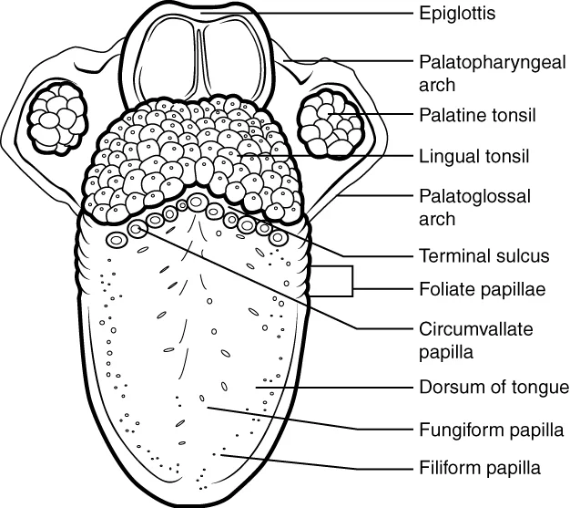
Long description
This superior view of the tongue shows the locations and types of lingual papillae.

Figure 1: This superior view of the tongue shows the locations and types of lingual papillae.
DefinitionSurface Anatomy
The tongue is divided into symmetrical halves by a medial septum and anchored by the lingual frenulum to the floor of the mouth.
Testing taste involves introducing salty, sour, bitter, or sweet stimuli to either side of the tongue. The patient should respond to the taste stimulus before retracting the tongue into the mouth. These sensations, along with certain taste stimuli, can stimulate the gag reflex. If the examiner moves a tongue depressor to contact the lateral wall of the fauces, this should elicit the gag reflex. Stimulation of either side of the fauces should elicit an equivalent response. [3]
Clinical pearlClinical Pearl
Lateral deficits in taste may be masked if stimuli dissolve in saliva and stimulate bilateral nerve branches.
The tongue is obviously important for tasting food, but it is also necessary for mastication, deglutition, and speech. Because it is so moveable, the tongue facilitates complex speech patterns and sounds. Loss of function of the tongue can interfere with speech and swallowing. Additionally, because the location of the hypoglossal nerve and nucleus is near the cardiovascular center, inspiratory and expiratory areas for respiration, and the vagus nuclei that regulate digestive functions, a tongue that protrudes incorrectly can suggest damage in adjacent structures. [3][4]
The tongue is a muscular organ situated within the oral cavity.
Figure 2Gray1013
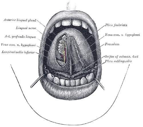
Long description
The mouth cavity. The apex of the tongue is turned upward, and on the right side a superficial dissection of its under surface has been made.

Figure 2: The mouth cavity.
Viewed from above, the anterior two-thirds form the oral part, while the posterior one-third forms the pharyngeal part. These parts are separated by a V-shaped groove called the sulcus terminalis. At the apex of this V lies the foramen cecum. The anterior oral part is mobile and rests against the teeth. The posterior pharyngeal part is fixed and forms the anterior wall of the oropharynx.
The palate forms the roof of the mouth. The anterior hard palate is bony, created by the maxillary and palatine bones. It serves as a rigid shelf against which the tongue pushes food. The posterior soft palate is composed mainly of skeletal muscle. A fleshy bead called the uvula drops from the center of the posterior edge of the soft palate. During swallowing, the soft palate and uvula move upward to prevent food entering the nasal cavity. Two muscular folds extend downward from the soft palate: [2]
DefinitionAnkyloglossia
A congenital anomaly where the lingual frenulum is too short or malformed, potentially impairing speech. [2]
The tongue contains many small salivary glands within its mucous membranes. These minor exocrine glands secrete saliva directly or through ducts. An average of 1 to 1.5 liters of saliva is secreted daily. Secretion increases during eating to moisten food and initiate carbohydrate breakdown. The lingual glands in the tongue help supply adequate saliva to all areas. [2]
| Type | Location | Function |
|---|---|---|
| Circumvallate | Posterior tongue | Taste |
| Foliate | Lateral posterior tongue | Taste |
| Filiform | Anterior tongue | Texture |
| Fungiform | Anterior tongue | Taste |
The tongue is a muscular hydrostat situated within the oral cavity, acting as the primary organ for taste and manipulation of food. It is not merely a surface structure but a complex entity defined by its deep attachments and superficial relations. To understand its position, one must visualise the tongue in relation to the bony and soft tissue boundaries of the mouth [4] [2]
Figure 3Gray1014
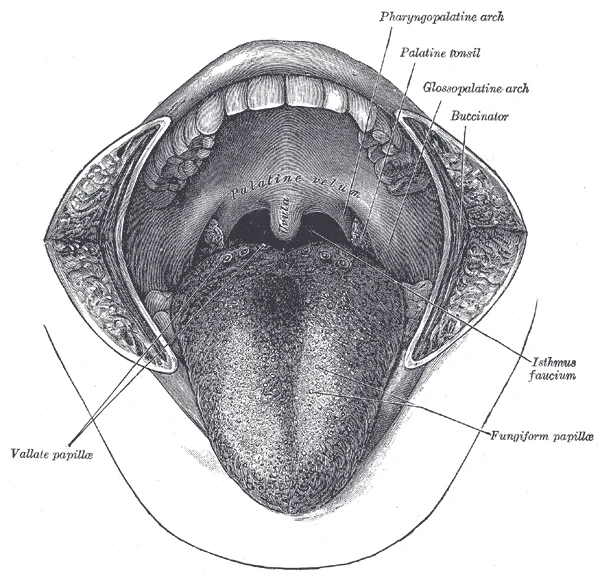
Long description
The mouth cavity. The cheeks have been slit transversely and the tongue pulled forward.

Figure 3: The mouth cavity.
The anterior two-thirds, or oral part, lies within the oral cavity proper. It rests upon the mylohyoid muscle, which forms the muscular floor of the mouth. Superiorly, it is bounded by the hard and soft palates. Posteriorly, it transitions into the pharyngeal part, or root, which lies in the oropharynx. The boundary between these parts is marked by the sulcus terminalis, a V-shaped groove on the dorsal surface. The apex of the V points posteriorly towards the foramen cecum. Lateral to the tongue are the buccal mucosa and the teeth. Inferiorly, the tongue is attached to the hyoid bone and the mandible by several extrinsic muscles. These muscles allow for the protrusion, retraction, and depression of the tongue. The intrinsic muscles, contained entirely within the tongue, alter its shape for speech and swallowing.
Figure 42406 Structures of the Mouth
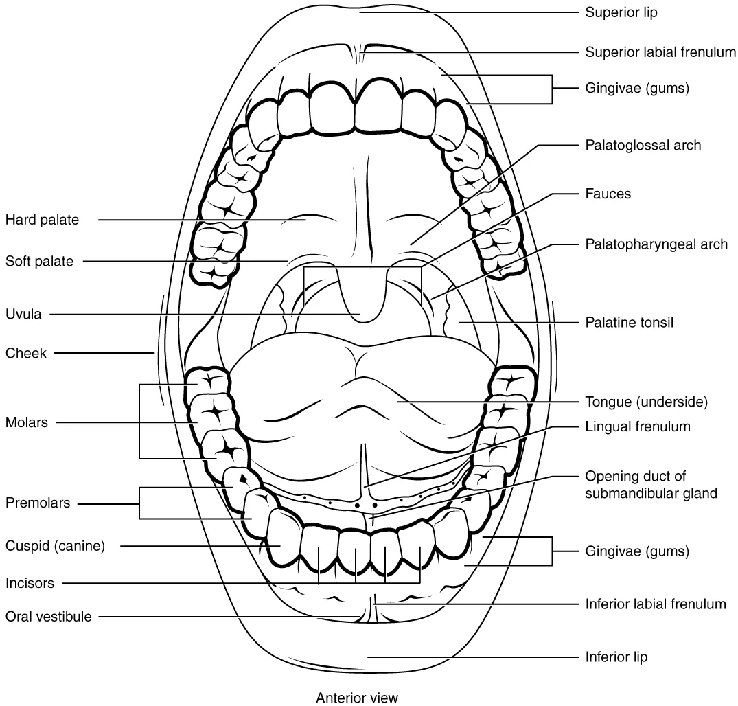
Long description
The mouth includes the lips, tongue, palate, gums, and teeth.

Figure 4: The mouth includes the lips, tongue, palate, gums, and teeth.
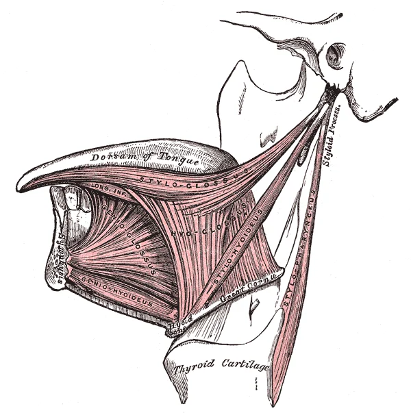
Figure 5Gray1019
Long description
Extrinsic muscles of the tongue. Left side.

Figure 5: Extrinsic muscles of the tongue (left side).
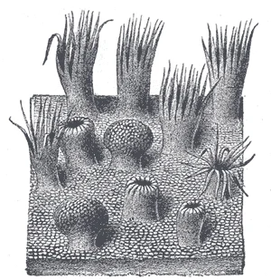
The tongue is covered by mucosa, which is stratified squamous epithelium. On the dorsal surface, this mucosa is modified into papillae, which house the taste buds. The taste buds are specialized chemoreceptors that interpret chemical stimuli from food. These receptors are located within the papillae. [1]
The tongue is covered by mucosa, which is stratified squamous epithelium. On the dorsal surface, this mucosa is modified into papillae, which house the taste buds. The taste buds are specialized chemoreceptors that interpret chemical stimuli from food. These receptors are located within the papillae, specifically the fungiform, foliate, and circumvallate papillae. The circumvallate papillae are arranged in a row just ant [1]
Figure 6Gray1018
Long description
Semidiagrammatic view of a portion of the mucous membrane of the tongue. Two fungiform papillæ are shown. On some of the filiform papillæ the epithelial prolongations stand erect, in one they are spread out, and in three they are folded in.

Figure 6: Semidiagrammatic view of a portion of the mucous membrane of the tongue (two fungiform papillæ are shown).
The anterior two-thirds of the tongue is distinct from the posterior one-third, which is separated by the sulcus terminalis.
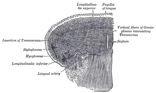
The interior of the tongue is composed primarily of skeletal muscle bundles arranged in three dimensions. These muscles are divided into intrinsic and extrinsic groups. The intrinsic muscles lie entirely within the tongue and alter its shape without moving its position. They include the superior longitudinal, inferior longitudinal, transverse, and vertical muscles. The extrinsic muscles originate outside the tongue and insert into it, thereby changing its position. These include the genioglossus, hyoglossus, styloglossus, and palatoglossus. The genioglossus is the largest extrinsic muscle; it originates from the mental spine of the mandible and fans out to insert into the body of the hyoid bone and the tongue itself. Its contraction protrudes the tongue, while its posterior fibres depress the centre of the tongue.
Figure 7Gray1020
Long description
Coronal section of tongue, showing intrinsic muscles. (Altered from Krause.)

Figure 7: Coronal section of tongue, showing intrinsic muscles (Altered from krause.).
DefinitionSulcus Terminalis
A V-shaped groove on the dorsal surface of the tongue that separates the anterior two-thirds from the posterior one-third.
The vascular and nervous supply is critical for function. The anterior portion receives general sensation from the lingual nerve, a branch of the mandibular division of the trigeminal nerve, and taste from the chorda tympani, a branch of the facial nerve. The posterior portion receives both general sensation and taste from the glossopharyngeal nerve. This arrangement allows for precise control of speech and swallowing. [1][3]
| Muscle | Origin | Insertion | Action | Nerve Supply |
|---|---|---|---|---|
| Genioglossus | Mental spine of mandible | Body of hyoid, tongue | Protrudes tongue | Hypoglossal |
| Hyoglossus | Greater horn of hyoid | Side of tongue | Depresses tongue | Hypoglossal |
| Styloglossus | Styloid process | Side of tongue | Elevates and retracts tongue | Hypoglossal |
| Palatoglossus | Palatine aponeurosis | Side of tongue | Elevates posterior tongue | Vagus |
Deglutition involves the coordinated action of these muscles. The voluntary phase begins with the tongue pushing the bolus against the palate. The suprahyoid muscles, including the digastric and mylohyoid, raise the hyoid bone and larynx, facilitating the movement of food into the pharynx. This process is aided by mucus and saliva, ensuring smooth passage. The autonomic nervous system controls the subsequent pharyngeal and esophageal phases, ensuring that the airway is protected by the epiglottis and soft palate.
The arterial supply of the tongue is derived primarily from the lingual artery, a branch of the external carotid artery. This vessel ensures robust perfusion to the muscular organ, which is essential for its complex functions in taste, speech, and deglutition. The lingual artery arises in the neck and courses anteriorly towards the tongue, giving off several important branches that supply different regions of the organ.
The deep lingual artery runs along the inferior surface of the tongue, extending to the tip, while the sublingual artery supplies the floor of the mouth and the sublingual gland.
The deep lingual artery, also known as the ranine artery, is the terminal branch of the lingual artery. It ascends between the genioglossus and hyoglossus muscles, running along the undersurface of the tongue to the tip. It anastomoses with the artery of the opposite side, ensuring collateral circulation. The sublingual artery runs forward between the mylohyoid and hyoglossus muscles, supplying the sublingual gland and the muscles of the floor of the mouth.
In addition to the lingual artery, the facial artery contributes to the blood supply of the tongue via its tonsillar and submental branches. The tonsillar branch supplies the palatine tonsil and the soft palate, while the submental branch runs along the inferior border of the mandible, supplying the submandibular gland and the muscles of the neck.
The arterial supply is crucial for the function of the tongue, which is involved in mechanical digestion and the initiation of swallowing. The tongue and the teeth begin mechanical digestion, and saliva begins chemical digestion. The rich blood supply supports the high metabolic demand of the tongue muscles and the sensory receptors involved in taste. [2]
Clinical pearlClinical Pearl
The deep lingual artery is a key landmark in oral surgery, as it is vulnerable during procedures involving the floor of the mouth.
| Branch | Origin | Course | Termination |
|---|---|---|---|
| Deep Lingual Artery | Lingual Artery | Runs along undersurface of tongue | Tip of tongue |
| Sublingual Artery | Lingual Artery | Between mylohyoid and hyoglossus | Floor of mouth |
| Tonsillar Branch | Facial Artery | To palatine tonsil | Soft palate |
With the exception of somatosensation, this list represents the special senses, or those systems of the body that are associated with specific organs such as the tongue or eye.
The venous drainage of the tongue is extensive and follows a hierarchical pattern, ultimately converging into major vessels that facilitate return to the heart. Understanding this system is critical for appreciating the spread of infection or malignancy, as the rich vascular network provides direct pathways to deep cervical structures.
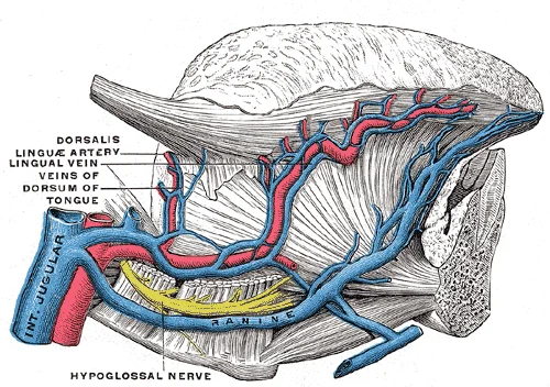
The central structure is the lingual vein, which runs along the inferior surface of the tongue. It is accompanied by the hypoglossal nerve, which lies lateral to the vein. Superiorly, the dorsal lingual veins drain the posterior third of the tongue. These veins converge to form the lingual vein, which then joins the internal jugular vein.
DefinitionLingual Vein
The primary vessel draining the tongue, formed by the union of the deep lingual and sublingual veins, terminating in the internal jugular vein.
It receives tributaries from the lateral margins of the tongue. As it progresses, it lies superficial to the hyoglossus muscle. This relationship is crucial during surgical procedures to avoid inadvertent injury. The vein then joins with the sublingual vein to form the lingual vein proper.
It typically joins the deep lingual vein near the posterior border of the mylohyoid muscle. This union creates the lingual vein, which then passes deep to the hyoglossus muscle.
| Structure | Course | Relations | Termination |
|---|---|---|---|
| Deep Lingual Vein | Runs along ventral surface | Superficial to hyoglossus muscle | Joins sublingual vein |
| Sublingual Vein | Runs along mylohyoid muscle | Deep to sublingual gland | Joins deep lingual vein |
| Lingual Vein | Passes deep to hyoglossus | Lateral to hypoglossal nerve | Internal jugular vein |
The lingual vein, once formed, travels posteriorly and laterally. It passes deep to the hyoglossus muscle and the styloglossus muscle. Within the sheath, it joins the internal jugular vein. This termination point is significant because it provides a direct route for metastatic spread to the cervical lymph nodes.
NoteClinical Relevance
Injury to the lingual vein during surgery can lead to significant bleeding due to its high pressure and lack of valves.
The dorsal lingual veins drain the posterior third of the tongue. They are smaller and more numerous than the deep lingual veins. They drain into the lingual vein or directly into the internal jugular vein. This dual drainage pattern ensures efficient removal of blood from the entire organ.
Venous Drainage Pathway
- Tongue CapillariesBlood collection from mucosa and muscle
- Question: Which region?
Anterior 2/3
- Deep Lingual Vein
- Joins Sublingual Vein
Posterior 1/3
- Dorsal Lingual Veins
- Lingual Vein FormationUnion of deep and sublingual veins
- Passes Deep to HyoglossusRelation to hypoglossal nerve
- Internal Jugular VeinFinal termination point
This hierarchical drainage ensures that blood from all parts of the tongue is efficiently returned to the systemic circulation. The close association with the hypoglossal nerve is a constant anatomical feature, requiring careful dissection to preserve nerve function while managing vascular structures.
The lymphatic drainage of the tongue is complex and varies significantly depending on the specific region of the organ. This anatomical arrangement is clinically critical because it dictates the pathways for metastatic spread in cases of tongue carcinoma. Understanding these pathways allows clinicians to predict which lymph node groups are at risk and guides surgical planning for neck dissections.
The central structure is the body of the tongue, drained by vessels that converge towards the submandibular region. Lateral to the midline, vessels from the lateral margins of the tongue course inferiorly and posteriorly. These vessels pass deep to the mylohyoid muscle and terminate in the submandibular lymph nodes.
The tip of the tongue drains primarily into the submental lymph nodes. These nodes are located in the submental triangle, anterior to the mandible. From here, efferent vessels pass to the submandibular nodes. The anterior two-thirds of the tongue, which constitutes the bulk of the oral tongue, drains mainly into the submandibular lymph nodes. These nodes are situated beneath the mandible and are easily palpable during clinical examination. The lateral margins of the tongue have a more complex drainage pattern. Vessels from the lateral border may drain into the submandibular nodes or pass directly to the deep cervical lymph nodes. This direct drainage pathway is significant because it allows cancer cells to bypass the submandibular nodes and spread directly to the neck.
The posterior third of the tongue, or the lingual tonsil region, drains into the deep cervical lymph nodes. These nodes are located along the internal jugular vein. Drainage from the posterior tongue can also cross the midline, leading to bilateral cervical lymphadenopathy. This bilateral potential is a key consideration in the staging and management of posterior tongue cancers.
The clinical implication of this drainage pattern is profound. A tumour in the tip of the tongue is likely to metastasise to the submental nodes first. A tumour in the anterior two-thirds is likely to involve the submandibular nodes. However, a tumour on the lateral border or posterior third may present with deep cervical node involvement, sometimes without obvious submandibular involvement. This variability necessitates careful imaging and physical examination of the entire neck. Palpation of the submental and submandibular triangles is essential. Deep cervical nodes are assessed along the sternocleidomastoid muscle. Early detection of nodal metastasis improves prognosis and guides the extent of surgical intervention.
Figure 8Gray559
Long description
Veins of the tongue. The hypoglossal nerve has been displaced downward in this preparation. (Testut after Hirschfeld.)

Figure 8: Veins of the tongue.
The tongue receives a complex innervation involving somatic, special sensory, and autonomic components. Understanding this requires distinguishing between general sensation, taste, and motor control.
DefinitionSomatic Sensation
General sensation (touch, pain, temperature) is supplied by the trigeminal nerve for the anterior two-thirds and the glossopharyngeal nerve for the posterior one-third. [1]
The anterior portion of the tongue receives general somatic afferent fibres from the lingual nerve, a branch of the mandibular division of the trigeminal nerve. These fibres carry sensations of touch, pain, and temperature. Taste sensation is relayed to the brain stem through fibers of the facial and glossopharyngeal nerves. The chorda tympani joins the lingual nerve and travels with it to the tongue. The posterior portion of the tongue receives both general somatic sensation and taste from [3]
DefinitionMotor Supply
All intrinsic and extrinsic muscles of the tongue are innervated by the hypoglossal nerve, except the palatoglossus.
Motor innervation to the tongue muscles is provided by the hypoglossal nerve. This nerve supplies all the intrinsic muscles and the extrinsic muscles (genioglossus, hyoglossus, and styloglossus). The palatoglossus muscle is an exception; it is innervated by the vagus nerve via the pharyngeal plexus. The hypoglossal nerve emerges from the medulla and exits the skull via the hypoglossal canal. It descends in the neck, crossing the hyoid bone, and enters the tongue. [3]
Referred pain from the tongue can occur due to the shared pathways of sensory nerves. Pain from the anterior tongue may be referred to the mandibular region, while pain from the posterior tongue may be referred to the ear. This is because the glossopharyngeal nerve also supplies the pharynx and middle ear.
Tongue Innervation
- Identify RegionAnterior vs Posterior 2/3
- Question: Anterior 2/3?
Yes
- General Sensation: Trigeminal (Lingual)
- Taste: Facial (Chorda Tympani)
No
- General Sensation & Taste: Glossopharyngeal
The chorda tympani joins the lingual nerve.
| Structure | Nerve | Function |
|---|---|---|
| Anterior 2/3 General Sensation | Trigeminal (Lingual) | Touch, Pain, Temperature |
| Anterior 2/3 Taste | Facial (Chorda Tympani) | Taste |
| Posterior 1/3 General Sensation | Glossopharyngeal | Touch, Pain, Temperature |
| Posterior 1/3 Taste | Glossopharyngeal | Taste |
| All Muscles (except Palatoglossus) | Hypoglossal | Motor |
| Palatoglossus | Vagus | Motor |
Link to the histology page.
Taste perception begins with chemical stimuli. The first two tastes, salty and sour, are triggered by the cations Na+ and H+. The other tastes result from food molecules binding to a G protein–coupled receptor. A G protein signal transduction system ultimately leads to depolarization of the gustatory cell. Other monosaccharides such as fructose, or artificial sweeteners such as aspartame (NutraSweet™), saccharine, or sucralose (Splenda™) also activate the sweet receptors. The affinity for each of these molecules varies, and some will taste sweeter than glucose because they bind to the G protein–coupled receptor differently. [1]
The tongue's movement is controlled by intrinsic and extrinsic muscles. The intrinsic muscles are entirely within the tongue and alter its shape. The extrinsic muscles originate outside the tongue and insert into it, changing its position. These muscles are innervated by the hypoglossal nerve (CN XII).
DefinitionTaste Bud
Specialized sensory receptors located within the papillae of the tongue that detect chemical stimuli.
Exam trapNerve Supply
Remember that the general sensation of the anterior two-thirds is via the lingual nerve (CN V3), while taste is via the chorda tympani (CN VII). Posterior one-third general and taste are via CN IX. [1]
The tongue is richly vascularized and innervated. The lingual artery supplies blood to the tongue. The hypoglossal nerve provides motor innervation to all intrinsic and extrinsic muscles except the palatoglossus, which is supplied by the vagus nerve (CN X).
| Muscle | Origin | Insertion | Action | Nerve Supply |
|---|---|---|---|---|
| Genioglossus | Mental spine of mandible | Tongue and hyoid | Protrudes tongue | Hypoglossal (CN XII) |
| Hyoglossus | Hyoid bone | Side of tongue | Depresses tongue | Hypoglossal (CN XII) |
| Styloglossus | Styloid process | Side of tongue | Elevates and retracts tongue | Hypoglossal (CN XII) |
| Palatoglossus | Palatine aponeurosis | Side of tongue | Elevates posterior tongue | Vagus (CN X) |
Swallowing involves the tongue pushing the bolus posteriorly. In the pharyngeal phase, stimulation of receptors in the oropharynx sends impulses to the deglutition center (a collection of neurons that controls swallowing) in the medulla oblongata. Impulses are then sent back to the uvula and soft palate, causing them to move upward and close off the nasopharynx. The laryngeal muscles also constrict to prevent aspiration of food into the trachea. At this point, deglutition apnea takes place, which means that breathing ceases for a very brief time. Contractions of the pharyngeal constrictor muscles move the bolus through the oropharynx and laryngopharynx. Relaxation of the upper esophageal sphincter then allows food to enter the esophagus. [2]
The tongue is also involved in speech. The precise movements of the tongue allow for the articulation of various sounds. The tip of the tongue is particularly important for alveolar sounds.
NoteClinical Note
Lesions of the hypoglossal nerve cause the tongue to deviate toward the side of the lesion upon protrusion.
The tongue arises from the primitive pharynx during the fourth and sixth weeks of gestation. This complex process involves the fusion of multiple embryological prominences, resulting in a structure with dual embryological origins that dictates its sensory and motor innervation. Understanding this developmental timeline is crucial for correlating anatomical variations with clinical presentations.
The anterior two-thirds of the tongue, known as the oral part, develops primarily from the first pharyngeal arch. Specifically, it forms from three swellings: a single median swelling called the tuberculum impar and two lateral swellings known as the lateral lingual swellings. The lateral lingual swellings overgrow the tuberculum impar and fuse in the midline to form the bulk of the anterior tongue. This region is covered by ectoderm, which explains its sensory innervation. The general sensation [1]
The posterior one-third of the tongue, or the pharyngeal part, develops from the second, third, and fourth pharyngeal arches. The copula, formed by the second arch, is largely overgrown by the third arch. The third arch contributes to the posterior third of the tongue, while the fourth arch contributes to the most posterior region near the epiglottis. This area is covered by endoderm, reflecting its origin from the foregut. Consequently, general sensation and taste for the posterior third are su [1]
The intrinsic and extrinsic muscles of the tongue derive from the occipital myotomes. These muscle buds migrate anteriorly into the tongue during development. All tongue muscles, except the palatoglossus, are innervated by the hypoglossal nerve (CN XII). The palatoglossus is innervated by the vagus nerve via the pharyngeal plexus, reflecting its functional association with the soft palate rather than the tongue proper. This distinct motor innervation pattern is a key diagnostic feature in neurological examinations. [1]
Developmental anomalies such as lingual thyroid occur when the thyroglossal duct fails to regress properly. The thyroid gland originates at the foramen cecum, the remnant of the thyroglossal duct, and descends to its final position in the neck. Failure of descent results in ectopic thyroid tissue at the base of the tongue. This condition must be considered in patients presenting with a midline neck mass or dysphagia. Surgical removal without confirming the presence of normal thyroid tissue elsewhere can lead to permanent hypothyroidism.
The tongue is a complex muscular organ situated within the oral cavity, visible on cross-sectional imaging as a broad, flat structure. In axial CT or MRI scans, it occupies the anterior two-thirds of the floor of the mouth. The midline raphe is often visible as a thin, hypointense line dividing the tongue into left and right halves. This raphe represents the fibrous septum that anchors the tongue to the hyoid bone and mandible. Surrounding the tongue is the buccal mucosa laterally and the floor of the mouth inferiorly. The genioglossus muscle, the primary protruder, forms the bulk of the anterior tongue and is easily identified by its fan-shaped origin from the superior mental spine of the mandible.
DefinitionTongue Anatomy
The tongue consists of intrinsic and extrinsic muscles, innervated by multiple cranial nerves, with sensory and motor functions critical for speech and swallowing.
The tongue is centrally located, surrounded by the teeth and alveolar ridges. The airway is patent and centrally positioned behind the tongue base.
The intrinsic muscles, including the superior and inferior longitudinal, transverse, and vertical muscles, alter the shape of the tongue. These muscles are innervated by the hypoglossal nerve (CN XII). The extrinsic muscles, including the genioglossus, hyoglossus, styloglossus, and palatoglossus, move the tongue as a whole. These muscles are also innervated by CN XII, except for palatoglossus, which is innervated by the vagus nerve (CN X).
| Muscle | Origin | Insertion | Action | Nerve Supply |
|---|---|---|---|---|
| Genioglossus | Superior mental spine | Hyoid bone, tongue | Protrudes, depresses | Hypoglossal (CN XII) |
| Hyoglossus | Hyoid bone | Side of tongue | Depresses, retracts | Hypoglossal (CN XII) |
| Styloglossus | Styloid process | Side of tongue | Elevates, retracts | Hypoglossal (CN XII) |
| Palatoglossus | Palatine aponeurosis | Side of tongue | Elevates posterior tongue | Vagus (CN X) |
Sensory innervation is divided. The anterior portion receives general sensation via the lingual nerve and taste via the chorda tympani. The posterior portion receives both general sensation and taste via the glossopharyngeal nerve. The epiglottis and extreme posterior tongue are innervated by the vagus nerve. This complex innervation is crucial for clinical examination and localizing lesions. [1]
The tongue is a muscular organ essential for mastication, deglutition, and phonation. It is divided into an anterior two-thirds, known as the oral part, and a posterior one-third, termed the pharyngeal part. The boundary between these regions is marked by the sulcus terminalis, a V-shaped groove. The apex of this 'V' is the foramen caecum, which represents the embryological origin of the thyroid gland. The oral part moves voluntarily, while the pharyngeal part is largely involuntary.
The extrinsic muscles alter the shape and position of the tongue. They originate outside the tongue and insert into it.
| Muscle | Origin | Insertion | Action | Nerve Supply |
|---|---|---|---|---|
| Genioglossus | Superior mental spine | Fibres of tongue | Protrudes tongue | Hypoglossal nerve |
| Hyoglossus | Greater horn of hyoid | Side of tongue | Depresses tongue | Hypoglossal nerve |
| Styloglossus | Styloid process | Side of tongue | Elevates and retracts tongue | Hypoglossal nerve |
| Palatoglossus | Palatine aponeurosis | Side of tongue | Elevates posterior tongue | Vagus nerve |
The genioglossus is the primary protruder of the tongue. When one side contracts, the tongue deviates towards the side of the lesion if the hypoglossal nerve is damaged.
The intrinsic muscles lie entirely within the tongue and change its shape without moving its position. They include the superior longitudinal, inferior longitudinal, transverse, and vertical muscles. These muscles are supplied by the hypoglossal nerve. They allow for the complex movements required for speech and swallowing.
Sensory innervation differs between the two parts. The anterior two-thirds receives general sensation from the lingual nerve, a branch of the mandibular nerve, and taste from the chorda tympani, a branch of the facial nerve. The posterior one-third receives both general sensation and taste from the glossopharyngeal nerve. The motor supply to all intrinsic and extrinsic muscles, except the palatoglossus, is from the hypoglossal nerve. The blood supply is primarily from the lingual artery, a branch of the external carotid artery. Venous drainage occurs via the lingual veins into the internal jugular vein.
The olfactory pathway is unique among sensory systems because it bypasses the thalamus, projecting directly from the olfactory bulb to the primary olfactory cortex in the inferior and medial areas of the temporal lobe. This direct route explains why smells can trigger emotional memories via the limbic system. The olfactory neurons extend axons through an olfactory foramen in the cribriform plate of the ethmoid bone to synapse in the olfactory bulb. [1]
The inner ear comprises a bony labyrinth within the temporal bone, containing the cochlea for hearing and the vestibule for balance. These functions are mediated by the vestibulocochlear nerve, which carries signals from both regions to the brain stem. The cochlea connects to the stapes via the oval window, where sound vibrations cause fluid waves that are transduced into neural signals. [1]
The vestibular system includes three semicircular canals oriented in horizontal and vertical planes to detect rotational head movements. The ampulla at the base of each canal contains hair cells whose stereocilia extend into the cupula. Head rotation causes fluid lag, deflecting the cupula opposite to the movement direction. [1]
Somatosensory transduction involves free nerve endings for pain and temperature. Nociceptors respond to mechanical, chemical, or thermal stimuli exceeding a specific threshold. Capsaicin binds to ion channels sensitive to temperatures above 37°C, mimicking heat pain. [1]
Visual processing begins with photoreceptors in the retina, consisting of rods and cones. Rods contain rhodopsin in stacked discs for low-light vision, while cones use membrane infoldings for colour vision. Three cone opsins are sensitive to different light wavelengths. [1]
Clinical testing of the vestibular system includes the Romberg test for equilibrium and assessment of the vestibulo-ocular reflex. Damage near the vestibular and cochlear nuclei may cause combined deficits due to their proximity. [3]
Eye movement examination assesses extraocular muscles; diplopia may result from failure of any muscle, not just the lateral rectus. Convergence tests medial recti function as eyes adduct to view near objects. Accommodation, controlled by parasympathetic fibres of the oculomotor nerve, adjusts lens shape for near focus. [3]
Motor testing of the neck involves flexion, extension, and shrugging against resistance to evaluate spinal nerve control. Asymmetry in lateral flexion suggests weakness on the affected side. [3]
The olfactory tract connects to the olfactory bulb on the ventral surface of the frontal lobe before splitting to various brain regions. [1]
Exam trapThalamic Bypass
Smell is the only sense not relayed through the thalamus before reaching the cortex.
Clinical pearlCapsaicin Threshold
Nociceptors activated by capsaicin respond to temperatures above 37°C.
DefinitionConvergence
Adduction of both eyes to focus on a near object.
NoteVestibular Anatomy
Three semicircular canals detect rotation in horizontal and vertical planes.
Exam trapDiplopia Cause
Diplopia can arise from failure of any extraocular muscle, not solely the lateral rectus.
Clinical pearlOlfactory Pathway
Axons pass through the cribriform plate to reach the olfactory bulb.
DefinitionAccommodation
Change in lens shape controlled by parasympathetic fibres to focus on near objects.
NoteInner Ear Structure
The cochlea and vestibule are embedded in the temporal bone.
Exam trapMotor Asymmetry
Weakness in neck lateral flexion indicates damage on the weaker side.
Clinical pearlPhotoreceptor Types
Rods use stacked discs for rhodopsin; cones use membrane infoldings.
DefinitionVestibulocochlear Nerve
Carries signals from both hearing and balance regions to the brain stem.
Students frequently confuse the functional anatomy of the extraocular muscles, particularly regarding the obliques. A common error is assuming the superior oblique elevates the eye because of its name. In reality, the angle of the tendon through the trochlea means that contraction of the superior oblique rotates the eye laterally. This lateral rotation is necessary because the eye is not perfectly aligned on the sagittal plane. The inferior oblique muscle originates from the floor of the orbit and inserts into the inferolateral surface of the eye. [1]
Another pitfall involves the structure of the retina and visual acuity. Students often assume the photoreceptors are the first layer light encounters. Note that the photoreceptors in the retina (rods and cones) are located behind the axons, RGCs, bipolar cells, and retinal blood vessels. A significant amount of light is absorbed by these structures before the light reaches the photoreceptor cells. However, at the exact center of the retina is a small area known as the fovea. At the fovea, the re [1]
Exam trapFoveal Convergence
Do not confuse the fovea with the peripheral retina. Toward the edges of the retina, several photoreceptors converge on RGCs (through the bipolar cells) up to a ratio of 50 to 1. This convergence reduces the accuracy of visual transduction. [1]
A third mistake relates to the mucosal lining of the upper gastrointestinal tract. Students may incorrectly identify the epithelium as keratinized. The mucosa of the esophagus is made up of an epithelial lining that contains non-keratinized, stratified squamous epithelium, with a layer of basal and parabasal cells. This epithelium protects against erosion from food particles. Furthermore, the outer layer is often misidentified as serosa. The most superficial layer of the esophagus is called the adventitia, not the serosa. In contrast to the stomach and intestines, the loose connective tissue of the adventitia is not covered by a fold of visceral peritoneum. [2]
DefinitionMastication
In anatomical terminology, chewing is called mastication. Muscles involved in chewing must be able to exert enough pressure to bite through and then chew food before it is swallowed. [4]
This knowledge is applied at the bedside during cranial nerve examinations. Testing eye movement is simply a matter of having the patient track the tip of a pen as it is passed through the visual field. This may appear similar to testing visual field deficits related to the optic nerve, but the difference is that the patient is asked to not move the eyes while the examiner moves a stimulus into the peripheral visual field. Here, the extent of movement is the point of the test. The examiner is watching for conjugate movements representing proper function of the related nuclei and the MLF. Failure of one eye to abduct while the other adducts in a horizontal movement is referred to as internuclear ophthalmoplegia. When this occurs, the patient will experience diplopia, or double vision, as the two eyes are temporarily pointed at different stimuli. [3]
Eye Movement Assessment
- StartAsk patient to track pen tip
- Question: Conjugate movement?
Yes
- Normal function
No
- Question: One eye fails to abduct?
Yes
- Internuclear ophthalmoplegia
No
- Other deficit
In optometry and general practice, testing vision relies on the tests that are common in an optometry office. The Snellen chart demonstrates visual acuity by presenting standard Roman letters in a variety of sizes. The result of this test is a rough generalization of the acuity of a person based on the normal accepted acuity, such that a letter that subtends a visual angle of 5 minutes of an arc at 20 feet can be seen. To have 20/60 vision, for example, means that the smallest letters that a per [3]
| Structure | Location | Key Feature |
|---|---|---|
| Fovea | Center of retina | No blood vessels; highest acuity |
| Peripheral Retina | Edges of retina | High convergence (50:1); low acuity |
| Photoreceptors | Behind axons/RGCs | Light absorption before reception |
In the operating theatre, understanding the muscles of mastication is vital for surgical approaches to the mandible. The masseter muscle is the main muscle used for chewing because it elevates the mandible (lower jaw) to close the mouth, and it is assisted by the temporalis muscle, which retracts the mandible. You can feel the temporalis move by putting your fingers to your temple as you chew. Knowledge of the esophageal layers is crucial during endoscopy. The muscularis layer changes according to location: In the upper third of the esophagus, the muscularis is skeletal muscle. In the middle third, it is both skeletal and smooth muscle. In the lower third, it is smooth muscle. [2] [4]
On imaging, the distinction between the fovea and peripheral retina explains visual field defects. The visual stimulus in the middle of the field of view falls on the fovea and is in the sharpest focus. Without moving your eyes off that word, notice that words at the beginning or end of the paragraph are not in focus. The images in your peripheral vision are focused by the peripheral retina, and have vague, blurry edges and words that not as clearly identified. This anatomical arrangement dictates how lesions in the optic nerve or retina present clinically. [1]
NoteColor Perception
If you think that you can see colors in the dark, it is most likely because your brain knows what color something is and is relying on that memory. [1]
In examinations, students must distinguish between the functions of the iris and the ciliary body. These two structures bend the lens, allowing it to focus light on the back of the eye. Overlaying the ciliary body, and visible in the anterior eye, is the iris—the colored part of the eye. The iris is a smooth muscle that opens or closes the pupil, which is the hole at the center of the eye that allows light to enter. The iris constricts the pupil in response to bright light and dilates the pupil in response to dim light. [1]
Clinical pearlUmami Taste
The taste known as umami is often referred to as the savory taste. Like sweet and bitter, it is based on the activation of G protein–coupled receptors by a specific molecule. The molecule that activates this receptor is the amino acid L-glutamate. Therefore, the umami flavor is often perceived while eating protein-rich foods. Not surprisingly, dishes that contain meat are often described as savory. [1]
This sensory knowledge also links to memory. This intimate connection between the olfactory system and the cerebral cortex is one reason why smell can be a potent trigger of memories and emotion. While not directly tongue anatomy, this sensory integration is often tested in clinical vignettes involving cranial nerves and cortical processing. [1]
The superior oblique rotates the eye laterally due to the trochlear angle. The inferior oblique originates from the orbit floor and inserts inferolaterally. Visual acuity is highest at the fovea due to lack of blood vessels. Photoreceptors are located behind retinal layers, causing light absorption. The esophageal mucosa uses non-keratinized stratified squamous epithelium. The esophageal outer layer is adventitia, not serosa. Mastication involves the masseter elevating and temporalis retracting the mandible. Internuclear ophthalmoplegia presents as failure of one eye to abduct.
Answer each one in your head first, then open the answer.
What is the primary action of the superior oblique muscle?
Show answerHide answer
Rotates the eye laterallyWhat type of epithelium lines the esophagus?
Show answerHide answer
Non-keratinized, stratified squamous epitheliumWhy is visual acuity greatest at the fovea?
Show answerHide answer
It lacks supporting cells and blood vesselsWhat is the outer layer of the esophagus called?
Show answerHide answer
AdventitiaWhat molecule activates the umami receptor?
Show answerHide answer
L-glutamate
- Betts JG, Young KA, Wise JA, et al. Sensory Perception. OpenStax, Anatomy and Physiology 2e (CC BY-NC-SA 4.0). Accessed .
- Betts JG, Young KA, Wise JA, et al. The Mouth, Pharynx, and Esophagus. OpenStax, Anatomy and Physiology 2e (CC BY-NC-SA 4.0). Accessed .
- Betts JG, Young KA, Wise JA, et al. The Cranial Nerve Exam. OpenStax, Anatomy and Physiology 2e (CC BY-NC-SA 4.0). Accessed .
- Betts JG, Young KA, Wise JA, et al. Axial Muscles of the Head, Neck, and Back. OpenStax, Anatomy and Physiology 2e (CC BY-NC-SA 4.0). Accessed .
Figure credits
- Figure 1: OpenStax College, via Wikimedia Commons, CC BY 3.0, CC BY 3.0.
- Figure 2: Henry Vandyke Carter, via Wikimedia Commons, Public domain, Public domain.
- Figure 3: Henry Vandyke Carter, via Wikimedia Commons, Public domain, Public domain.
- Figure 4: OpenStax College, via Wikimedia Commons, CC BY 3.0, CC BY 3.0.
- Figure 5: Henry Vandyke Carter, via Wikimedia Commons, Public domain, Public domain.
- Figure 6: Henry Vandyke Carter, via Wikimedia Commons, Public domain, Public domain.
- Figure 7: Henry Vandyke Carter, via Wikimedia Commons, Public domain, Public domain.
- Figure 8: Henry Vandyke Carter, via Wikimedia Commons, Public domain, Public domain.
Educational content for medical students and doctors. It is not a substitute for clinical judgement or local guidelines.