Pharynx
Anatomical and functional overview of the pharyngeal region and its neural supply.
The pharynx is a fibromuscular tube that serves as a common pathway for both the respiratory and digestive systems. A short tube of skeletal muscle lined with a mucous membrane, the pharynx runs from the posterior oral and nasal cavities to the opening of the esophagus and larynx. Anteriorly, it communicates with the nasal cavities, oral cavity, and larynx. The wall of the oropharynx is histologically similar to that of the oral cavity, featuring a stratified squamous epithelium endowed with mucus-producing glands. < [1]
The pharyngeal wall consists of four layers: mucosa, submucosa, muscular layer, and fibrous membrane. The muscular layer is divided into two functional groups: the elevator muscles and the constrictor muscles. During swallowing, the elevator skeletal muscles of the pharynx contract, raising and expanding the pharynx to receive the bolus of food. Once received, these muscles relax and the constrictor muscles of the pharynx contract, forcing the bolus into the esophagus and initiating peristalsis. This coordinated action ensures efficient transport of food while protecting the airway. [1]
During deglutition, the soft palate rises to close off the nasopharynx, preventing food entry into the nasal cavity. The larynx elevates, and the epiglottis folds over the glottis to protect the airway. The esophagus includes an upper esophageal sphincter made of skeletal muscle, which regulates the movement of food from the pharynx to the esophagus. Cells in the esophageal wall secrete mucus that eases the passage of the food bolus. [1]
The pharyngeal nerve plexus, or upper pharyngeal nerves, originate from the glossopharyngeal and vagus nerves. After diverging from the glossopharyngeal and vagus nerves at the level of the hyoid bone, the pharyngeal nerves entered the constrictor pharyngis medius muscle. According to Ohtsuka et al. and Oda et al. , in these nerves, the lingual branches of the glossopharyngeal nerve are notably thick, and they follow a downward course through a slit between the constrictor pharyngis superior and medius muscles (CPS and CPM). Likewise, many atlases of human anatomy () display the descending courses of the other, thinner, pharyngeal nerves along the [1][2]
DefinitionPharyngeal Plexus
An intramuscular nerve network formed by branches of the glossopharyngeal and vagus nerves, primarily supplying the pharyngeal muscles rather than the mucosa.
This developmental change explains why adult anatomical atlases often depict nerves running along the posterior surface, whereas fetal studies show an intramuscular course. [2]
| Muscle Group | Primary Action | Nerve Supply |
|---|---|---|
| Elevators | Raise and expand pharynx | Glossopharyngeal, Vagus |
| Constrictors | Force bolus into esophagus | Glossopharyngeal, Vagus |
Figure 1Gray1031
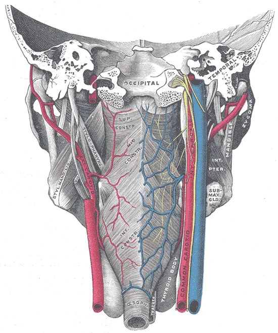
Long description
Muscles of the pharynx, viewed from behind, together with the associated vessels and nerves. (Modified after Testut.)

Figure 1: Muscles of the pharynx, viewed from behind, together with the associated vessels and nerves (Modified after testut.).
Figure 2Gray1030
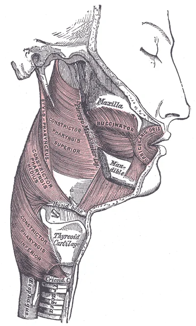
Long description
Muscles of the pharynx and cheek. Labelled illustration from Wikimedia Commons, used under its open licence.

Figure 2: Muscles of the pharynx and cheek.
The pharynx is a muscular tube involved in both digestion and respiration.
The pharynx is divided into three parts: the nasopharynx, oropharynx, and laryngopharynx. The nasopharynx lies posterior to the nasal cavities. The oropharynx lies posterior to the oral cavity. The laryngopharynx lies posterior to the larynx. The constrictor muscles form the lateral and posterior walls of the pharynx. These muscles are the superior, middle, and inferior pharyngeal constrictors. They overlap like flower pots. Contraction of these muscles propels the bolus downward.
The tonsils are lymphoid tissues located in the pharynx. The palatine tonsils lie in the oropharynx. The pharyngeal tonsil lies in the nasopharynx. The lingual tonsil lies at the base of the tongue. Together, these form the Waldeyer ring. This ring provides immune surveillance. [1]
The constrictor muscles originate from various bony and soft tissue landmarks. The superior pharyngeal constrictor originates from the pterygomandibular raphe and the mandible. The middle pharyngeal constrictor originates from the hyoid bone. The inferior pharyngeal constrictor originates from the thyroid and cricoid cartilages. The upper esophageal sphincter is continuous with the inferior pharyngeal constrictor. It controls the movement of food from the pharynx into the esophagus. [1]
The nerves supply these muscles. The pharyngeal nerve plexus provides innervation. The glossopharyngeal nerve is involved. The constrictor pharyngis superior muscle is innervated by this plexus. The levator veli palatini muscle is also innervated by this plexus. The pharyngeal nerves exhibit a curved or recurrent course. They originate from the nerve trunks at the supero-inferior level including the hyoid bone. They turn upward and reach the superior level, including the pharyngotympanic tube. T [2]
DefinitionDeglutition
Deglutition is another word for swallowing—the movement of food from the mouth to the stomach. The entire process takes about 4 to 8 seconds for solid or semisolid food, and about 1 second for very soft food and liquids. [1]
In the pharyngeal phase, stimulation of receptors in the oropharynx sends impulses to the deglutition center. Impulses are then sent back to the uvula and soft palate, causing them to move upward and close off the nasopharynx. The laryngeal muscles also constrict to prevent aspiration of food into the trachea. At this point, deglutition apnea takes place, which means that breathing ceases for a very brief time. Contractions of the pharyngeal constrictor muscles move the bolus through the oropharynx and laryngopharynx. Relaxation of the upper esophageal sphincter then allows food to enter the esophagus. [1]
| Muscle | Origin | Insertion | Action | Nerve Supply |
|---|---|---|---|---|
| Superior | Pterygomandibular raphe | Pharyngeal tubercle | Constricts upper pharynx | Pharyngeal plexus |
| Middle | Hyoid bone | Pharyngeal raphe | Constricts middle pharynx | Pharyngeal plexus |
| Inferior | Thyroid/Cricoid cartilage | Pharyngeal raphe | Constricts lower pharynx | Pharyngeal plexus |
The pharynx is a fibromuscular tube that serves as a common pathway for both the respiratory and digestive systems. It extends from the base of the skull to the inferior border of the cricoid cartilage, where it continues as the oesophagus. Understanding its spatial relationships is critical for clinical procedures and surgical approaches. [1]
Figure 3Gray994
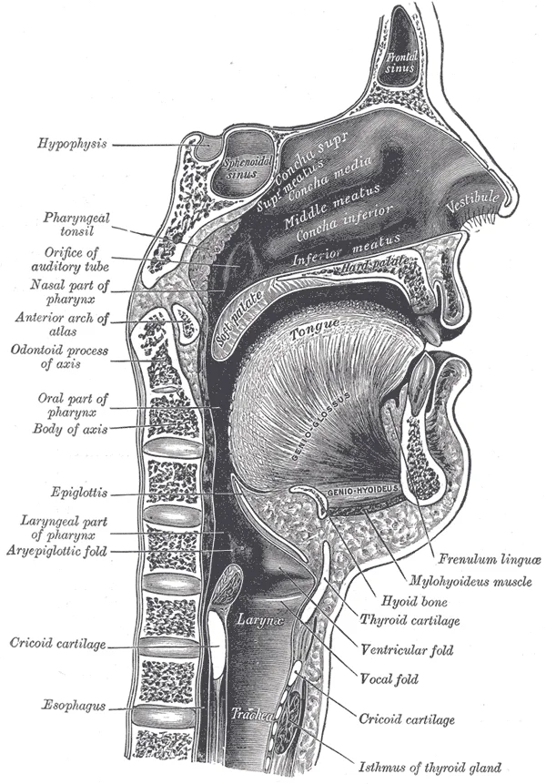
Long description
Sagittal section of nose, mouth, pharynx, and larynx.

Figure 3: Sagittal section of nose, mouth, pharynx, and larynx.
The inferior constrictor originates from the thyroid and cricoid cartilages. These muscles overlap like flower pots, creating a funnel shape that widens superiorly and narrows inferiorly. [1]
Anteriorly, the pharynx is related to the nasal cavity, oral cavity, and larynx. The nasopharynx lies posterior to the nasal cavity and is separated from it by the posterior nasal apertures. The oropharynx lies posterior to the oral cavity and is separated from it by the fauces. The laryngopharynx lies posterior to the larynx and is continuous with the oesophagus. The epiglottis forms the anterior boundary of the laryngeal inlet, directing food away from the respiratory tract.
Laterally, the pharynx is related to the carotid sheath, which contains the common carotid artery, internal jugular vein, and vagus nerve. The stylopharyngeus muscle passes between the superior and middle constrictors, providing structural support. The glossopharyngeal nerve and tonsillar branches of the external carotid artery are also found in this region. The parotid gland lies lateral to the mandibular ramus, near the angle of the jaw.
Posteriorly, the pharynx is related to the prevertebral fascia and the cervical vertebrae. This relationship is important for the spread of infection from the pharynx to the deep neck spaces. The alar fascia separates the prevertebral space from the danger space, which extends from the skull base to the diaphragm. Infection in the danger space can lead to mediastinitis, a life-threatening condition.
Foreign bodies often lodge in the piriform fossa due to its dependent position. The recurrent laryngeal nerve lies deep to the mucosa in this region, making it vulnerable during surgical procedures. [1]
DefinitionPiriform Fossa
A shallow depression on either side of the laryngeal inlet, bounded by the aryepiglottic fold and thyroid cartilage, where foreign bodies commonly lodge.
| Muscle | Origin | Insertion | Action | Nerve Supply |
|---|---|---|---|---|
| Superior Constrictor | Pterygomandibular raphe, mandible, tongue | Pharyngeal tubercle, median pharyngeal raphe | Constricts pharynx during swallowing | Pharyngeal plexus |
| Middle Constrictor | Styloid process, hyoid bone | Median pharyngeal raphe | Constricts pharynx during swallowing | Pharyngeal plexus |
| Inferior Constrictor | Thyroid and cricoid cartilages | Median pharyngeal raphe | Constricts pharynx during swallowing | Pharyngeal plexus, recurrent laryngeal nerve |
| Stylopharyngeus | Styloid process | Pharyngeal wall | Elevates pharynx during swallowing | Glossopharyngeal nerve |
| Salpingopharyngeus | Cartilage of auditory tube | Pharyngeal wall | Elevates pharynx during swallowing | Pharyngeal plexus |
The nasopharynx contains the pharyngeal tonsils, also known as adenoids, in its roof and posterior wall. These lymphoid tissues are part of Waldeyer's ring, which includes the palatine and lingual tonsils. The pharyngeal tonsils can hypertrophy in children, leading to nasal obstruction and sleep apnoea. The auditory tubes open into the lateral walls of the nasopharynx, equalising pressure between the middle ear and the atmosphere.
The oropharynx begins inferior to the nasopharynx and is continuous below with the laryngopharynx (). The inferior border of the laryngopharynx connects to the esophagus, whereas the anterior portion connects to the larynx, allowing air to flow into the bronchial tree. [1]
Figure 4Gray1029
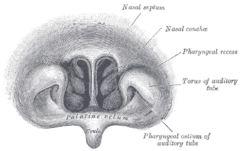
Long description
Front of nasa part of pharynx, as seen with the laryngoscope.

Figure 4: Front of nasa part of pharynx, as seen with the laryngoscope.
The nasopharynx is located at the top, behind the nasal cavity. Below it lies the oropharynx, which is continuous with the laryngopharynx at the bottom. The laryngopharynx connects anteriorly to the larynx and inferiorly to the esophagus. The palatine tonsils are situated between the palatoglossal and palatopharyngeal arches. The lingual tonsils are located at the base of the tongue. The glottis is the opening to the larynx, covered by the epiglottis during swallowing. The trachea and bronchi are below the larynx. The esophagus is posterior to the trachea. The nasal cavity is superior to the nasopharynx. The oral cavity is anterior to the oropharynx. The structures are arranged to facilitate both respiration and digestion. [1]
The pharyngeal arches, also known as branchial arches, are the fundamental structural units that give rise to the muscles, bones, cartilages, and nerves of the head and neck. [1]
Usually during swallowing, the soft palate and uvula rise reflexively to close off the entrance to the nasopharynx. At the same time, the larynx is pulled superiorly and the cartilaginous epiglottis, its most superior structure, folds inferiorly, covering the glottis (the opening to the larynx); this process effectively blocks access to the trachea and bronchi. When the food “goes down the wrong way,” it goes into the trachea. When food enters the trachea, the reaction is to cough, which usually forces the food up and out of the trachea, and back into the pharynx. [1]
The arterial network of the pharynx is complex, deriving from both the external and internal carotid systems. Understanding this supply is critical for surgical interventions, particularly those involving the tonsillar region. The primary source for the posterior pharyngeal wall is the ascending pharyngeal artery, a branch of the external carotid artery. This vessel ascends between the internal carotid artery and the side of the pharynx, supplying the superior constrictor muscle and the mucous membrane of the nasopharynx and oropharynx.
In the centre lies the palatine tonsil, situated in the tonsillar fossa between the palatoglossal and palatopharyngeal arches. The blood supply to the palatine tonsil is predominantly from the tonsillar branch of the facial artery. This branch descends to the anterior surface of the tonsil, piercing the superior constrictor muscle to reach the organ. Additional contributions come from the ascending palatine artery, also a branch of the facial artery, which supplies the upper pole. The dorsal lingual branches of the lingual artery supply the base of the tongue and the lower part of the tonsil. The lesser palatine arteries, branches of the descending palatine artery from the maxillary artery, contribute to the posterior and lateral aspects. The ascending pharyngeal artery provides minor branches to the posterior wall.
The Waldeyer ring is a circular arrangement of lymphoid tissue in the pharynx. It includes the pharyngeal tonsil (adenoids) in the roof of the nasopharynx, the tubal tonsils near the Eustachian tube openings, the palatine tonsils in the oropharynx, and the lingual tonsil at the base of the tongue. The arterial supply to this ring is multifactorial. The pharyngeal tonsil receives blood from the ascending pharyngeal artery and the dorsal lingual branches. The tubal tonsils are supplied by the ascending palatine artery and the ascending pharyngeal artery. The palatine tonsils, as noted, are mainly supplied by the tonsillar branch of the facial artery. The lingual tonsils receive blood from the dorsal lingual branches of the lingual artery and the ascending palatine artery.
Clinically, the rich vascular supply of the palatine tonsil explains the risk of significant haemorrhage during tonsillectomy. The tonsillar branch of the facial artery is the main vessel at risk. The ascending palatine artery may also be injured. The lingual artery branches are less commonly involved but can cause bleeding if the dissection extends too far inferiorly. The proximity of the internal carotid artery, which lies lateral to the pharyngeal wall, necessitates careful surgical technique to avoid catastrophic vascular injury. The ascending pharyngeal artery, being smaller, is less likely to cause major haemorrhage but can contribute to post-operative oozing. The tonsillar fossa is bounded anteriorly by the palatoglossal arch and posteriorly by the palatopharyngeal arch. The superior constrictor muscle forms the floor of the fossa, separating the tonsil from the pharyngeal wall and the underlying vessels. [1]
The venous drainage of the pharynx is complex, reflecting its rich vascular supply and critical anatomical position. Understanding this system requires tracing the flow from the mucosal plexuses to the major venous trunks of the neck. This plexus receives tributaries from the nasal cavity, the palate, and the tonsils. [1]
The plexus acts as a reservoir, collecting deoxygenated blood from the surrounding structures. It is closely related to the stylopharyngeus muscle and the glossop [1]
The illustration highlights how the lymphatic channels from the nasopharynx drain superiorly and laterally, while those from the
The proximity of the pharyngeal veins to the carotid sheath is clinically important. Infections can spread laterally into the deep cervical fascia. This can lead to parapharyngeal abscess formation. The internal jugular vein is at risk of thrombophlebitis. This condition, known as Lemierre's syndrome, often follows a tonsillar infection. The causative organism is typically Fusobacterium necrophorum. [1]
The internal jugular vein runs vertically in the carotid sheath. The glossopharyngeal nerve is located posterior to the plexus. The stylopharyngeus muscle lies lateral to the plexus.
| Structure | Origin | Termination |
|---|---|---|
| Superior Pharyngeal Vein | Pharyngeal Plexus | Facial Vein / Internal Jugular Vein |
| Middle Pharyngeal Vein | Pharyngeal Plexus | Internal Jugular Vein |
| Inferior Pharyngeal Vein | Lower Pharynx | Internal Jugular Vein / Brachiocephalic Vein |
The pharynx possesses a rich and interconnected lymphatic network that serves as a critical pathway for immune surveillance and fluid return. This system is not a single uniform channel but rather a complex plexus of vessels that vary in density and orientation depending on the specific sub-region of the pharynx being examined. Understanding this drainage is vital for medical students because it explains why infections in the oropharynx can spread rapidly to deep neck spaces, potentially leading
The illustration highlights how the lymphatic channels from the nasopharynx drain superiorly and laterally, while those from the
The clinical significance of this drainage pattern is profound. Because the lymphatic system of the pharynx is so extensive, it acts as a filter for pathogens and malignant cells. In the context of head and neck oncology, the pattern of lymphatic spread is a primary determinant of staging and surgical planning. For instance, a tumour in the tonsillar fossa will typically drain first to the jugulodigastric node, which is the first-echelon node for this region. If the disease progresses, it will involve the second-echelon nodes, such as the jugulo-omohyoid node. This stepwise progression is a fundamental concept in head and neck surgery.
Furthermore, the lymphatic drainage of the pharynx is intimately related to the venous drainage. The pharyngeal plexus of veins runs parallel to the lymphatic vessels, and both systems terminate in the internal jugular vein. This anatomical relationship is crucial during surgical procedures, as damage to these structures can lead to significant haemorrhage or chyle leaks. The pharyngeal plexus is also a site of potential venous stasis, which can contribute to the development of varices in patients with portal hypertension, although this is more commonly associated with the lower oesophagus.
In summary, the lymphatic drainage of the pharynx is a complex, multi-layered system that follows predictable anatomical pathways. Mastery of this topic requires not only memorising the names of the nodes but also understanding the functional implications of their location and connectivity. This knowledge is essential for interpreting clinical findings, planning investigations, and managing patients with head and neck pathologies. The interplay between lymphatic and venous systems in this region underscores the importance of a holistic anatomical understanding for safe and effective clinical practice.
The pharynx receives a complex innervation that integrates somatic motor control for swallowing with autonomic regulation and sensory feedback. This dual supply ensures coordinated function during deglutition and protects the airway. The primary somatic motor supply to the pharyngeal constrictors and most pharyngeal muscles derives from the pharyngeal plexus.
DefinitionPharyngeal Plexus
A network of nerves on the middle constrictor muscle formed by vagal and glossopharyngeal contributions, providing motor and sensory innervation to the pharynx.
Sensory innervation is critical for the gag reflex and swallowing initiation. The glossopharyngeal nerve supplies general sensation to the oropharynx, including the tonsillar fossa and the posterior third of the tongue. The vagus nerve supplies sensation to the laryngopharynx and the lower part of the oropharynx. The internal laryngeal branch of the superior laryngeal nerve, a branch of the vagus, provides sensation to the mucosa above the vocal cords. This sensory input triggers the afferent limb of the gag reflex, while the efferent limb is mediated by the vagus nerve. [1]
Referred pain is a significant clinical feature. Pain from the tonsils, often due to tonsillitis, is frequently referred to the ear. This occurs because both the tonsillar region and the external ear share sensory innervation from the glossopharyngeal nerve and the auriculotemporal branch of the trigeminal nerve. Understanding this pathway helps in diagnosing otalgia without primary ear pathology.
| Structure | Nerve Supply | Function |
|---|---|---|
| Superior Constrictor | Pharyngeal Plexus (Vagus) | Motor: Swallowing |
| Middle Constrictor | Pharyngeal Plexus (Vagus) | Motor: Swallowing |
| Inferior Constrictor | Pharyngeal Plexus (Vagus) | Motor: Swallowing |
| Stylopharyngeus | Glossopharyngeal Nerve | Motor: Elevates pharynx |
| Oropharyngeal Mucosa | Glossopharyngeal Nerve | Sensation |
| Laryngopharyngeal Mucosa | Vagus Nerve | Sensation |
Autonomic innervation is provided by the sympathetic and parasympathetic fibres that travel with the pharyngeal plexus. These fibres regulate blood flow and glandular secretion in the pharyngeal mucosa. The sympathetic fibres originate from the superior cervical ganglion, while parasympathetic fibres arise from the inferior salivatory nucleus via the glossopharyngeal nerve and the superior salivatory nucleus via the facial nerve. These autonomic inputs ensure adequate lubrication and vascular tone during swallowing.
The Waldeyer’s ring, a circular arrangement of lymphoid tissue, relies on this rich sensory and motor supply for its protective function. The tonsils, a key component, are innervated by the glossopharyngeal nerve, which explains the severe pain associated with tonsillar inflammation. The coordination of these nerves allows for the seamless transition of food from the oral cavity to the oesophagus, preventing aspiration and ensuring efficient digestion.
The pharynx serves as a critical junction for both the respiratory and digestive systems, lined by distinct epithelial types that reflect its dual functional demands. Understanding the histological architecture is essential for grasping how the pharynx withstands mechanical stress while maintaining barrier integrity.
The nasopharynx is the uppermost part of the pharynx, located posterior to the nasal cavity. It is lined by pseudostratified ciliated columnar epithelium, also known as respiratory epithelium. This epithelium contains goblet cells that secrete mucus to trap particulate matter. The underlying lamina propria contains seromucous glands that further lubricate the surface. This mucociliary escalator is vital for protecting the lower respiratory tract.
The oropharynx extends from the soft palate to the epiglottis. Its lining is non-keratinised stratified squamous epithelium, which provides protection against abrasion from food boluses. This epithelium is thicker than that of the nasopharynx to withstand mechanical stress. The lamina propria is rich in lymphoid tissue, forming the palatine and lingual tonsils. These lymphoid aggregates are part of Waldeyer's ring and play a significant role in immune surveillance. The presence of lymphoid follicles allows for the trapping and destruction of pathogens ingested or inhaled. [1]
The laryngopharynx, or hypopharynx, lies posterior to the larynx and extends to the lower border of the cricoid cartilage. It is also lined by non-keratinised stratified squamous epithelium. This region serves as a common pathway for both air and food. The epithelium here is particularly robust due to the constant passage of food. The transition to the oesophagus occurs at the level of the cricopharyngeus muscle, which acts as a sphincter to prevent reflux.
Clinical pearlClinical Correlation
Changes in epithelial type can indicate metaplasia due to chronic irritation, such as smoking, which may predispose to malignancy.
The histological differences between these regions highlight the specialized adaptations of the pharynx. The nasopharynx prioritizes air filtration and humidification, while the oropharynx and laryngopharynx prioritize protection and lubrication. This structural diversity ensures efficient function in both respiration and digestion.
The first arch, or mandibular arch, gives rise to the muscles of mastication, mylohyoid, anterior belly of digastric, tensor tympani, and tensor veli palatini. The second arch, or hyoid arch, contributes to the muscles of facial expression, stapedius, stylohyoid, and posterior belly of digastric. These are innervated by the facial nerve (CN VII). The third arch forms the stylopharyngeus muscle, supplied by t [1]
The second pouch contributes to the palatine tonsils. [1]
The pharyngeal clefts, or grooves, are ectodermal depressions between the arches. The first cleft forms the external auditory meatus. The other clefts normally obliterate, but failure of obliteration can lead to branchial cysts or fistulas. The membrane between the first cleft and first pouch becomes the tympanic membrane. Understanding these developmental origins is essential for recognizing congenital anomalies and their clinical presentations. [1]
DefinitionPharyngeal Arch
A series of mesenchymal swellings on the lateral sides of the embryonic head and neck region, each consisting of a cartilaginous bar, muscle mass, artery, and nerve.
| Arch | Cartilage | Muscle | Nerve | Artery |
|---|---|---|---|---|
| 1st | Meckel's cartilage | Muscles of mastication | CN V3 | Maxillary artery |
| 2nd | Reichert's cartilage | Facial expression | CN VII | Stapedial artery |
| 3rd | Greater horn of hyoid | Stylopharyngeus | CN IX | Carotid artery |
| 4th | Thyroid cartilage | Pharyngeal constrictors | CN X (SLN) | Aortic arch (L) |
| 6th | Cricoid cartilage | Intrinsic laryngeal | CN X (RLN) | Pulmonary artery |
The pharynx is a funnel-shaped muscular tube that connects the nasal and oral cavities to the larynx and oesophagus. It extends from the base of the skull to the lower border of the cricoid cartilage at the sixth cervical vertebra. Radiologically, the pharynx is divided into three distinct regions: the nasopharynx, the oropharynx, and the hypopharynx. These regions are separated by anatomical landmarks that are clearly visible on sagittal and coronal imaging planes.
The nasopharynx lies posterior to the nasal cavity and superior to the soft palate. It extends from the base of the skull to the level of the soft palate. The opening of the Eustachian tube is located on the lateral wall of the nasopharynx, just posterior to the torus tubarius. The adenoid pad, or pharyngeal tonsil, is situated on the posterior wall. In children, this tissue is often prominent and can be seen as a soft tissue mass in the nasopharynx on lateral neck radiographs. The nasopharynx is the site of the fossa of Rosenmüller, a recess located posterior to the Eustachian tube opening. This area is clinically significant because it is a common site for nasopharyngeal carcinoma.
The oropharynx extends from the level of the soft palate to the epiglottis. It includes the fauces, the palatine arches, the tongue, and the palatine tonsils. The palatine arches are formed by the palatoglossal and palatopharyngeal muscles. The palatine tonsils are located in the tonsillar fossa, which is the space between the palatoglossal and palatopharyngeal arches. On imaging, the tonsils appear as soft tissue masses in the lateral oropharynx. The base of the tongue forms the anterior boundary of the oropharynx. The epiglottis marks the inferior limit of this region. The oropharynx is the most common site for foreign body impaction, particularly in children.
The hypopharynx, or laryngopharynx, extends from the epiglottis to the lower border of the cricoid cartilage. It is divided into three parts: the valleculae, the piriform fossae, and the postcricoid region. The valleculae are the recesses located between the base of the tongue and the epiglottis. The piriform fossae are the funnel-shaped depressions located on either side of the laryngeal inlet. They are bounded anteriorly by the aryepiglottic fold and posteriorly by the thyroid cartilage. The postcricoid region is the area posterior to the larynx. The hypopharynx is the most common site for squamous cell carcinoma in adults. The piriform fossa is a critical landmark for identifying the laryngeal inlet on imaging.
Radiological anatomy of the pharynx is best understood using sagittal and coronal views. The image displays the nasopharynx at the top, with the adenoid pad visible on the posterior wall. The torus tubarius is seen as a bony prominence on the lateral wall. The soft palate separates the nasopharynx from the oropharynx. The oropharynx contains the palatine tonsils, which are visible as soft tissue masses on the lateral sides. The base of the tongue forms the anterior boundary of the oropharynx. The epiglottis is clearly visible as a curved structure at the inferior limit of the oropharynx. The hypopharynx is seen below the epiglottis, with the piriform fossae visible as depressions on either side of the laryngeal inlet. The cricoid cartilage forms the inferior boundary of the pharynx. The airway is patent throughout the pharynx, with no evidence of obstruction. The soft tissues are normal in thickness and density. The bony structures, including the cervical vertebrae, are intact and aligned. [1]
| Region | Superior limit | Inferior limit | Key structures |
|---|---|---|---|
| Nasopharynx | Base of skull | Soft palate | Adenoids, Eustachian tube |
| Oropharynx | Soft palate | Epiglottis | Palatine tonsils, tongue |
| Hypopharynx | Epiglottis | Cricoid cartilage | Piriform fossae, valleculae |
The pharynx is a muscular tube connecting the nasal and oral cavities to the larynx and oesophagus. It is divided into three distinct regions: the nasopharynx, oropharynx, and laryngopharynx. The nasopharynx extends from the base of the skull to the soft palate. It contains the torus tubarius, which is the swelling formed by the opening of the auditory tube, and the pharyngeal recess, or fossa of Rosenmuller, located posterior to the torus tubarius. This recess is a common site for nasopharyngeal carcinoma. The adenoid tissue, or pharyngeal tonsil, lies in the roof and posterior wall of this region. It is bounded laterally by the palatoglossal arches, which contain the palatoglossus muscles.
The pharyngeal constrictor muscles form a funnel-shaped wall. The superior constrictor originates from the pterygomandibular raphe, the pterygoid hamulus, the posterior end of the mandibular alveolar process, and the side of the tongue. It inserts into the pharyngeal raphe. The middle constrictor arises from the hyoid bone and inserts into the raphe. The inferior constrictor originates from the thyroid and cricoid cartilages and inserts into the raphe and the posterior edge of the epiglottis. These muscles are innervated by the pharyngeal plexus, derived from the vagus nerve.
| Muscle | Origin | Insertion | Action | Nerve Supply |
|---|---|---|---|---|
| Superior Constrictor | Pterygomandibular raphe, pterygoid hamulus, mandible, tongue | Pharyngeal raphe | Narrows pharynx | Pharyngeal plexus |
| Middle Constrictor | Hyoid bone | Pharyngeal raphe | Narrows pharynx | Pharyngeal plexus |
| Inferior Constrictor | Thyroid and cricoid cartilages | Pharyngeal raphe, epiglottis | Narrows pharynx | Pharyngeal plexus |
The pharyngeal recess is a blind pouch behind the auditory tube opening. It is clinically significant because it is a common site for nasopharyngeal carcinoma, which may present with unilateral hearing loss due to obstruction of the auditory tube. The prevertebral space, located between the pharynx and the prevertebral fascia, is a potential space for abscess formation. Retropharyngeal abscesses can compress the airway and cause neck swelling. The tonsillar fossa, located in the oropharynx, contains the palatine tonsil. Tonsillitis and peritonsillar abscess are common infections in this region. The laryngopharynx is the most common site for squamous cell carcinoma in adults, often presenting with hoarseness or dysphagia.
The pharynx serves as a common pathway for both the respiratory and digestive systems, divided into three distinct regions: the nasopharynx, oropharynx, and laryngopharynx. [1]
The nasopharynx lies posterior to the nasal cavities and superior to the soft palate. It is lined by respiratory epithelium and contains the pharyngeal tonsils, also known as adenoids, in its posterior wall. The opening of the auditory tube (Eustachian tube) is located on the lateral wall, allowing pressure equalisation between the middle ear and the nasopharynx. A common trap is confusing the location of the adenoids with the palatine tonsils, which are situated in the oropharynx.
The oropharynx extends from the soft palate to the upper border of the epiglottis. It contains the palatine tonsils, located between the palatoglossal and palatopharyngeal arches. The posterior wall is formed by the prevertebral fascia and the superior constrictor muscle. Clinically, this region is a common site for infections and malignancies. Students often forget that the oropharynx is continuous with the oral cavity anteriorly.
The laryngopharynx extends from the upper border of the epiglottis to the lower border of the cricoid cartilage. It is divided into the piriform fossae laterally and the postcricoid region posteriorly. The piriform fossae are recesses where foreign bodies frequently lodge. The laryngopharynx opens anteriorly into the larynx and inferiorly into the oesophagus. Understanding the boundaries is crucial for endoscopic procedures.
The pharyngeal constrictor muscles overlap like a flower pot, contracting sequentially during swallowing to propel the bolus downwards. The stylopharyngeus, palatopharyngeus, and salpingopharyngeus muscles elevate the pharynx during swallowing and speech.
| Muscle | Action | Nerve Supply |
|---|---|---|
| Superior Constrictor | Constricts pharynx | Pharyngeal plexus |
| Middle Constrictor | Constricts pharynx | Pharyngeal plexus |
| Inferior Constrictor | Constricts pharynx | Pharyngeal plexus |
| Stylopharyngeus | Elevates pharynx | Glossopharyngeal nerve |
| Palatopharyngeus | Elevates pharynx | Pharyngeal plexus |
Peritonsillar abscess is a common complication of tonsillitis, presenting with a 'hot potato' voice and trismus. The abscess typically forms between the palatine tonsil and the superior constrictor muscle. Knowledge of the vascular supply, primarily from the facial and ascending pharyngeal arteries, is essential for managing haemorrhage.
Exam trapNerve Supply Trap
Most pharyngeal muscles are innervated by the pharyngeal plexus (vagus nerve), except the stylopharyngeus, which is supplied by the glossopharyngeal nerve.
Clinical pearlSwallowing Mechanism
During swallowing, the soft palate elevates to close off the nasopharynx, preventing food entry into the nasal cavity.
Students frequently confuse the anatomical boundaries of the oral cavity with those of the pharynx, leading to errors in clinical localisation. A critical distinction is that the oral vestibule is the pocket-like space framed on the inside by the gums and teeth, and on the outside by the cheeks and lips[1]. Many learners incorrectly include this vestibule within the oral cavity proper. The oral cavity proper is distinct; it runs from the gums and teeth to the fauces, which is the opening between the oral cavity and the throat (oropharynx)[1]. Understanding this separation is vital for accurate examination of lesions or foreign bodies.
Another frequent error involves the innervation and structure of the tongue. Students often overlook that the tongue is attached to multiple bony structures, specifically the mandible, the styloid processes of the temporal bones, and the hyoid bone[1]. A unique feature of the hyoid bone is that it only distantly or indirectly articulates with other bones[1]. Confusion also arises regarding the papillae. Fungiform papillae are mushroom-shaped and contain taste buds, whereas filiform papillae are long and thin, lacking taste buds but possessing touch receptors that help move food[1]. Mistaking the function of filiform papillae as gustatory is a common trap; they create an abrasive surface for mechanical grooming rather than taste perception[1].
Exam trapFrenulum Confusion
Do not confuse the labial frenulum, which attaches the inner lip to the gum, with the lingual frenulum, which tethers the tongue to the floor of the mouth[1].
Clinical anomalies such as ankyloglossia, or "tongue tie," are often misunderstood as purely cosmetic. In reality, severe ankyloglossia involves a lingual frenulum that is too short or malformed, which can impair speech and requires surgical correction[1]. Students may also underestimate the volume of saliva produced. An average of 1 to 1.5 liters of saliva is secreted each day by minor and major glands, a volume essential for moistening food and initiating carbohydrate breakdown[1]. Finally, the vascular nature of the lips is frequently ignored. The lips have a huge representation on the cerebral cortex and are very vascular with a thin layer of keratin, explaining their red colour and high sensitivity[1].
At the bedside, knowledge of the pharynx and oral anatomy is essential for assessing airway patency and swallowing function. The fauces serve as the gateway from the oral cavity to the oropharynx, and obstruction here can compromise breathing[1]. During examination, clinicians inspect the gingivae, which are soft tissues lining the alveolar processes and surrounding the necks of the teeth[1]. Periodontal health is assessed by checking the periodontal ligament that holds teeth in their sockets within the maxilla and mandible[1]. Salivary function is evaluated by observing the secretion from minor glands in the lips, cheeks, palate, and tongue, which ensure adequate moisture for speech and digestion[1].
In the operating theatre, anatomical precision is required for procedures involving the tongue or frenulum. Correction of severe ankyloglossia requires careful incision of the lingual frenulum to restore tongue mobility without damaging underlying structures[1]. Dental procedures involve the alveolar processes, where teeth are secured. Understanding the transition from skin to mucous membrane at the lips and cheeks is crucial for suturing and wound management[1]. The buccinator muscle, located between the skin and mucous membrane of the cheeks, must be respected during facial surgeries to maintain oral competence[1].
Clinical pearlAirway Landmark
The fauces is the key landmark separating the oral cavity proper from the oropharynx, critical for intubation and airway management[1].
On imaging, such as CT or MRI, distinguishing the oral vestibule from the oral cavity proper helps localise abscesses or tumours. The vestibule is lateral to the teeth, while the cavity proper is medial[1]. The tongue’s attachment to the hyoid and styloid processes provides landmarks for assessing deep neck space infections. The hyoid’s unique indirect articulation makes it a stable reference point in imaging analysis[1]. Salivary gland pathology is also evaluated; the major glands secrete the majority of saliva into ducts opening into the mouth, while minor glands are distributed throughout the mucosa[1].
In examinations, students must accurately describe the layers of the lips. The outer covering is skin, transitioning to mucous membrane, with the orbicularis oris muscle regulating the opening[1]. The non-keratinized stratified squamous epithelium of the cheeks contrasts with the keratinised skin externally[1]. Understanding the neuromuscular control of deglutition is also tested. The entry of food into the esophagus marks the esophageal phase, controlled by the medulla oblongata, where peristalsis propels the bolus via circular and longitudinal muscle contractions[1]. This coordinated action ensures food moves toward the stomach, facilitated by mucus from esophageal glands[1].
The oral vestibule is framed by the cheeks/lips externally and gums/teeth internally[1]. The oral cavity proper runs from the gums/teeth to the fauces[1]. The fauces is the opening between the oral cavity and oropharynx[1]. The tongue attaches to the mandible, styloid processes, and hyoid bone[1]. The hyoid bone only distantly or indirectly articulates with other bones[1]. Fungiform papillae contain taste buds; filiform papillae have touch receptors[1]. Severe ankyloglossia impairs speech and requires surgical correction[1]. 1 to 1.5 liters of saliva are secreted daily[1].
Answer each one in your head first, then open the answer.
What structures frame the oral vestibule?
Show answerHide answer
The gums and teeth on the inside, and the cheeks and lips on the outside[1]Which bone only indirectly articulates with other bones?
Show answerHide answer
The hyoid bone[1]What is the function of filiform papillae?
Show answerHide answer
They have touch receptors that help move food around[1]What is the medical term for tongue tie?
Show answerHide answer
Ankyloglossia[1]How much saliva is secreted on average each day?
Show answerHide answer
1 to 1.5 liters[1]
- Betts JG, Young KA, Wise JA, et al. The Mouth, Pharynx, and Esophagus. OpenStax, Anatomy and Physiology 2e (CC BY-NC-SA 4.0). Accessed .
- Abe S, Fukuda M, Yamane S, et al. Fetal anatomy of the upper pharyngeal muscles with special reference to the nerve supply: is it an enteric plexus or simply an intramuscular nerve?. Anatomy & cell biology 2013;46(2):141-148. doi:10.5115/acb.2013.46.2.141. Accessed .
Figure credits
- Figure 1: Henry Vandyke Carter, via Wikimedia Commons, Public domain, Public domain.
- Figure 2: Henry Vandyke Carter, via Wikimedia Commons, Public domain, Public domain.
- Figure 3: Henry Vandyke Carter, via Wikimedia Commons, Public domain, Public domain.
- Figure 4: Henry Vandyke Carter, via Wikimedia Commons, Public domain, Public domain.
Educational content for medical students and doctors. It is not a substitute for clinical judgement or local guidelines.