Larynx
Anatomy of the larynx, its cartilages, muscles, and role in airway and speech.
Figure 1Gray952
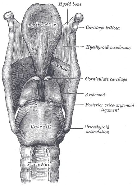
Long description
Ligaments of the larynx. Posterior view.

Figure 1: Ligaments of the larynx (posterior view).
Figure 2Gray951
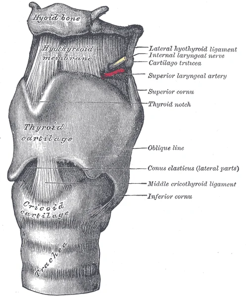
Long description
The ligaments of the larynx. Antero-lateral view.

Figure 2: The ligaments of the larynx (antero-lateral view).
The larynx is a cartilaginous structure situated inferior to the laryngopharynx, connecting the pharynx to the trachea. It serves to regulate the volume of air entering and leaving the lungs. The laryngopharynx lies posterior to the larynx and continues the route for ingested material and air until the digestive and respiratory systems diverge. Anteriorly, the laryngopharynx opens into the larynx, while posteriorly it enters the esophagus. [1]
The structure is formed by several cartilage pieces. Three large cartilages form the major framework: the thyroid cartilage anteriorly, the epiglottis superiorly, and the cricoid cartilage inferiorly. The thyroid cartilage is the largest piece, consisting of the laryngeal prominence or “Adam’s apple,” which is more prominent in males. The thick cricoid cartilage forms a complete ring with a wide posterior region and a thinner anterior region. [1]
Three smaller, paired cartilages attach to the epiglottis and vocal cords: the arytenoids, corniculates, and cuneiforms. These structures help move the vocal cords to produce speech. [1]
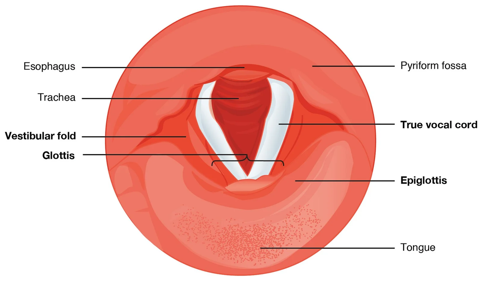
The epiglottis is a flexible piece of elastic cartilage attached to the thyroid cartilage. It covers the opening of the trachea. In the closed position, the unattached end rests on the glottis. The glottis is composed of the vestibular folds, the true vocal cords, and the space between them. The vestibular fold, or false vocal cord, is a folded section of mucous membrane. The true vocal cord is a white, membranous fold attached by muscle to the thyroid and arytenoid cartilages. The inner edges are free, allowing oscillation to produce sound. Folds in males are larger than in females, creating a deeper voice. [1]
Swallowing causes the pharynx and larynx to lift upward. This allows the epiglottis to swing downward, closing the opening to the trachea. These movements prevent food and beverages from entering the trachea. [1]
The trachea extends from the larynx toward the lungs. It is formed by 16 to 20 stacked, C-shaped pieces of hyaline cartilage. These are connected by dense connective tissue and the fibroelastic membrane. The trachea is lined with pseudostratified ciliated columnar epithelium, continuous with the larynx. The esophagus borders the trachea posteriorly. [1]
DefinitionRima Glottidis
The space between the true vocal cords, crucial for phonation and airway patency.
| Cartilage | Type | Key Feature |
|---|---|---|
| Thyroid | Hyaline | Largest; Adam's apple |
| Cricoid | Hyaline | Complete ring; narrow anteriorly |
| Epiglottis | Elastic | Covers trachea during swallowing |
| Arytenoid | Hyaline | Paired; attaches vocal cords |
Exam trapAirway Emergency
In complete upper airway obstruction, a cricothyroidotomy may be required to bypass the obstruction by entering the trachea below the cricoid cartilage.
Figure 3Gray994
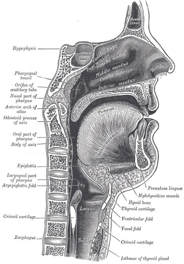
Long description
Sagittal section of nose, mouth, pharynx, and larynx.

Figure 3: Sagittal section of nose, mouth, pharynx, and larynx.
The central landmark is the thyroid cartilage, the largest cartilaginous structure, which forms the anterior and lateral walls. Its most prominent feature is the laryngeal prominence, commonly known as the Adam’s apple, which is more pronounced in males. Superior to the thyroid cartilage lies the hyoid bone, a U-shaped bone that does not articulate with any other bone but serves as an anchor for muscles. The epiglottis, a leaf-shaped elastic cartilage, is attached to the thyroid cartilage and projects upwards behind the tongue. Inferiorly, the cricoid cartilage forms a complete ring, resembling a signet ring, with the narrower anterior arch and the broader posterior lamina. The cricothyroid joint connects the cricoid and thyroid cartilages, allowing for tension adjustment of the vocal cords. The trachea extends inferiorly from the cricoid cartilage, continuing the airway into the thorax.
Figure 42306 The Larynx
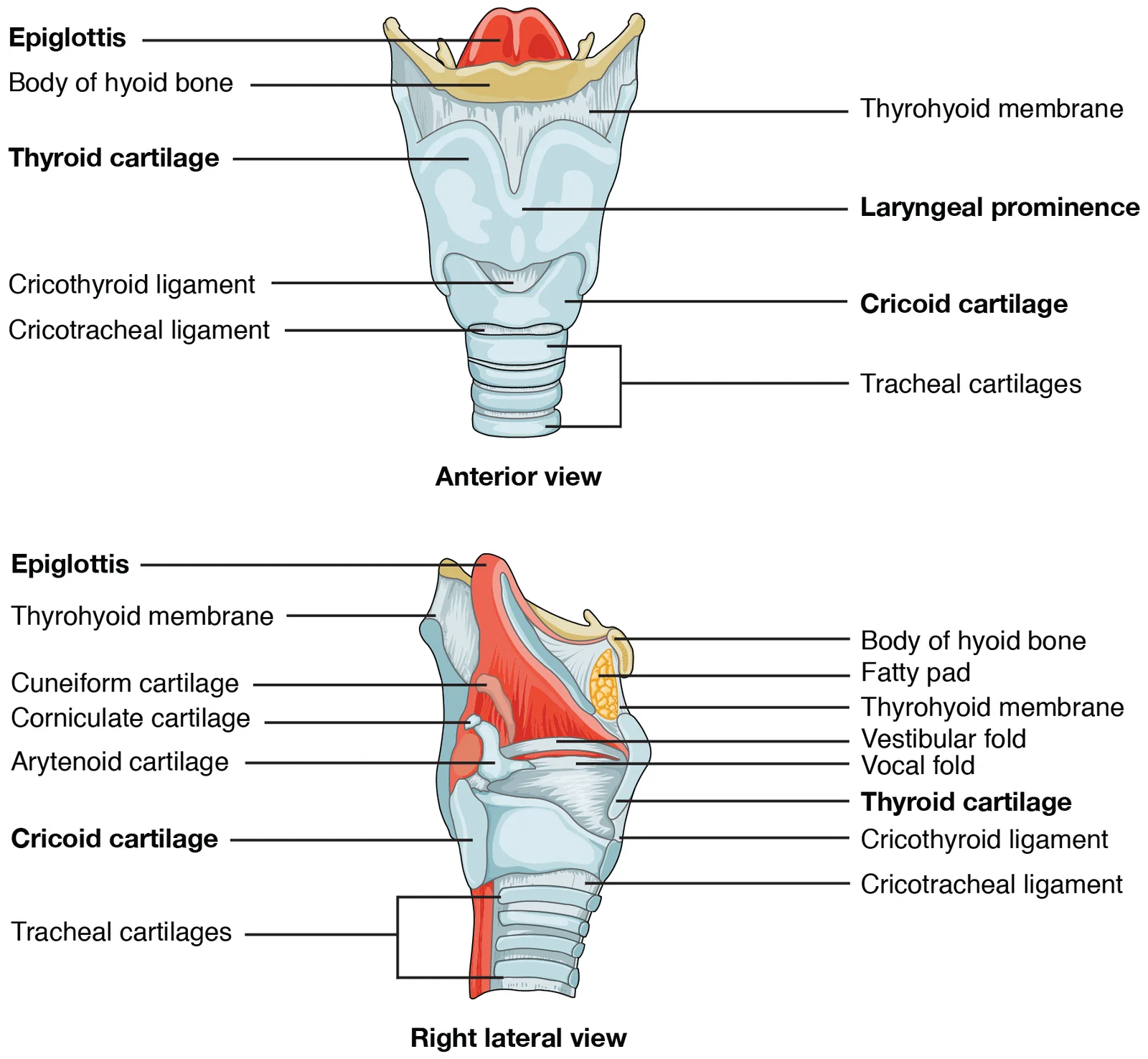
Long description
The larynx extends from the laryngopharynx and the hyoid bone to the trachea.

Figure 4: The larynx extends from the laryngopharynx and the hyoid bone to the trachea.
The larynx serves as the major entrance and exit for the respiratory system, positioned between the pharynx and the trachea. Its primary functions include phonation, protection of the lower airway during swallowing, and regulation of airflow. The internal lining of the larynx varies by region. The superior portion, continuous with the laryngopharynx, is lined with stratified squamous epithelium, which provides durability against mechanical stress. As one moves inferiorly, this transitions into pseudostratified ciliated columnar epithelium containing goblet cells. This specialized epithelium produces mucus to trap debris and pathogens as they enter the trachea. The cilia beat the mucus upward towards the laryngopharynx, where it can be swallowed down the esophagus, ensuring the lower airways remain clear. [1]
The muscles of the larynx are divided into extrinsic and intrinsic groups. Extrinsic muscles connect the larynx to surrounding structures, primarily elevating or depressing the larynx during swallowing and speech. Intrinsic muscles, located entirely within the larynx, control the movement of the vocal cords. These muscles are responsible for abducting and adducting the vocal folds, as well as adjusting their tension and length to modulate pitch.
| Muscle | Action | Nerve Supply |
|---|---|---|
| Cricothyroid | Tenses vocal cords | External branch of superior laryngeal nerve |
| Posterior cricoarytenoid | Abducts vocal cords | Recurrent laryngeal nerve |
| Lateral cricoarytenoid | Adducts vocal cords | Recurrent laryngeal nerve |
| Transverse arytenoid | Adducts vocal cords | Recurrent laryngeal nerve |
| Oblique arytenoid | Adducts vocal cords | Recurrent laryngeal nerve |
Damage to the recurrent laryngeal nerve can result in vocal cord paralysis, leading to hoarseness or airway obstruction. The blood supply to the larynx is derived from the superior and inferior laryngeal arteries, branches of the superior and inferior thyroid arteries respectively. Venous drainage follows the arterial supply, ultimately emptying into the internal jugular vein. Lymphatic drainage varies by location; structures above the vocal cords drain to the deep cervical nodes, while those below drain to the pretracheal and paratracheal nodes. This anatomical distinction is crucial in the staging of laryngeal cancers, as metastasis patterns differ significantly between supraglottic and glottic lesions. [1]
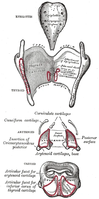
Figure 5Gray950
Long description
The cartilages of the larynx. Posterior view.

Figure 5: The cartilages of the larynx (posterior view).
The larynx is a complex tubular structure situated in the anterior neck, serving as the conduit for air and the organ of phonation. To understand its spatial arrangement, one must consider its relations to surrounding structures, which define its clinical accessibility and potential for compression. In this view, the larynx is shown in its anatomical position within the neck. The hyoid bone lies superior to the larynx, connected by the thyrohyoid membrane. The thyroid cartilage forms the largest anterior component, with its laminae meeting at the anterior thyroid notch. Posterior to the thyroid cartilage lies the cricoid cartilage, which is the only complete ring of cartilage in the respiratory tract. The cricoid cartilage sits inferiorly, articulating with the trachea below. Anteriorly, the larynx is covered by the strap muscles (infrahyoid muscles), specifically the sternothyroid and thyrohyoid muscles, which lie superficial to the thyroid cartilage. Posteriorly, the larynx relates to the prevertebral fascia and the cervical vertebrae. Laterally, the larynx is bounded by the carotid sheath, which contains the common carotid artery, internal jugular vein, and vagus nerve. The recurrent laryngeal nerves run in the tracheoesophageal groove, posterior to the larynx and thyroid gland, making them vulnerable during thyroid surgery. Superiorly, the larynx is continuous with the pharynx via the laryngeal inlet, guarded by the epiglottis. The epiglottis is a leaf-shaped cartilage that projects upwards and backwards, covering the inlet during swallowing to prevent aspiration. The vestibular folds (false vocal cords) and vocal folds (true vocal cords) are located within the laryngeal cavity. The vocal folds are attached anteriorly to the thyroid cartilage and posteriorly to the arytenoid cartilages. The arytenoid cartilages are pyramidal in shape and sit on the superior border of the cricoid cartilage. They play a crucial role in opening and closing the glottis. The larynx is supported by several ligaments, including the cricothyroid ligament, which connects the cricoid and thyroid cartilages. The intrinsic muscles of the larynx, such as the posterior cricoarytenoid, lateral cricoarytenoid, and thyroarytenoid, act on these cartilages to modulate vocal cord tension and position. The extrinsic muscles, including the sternothyroid, sternohyoid, omohyoid, and thyrohyoid, move the larynx as a whole during swallowing and speech. The blood supply to the larynx comes from the superior and inferior laryngeal arteries, branches of the superior and inferior thyroid arteries, respectively. Venous drainage follows the arterial supply, emptying into the superior and middle thyroid veins, which drain into the internal jugular vein. Lymphatic drainage from the larynx is divided into supraglottic and glottic regions. The supraglottic region drains to the deep cervical lymph nodes, while the glottic region has sparse lymphatic drainage, which explains the delayed metastasis in glottic cancers. The innervation of the larynx is primarily via the vagus nerve (CN X). Understanding these relations is essential for surgical approaches, such as tracheostomy, which is typically performed between the second and third tracheal rings, below the cricoid cartilage. [1]
Figure 62307 Cartilages of the Larynx
Long description
The true vocal cords and vestibular folds of the larynx are viewed inferiorly from the laryngopharynx.

Figure 6: The true vocal cords and vestibular folds of the larynx are viewed inferiorly from the laryngopharynx.
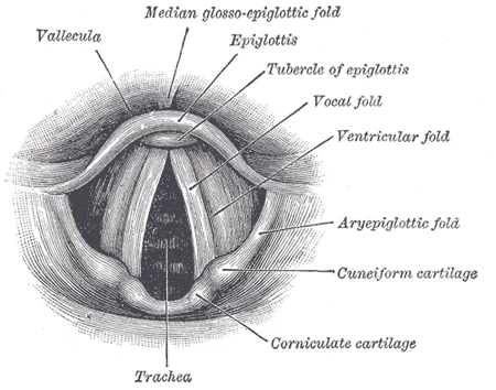
Figure 7Gray956
Long description
Laryngoscopic view of interior of larynx.

Figure 7: Laryngoscopic view of interior of larynx.
The larynx is a tubular organ connecting the pharynx to the trachea. Its internal cavity is divided into three regions: the laryngeal vestibule, the ventricle, and the infraglottic cavity. The vestibule lies above the vocal folds. The ventricle is a lateral recess between the true and false vocal folds. The infraglottic cavity extends from the vocal folds to the lower border of the cricoid cartilage.
DefinitionVocal Folds
The true vocal folds are the primary structures for phonation, located inferior to the false vocal folds.
The mucosa of the larynx is lined by respiratory epithelium, which is pseudostratified ciliated columnar epithelium containing mucus-producing goblet cells. This epithelium lines the trachea and the bronchi, providing a protective barrier. [1]
| Cartilage | Type | Key Feature |
|---|---|---|
| Thyroid | Hyaline | Largest cartilage; forms the anterior wall. |
| Cricoid | Hyaline | Complete ring; sits below the thyroid. |
| Epiglottis | Elastic | Leaf-shaped; covers the laryngeal inlet during swallowing. |
| Arytenoid | Hyaline | Pyramidal; sits on the cricoid; anchors vocal folds. |
The laryngeal skeleton consists of nine cartilages. The thyroid cartilage is the largest and forms the anterior wall. The cricoid cartilage is a complete ring that sits below the thyroid cartilage. The epiglottis is a leaf-shaped elastic cartilage that covers the laryngeal inlet during swallowing. The arytenoid cartilages are pyramidal and sit on the superior border of the cricoid cartilage. They anchor the vocal folds and allow for their movement.
The muscles of the larynx are innervated by the recurrent laryngeal nerve, a branch of the vagus nerve. The posterior cricoarytenoid muscles are the only abductors of the vocal folds. The lateral cricoarytenoid muscles adduct the vocal folds. The thyroarytenoid muscles relax the vocal folds. The cricothyroid muscles tense the vocal folds.
Clinical pearlClinical Pearl
Damage to the recurrent laryngeal nerve can cause vocal cord paralysis, leading to hoarseness or airway obstruction.
The blood supply to the larynx comes from the superior and inferior laryngeal arteries. The venous drainage follows the arterial supply.
The lymphatic drainage of the larynx is divided into supraglottic and glottic regions. The supraglottic region drains to the deep cervical lymph nodes. The glottic region has sparse lymphatic drainage, which explains why glottic cancers often present late. The infraglottic region drains to the pretracheal and paratracheal lymph nodes.
Exam trapCommon Trap
Do not confuse the false vocal folds (vestibular folds) with the true vocal folds. The false vocal folds are superior and do not produce sound.
The larynx plays a crucial role in phonation, respiration, and protection of the lower airway. The vocal folds vibrate to produce sound. The laryngeal inlet closes during swallowing to prevent aspiration. The cough reflex, mediated by the carina and laryngeal nerves, protects the airway from foreign bodies.
The arterial supply to the larynx is primarily derived from the superior and inferior thyroid arteries, with contributions from the laryngeal branches of the superior thyroid artery and the cricothyroid artery. The superior thyroid artery, a branch of the external carotid artery, descends to the upper pole of the thyroid gland and gives rise to the superior laryngeal artery. This artery pierces the thyrohyoid membrane and divides into anterior and posterior branches, which supply the structures of the larynx above the vocal cords. [1]
The inferior thyroid artery, a branch of the thyrocervical trunk, ascends in the neck to reach the posterior aspect of the thyroid gland. From this artery, the inferior laryngeal artery arises, ascending within the tracheoesophageal groove to supply the larynx below the vocal cords. The cricothyroid artery, a branch of the superior thyroid artery, supplies the cricothyroid muscle and anastomoses with the branches of the inferior laryngeal artery. [1]
DefinitionLaryngeal arterial supply
The larynx receives its blood supply primarily from the superior and inferior thyroid arteries, with anastomoses ensuring adequate collateral circulation.
The inferior thyroid artery arises from the thyrocervical trunk and sends the inferior laryngeal artery upward in the tracheoesophageal groove.
| Artery | Origin | Branches | Distribution |
|---|---|---|---|
| Superior laryngeal artery | Superior thyroid artery | Anterior and posterior branches | Structures above vocal cords |
| Inferior laryngeal artery | Inferior thyroid artery | Ascending branches | Structures below vocal cords |
| Cricothyroid artery | Superior thyroid artery | Anastomotic branches | Cricothyroid muscle and adjacent tissues |
The larynx drains through two main venous pathways that follow the arterial supply. The superior laryngeal veins accompany the superior laryngeal arteries and pierce the thyrohyoid membrane to join the superior thyroid veins. These in turn empty into the internal jugular vein. The inferior laryngeal veins run with the inferior laryngeal arteries and drain into the inferior thyroid veins, which descend in front of the trachea to enter the brachiocephalic veins. Both routes ultimately return blood to the superior vena cava.
The brachiocephalic veins form behind the sternoclavicular joints, accepting the inferior thyroid veins.
| Pathway | Veins | Course | Termination |
|---|---|---|---|
| Superior | Superior laryngeal vein, superior thyroid vein | Pierce thyrohyoid membrane, run lateral to thyroid cartilage | Internal jugular vein |
| Inferior | Inferior laryngeal vein, inferior thyroid veins | Descend anterior to trachea | Brachiocephalic veins |
The superior pathway lies close to the internal branch of the superior laryngeal nerve as both pierce the thyrohyoid membrane. The inferior pathway relates to the recurrent laryngeal nerve near the inferior thyroid artery. These relationships are important during thyroid and parathyroid surgery where venous bleeding can obscure the nerves. The venous plexus around the trachea communicates freely, allowing collateral drainage if one route is obstructed.
The lymphatic drainage of the larynx is not uniform; it varies significantly depending on the vertical level of the mucosa relative to the vocal folds. This anatomical distinction is critical because it dictates the patterns of metastatic spread in laryngeal carcinoma and influences surgical planning. The larynx is broadly divided into three regions: the supraglottis, the glottis, and the subglottis. Each region has distinct lymphatic pathways that drain into specific cervical lymph node groups.
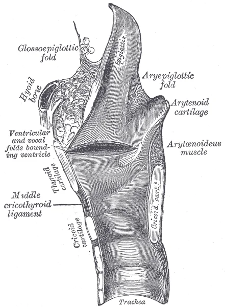
Figure 8Gray953
Long description
Sagittal section of the larynx and upper part of the trachea.

Figure 8: Sagittal section of the larynx and upper part of the trachea.
Specifically, vessels from the left and right sides of the supraglottis drain into the jugulodigastric nodes located at the angle of the mandible.
DefinitionSupraglottic Drainage
The supraglottis drains primarily into the upper deep cervical lymph nodes, particularly the jugulodigastric nodes.
The glottis, which comprises the true vocal cords, presents a unique anatomical feature: it is virtually devoid of lymphatic vessels. This absence of lymphatic drainage explains why early glottic cancers rarely metastasize to regional lymph nodes. No lymphatic vessels are shown emerging from this specific region, contrasting sharply with the dense network seen in the supraglottis above and the subglottis below. This avascular lymphatic zone acts as a barrier to early nodal spread.
NoteGlottic Avascularity
The true vocal cords lack significant lymphatic drainage, reducing the risk of early nodal metastasis in glottic cancer.
Below the vocal folds, the subglottis drains into the pretracheal and paratracheal lymph nodes. In the diagram, vessels from the inferior aspect of the larynx are shown descending towards the trachea. These vessels join the lymphatic chain that runs along the trachea, eventually draining into the deep cervical nodes at the root of the neck. The subglottic region also has connections to the anterior mediastinal nodes, although this is less prominent in standard cervical dissections. Understanding these distinct pathways allows clinicians to predict the spread of disease and tailor neck dissections accordingly. The supraglottis drains to upper deep cervical nodes, the glottis to none, and the subglottis to lower deep cervical and paratracheal nodes.
Figure 9Gray954
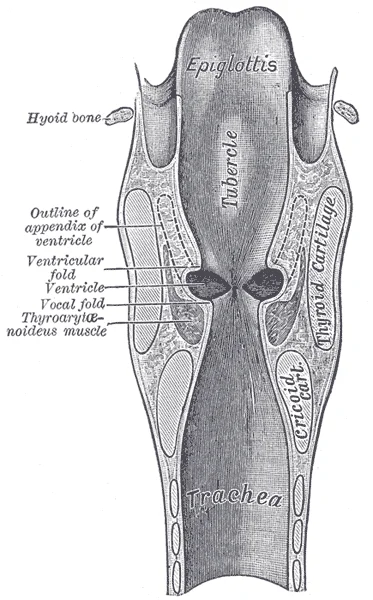
Long description
Coronal section of larynx and upper part of trachea.

Figure 9: Coronal section of larynx and upper part of trachea.
The larynx receives its innervation from branches of the vagus nerve (cranial nerve X), which provides both motor and sensory components. Understanding this supply is critical for clinical procedures such as cricothyroidotomy, where knowledge of the anatomical relationships prevents injury to these vital structures. The nerve supply can be divided into somatic motor, somatic sensory, and autonomic components, with significant implications for referred pain patterns.
The external laryngeal nerve is a branch of the superior laryngeal nerve. It descends alongside the superior thyroid artery to supply the cricothyroid muscle. This muscle is the only tensor of the vocal cords, playing a key role in pitch modulation. Injury to this nerve results in voice fatigue and an inability to hit high notes, as the cricothyroid muscle cannot tense the vocal fold properly.
The recurrent laryngeal nerve is a branch of the vagus nerve that loops around the subclavian artery on the right and the aortic arch on the left. It ascends in the tracheoesophageal groove to enter the larynx posteriorly. It supplies all intrinsic muscles of the larynx except the cricothyroid. It also provides sensory innervation to the laryngeal mucosa below the vocal cords. Damage to this nerve causes vocal cord paralysis, leading to hoarseness or airway obstruction. In this view, the recurrent laryngeal nerve is shown ascending in the tracheoesophageal groove, lying posterior to the thyroid gland and anterior to the esophagus. It enters the larynx deep to the inferior constrictor muscle. The labelled structures include the recurrent laryngeal nerve, the inferior thyroid artery, and the cricoid cartilage. The nerve lies medial to the inferior thyroid artery in most cases, a relationship crucial for thyroid surgery. The caption for this figure states: "Nerve supply of the larynx showing the recurrent laryngeal nerve in the tracheoesophageal groove."
Figure 10Gray957
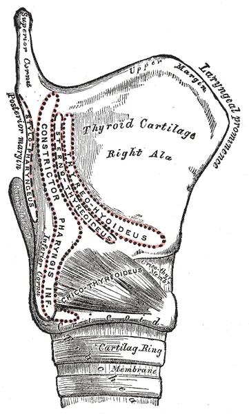
Long description
Side view of the larynx, showing muscular attachments.

Figure 10: Side view of the larynx, showing muscular attachments.
The larynx also receives autonomic innervation via the superior laryngeal nerve, which carries parasympathetic fibres to the glands of the laryngeal mucosa. Sensory innervation above the vocal cords is provided by the internal laryngeal nerve, a branch of the superior laryngeal nerve. This nerve pierces the thyrohyoid membrane to enter the larynx. It supplies the mucosa of the larynx above the vocal folds, including the epiglottis and aryepiglottic folds. Stimulation of this area triggers the cough reflex. Referred pain from the larynx can be felt in the neck or ear due to the shared innervation pathways. The cartilages, membranes, and muscles of the larynx are all intimately related to these nerve pathways. For instance, the cricothyroid membrane is a landmark for emergency airway access, lying just inferior to the thyroid cartilage and superior to the cricoid cartilage. Knowledge of the nerve supply ensures that surgical interventions do not compromise laryngeal function.
The laryngeal mucosa shows a clear regional transition that reflects its dual respiratory and protective roles. The ventral surface of the epiglottis and the upper part of the laryngeal inlet are covered by stratified squamous non‑keratinised epithelium, which resists mechanical abrasion during swallowing. The remainder of the laryngeal cavity — including the false vocal folds, the ventricle, the true vocal folds, and the subglottic region down to the lower border of the cricoid cartilage — is lined by pseudostratified ciliated columnar epithelium with goblet cells, the typical respiratory epithelium that moves mucus toward the pharynx. At the vocal fold itself the epithelium is tightly bound to the underlying vocal ligament (the deep layer of the lamina propria) and contains very few goblet cells, allowing rapid vibration without mucus interference.
Beneath the epithelium the lamina propria is organised into three distinct layers. The superficial layer (Reinke’s space) is loose connective tissue rich in hyaluronic acid and elastin, giving the vocal fold its pliability. The intermediate layer contains more elastin and collagen fibres, providing tensile strength. The deep layer is dense collagenous tissue that forms the vocal ligament, anchoring the mucosa to the thyroid and arytenoid cartilages. This layered arrangement is the histological basis for the mucosal wave that underlies phonation.
Seromucous glands are abundant in the supraglottic and subglottic regions and in the ventricles (laryngeal saccules), but are absent from the true vocal folds. Their secretions keep the respiratory epithelium moist and trap inhaled particles. Lymphoid nodules are scattered throughout the lamina propria, especially in the ventricular and subglottic mucosa, forming part of the Waldeyer’s ring equivalent in the larynx.
The cartilaginous framework consists of hyaline cartilage (thyroid, cricoid, and the basal parts of the arytenoids) which may undergo calcification or ossification with age, and elastic cartilage (epiglottis, corniculate, cuneiform, and the vocal processes of the arytenoids) which remains flexible throughout life. The vocalis muscle (the medial thyroarytenoid) lies deep to the vocal ligament and is composed of skeletal muscle fibres with a high proportion of fast‑twitch type II fibres, suited to rapid, fine tension adjustments during voice production.
DefinitionVocal fold layered structure
The vocal fold mucosa comprises epithelium, a three‑layered lamina propria (superficial, intermediate, deep), and the vocalis muscle; the superficial layer (Reinke’s space) is essential for the mucosal wave.
| Region | Epithelium type | Key feature |
|---|---|---|
| Epiglottis (lingual surface) & upper inlet | Stratified squamous non‑keratinised | Protects against swallowing trauma |
| False vocal folds, ventricle, true vocal folds, subglottis | Pseudostratified ciliated columnar with goblet cells | Mucociliary clearance |
| True vocal fold surface | Pseudostratified ciliated columnar, few goblet cells | Tightly bound to vocal ligament for vibration |
Histological zones from inlet to trachea
- Laryngeal inlet (epiglottis, aryepiglottic folds)Stratified squamous non‑keratinised epithelium
- Supraglottis (false folds, ventricle)Respiratory epithelium with seromucous glands
- Glottis (true vocal folds)Respiratory epithelium, no glands, three‑layered lamina propria, vocalis muscle deep
- Subglottis (below vocal folds to cricoid)Respiratory epithelium, seromucous glands, lymphoid nodules
- TracheaContinuous respiratory epithelium with C‑shaped hyaline cartilage rings
The larynx arises from the ventral wall of the primitive foregut, specifically from the fourth and sixth pharyngeal arches. This embryological origin is fundamental to understanding its dual innervation and muscular composition. The fourth arch gives rise to the epiglottic cartilages and the cricoid cartilage, while the sixth arch forms the arytenoid, corniculate, and cuneiform cartilages. The mucosal lining is derived from endoderm, whereas the connective tissue and muscles originate from splanchnic mesenchyme.
DefinitionPharyngeal Arches
Concentrated masses of mesenchyme located in the head and neck region, each contributing specific skeletal, muscular, and neural elements to the developing larynx.
The laryngeal cartilages begin as paired condensations of mesenchyme. The thyroid cartilage, the largest single cartilage, forms from the fusion of two lateral plates. It provides the anterior and lateral framework, protecting the vocal folds. The cricoid cartilage, shaped like a signet ring, sits inferior to the thyroid. Its posterior lamina is significantly higher than its anterior arch, a key anatomical landmark for surgical access. The arytenoid cartilages rest on the superior border of the cricoid lamina. These pyramidal structures are crucial for vocal fold movement, as the vocal ligaments attach to their vocal processes.
| Structure | Origin | Key Relation |
|---|---|---|
| Thyroid | Fourth arch | Anterior framework |
| Cricoid | Sixth arch | Inferior to thyroid |
| Arytenoids | Sixth arch | On cricoid lamina |
The intrinsic muscles of the larynx are derived from the sixth pharyngeal arch. Consequently, they are all innervated by the recurrent laryngeal nerve, a branch of the vagus nerve. The exception is the cricothyroid muscle, derived from the fourth arch, which is innervated by the external branch of the superior laryngeal nerve. This distinction is vital for understanding sensory and motor deficits in laryngeal pathology. The extrinsic muscles, which move the larynx as a whole during swallowing and phonation, are derived from the occipital myotomes and are innervated by the cervical plexus and the hypoglossal nerve.
Clinical pearlInnervation Rule
All intrinsic laryngeal muscles are supplied by the recurrent laryngeal nerve, except the cricothyroid, which is supplied by the external laryngeal nerve.
The laryngeal inlet is guarded by the epiglottis, a leaf-shaped elastic cartilage. It prevents aspiration by deflecting food towards the oesophageal opening during deglutition. The vestibular folds (false vocal cords) and vocal folds (true vocal cords) are mucosal folds containing ligaments and muscle. The space between the vocal folds is the rima glottidis, the narrowest part of the adult laryngeal cavity. Understanding these structural relationships allows for precise localisation of lesions and effective surgical planning.
Figure 11Gray955
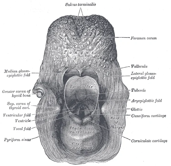
Long description
The entrance to the larynx, viewed from behind.

Figure 11: The entrance to the larynx, viewed from behind.
The larynx appears on cross‑sectional imaging as a midline airway structure surrounded by a framework of cartilages, fat‑filled spaces and mucosal folds. On axial CT or MRI the cartilaginous skeleton is identified by its cortical margins, while the airway lumen is air‑filled and the surrounding soft tissues show intermediate attenuation or signal. The thyroid cartilage forms the anterior and lateral walls, its two laminae meeting in the midline to create the laryngeal prominence. Posteriorly the cricoid cartilage sits as a complete ring with a broad lamina; the paired arytenoid cartilages rest on the superolateral aspects of the cricoid lamina, each displaying a vocal process directed anteromedially and a muscular process laterally. The epiglottis curves posteriorly behind the tongue base, separating the valleculae from the laryngeal inlet. Superior to the true cords the false vocal cords (vestibular folds) appear as mucosal folds; the true vocal cords are seen as thin, sharp air‑soft tissue interfaces enclosing the rima glottidis. The paraglottic space lies lateral to the true cords, bounded by the thyroid cartilage laterally and the cricothyroid muscle anterolaterally. The pre‑epiglottic space is anterior to the epiglottis and posterior to the thyrohyoid membrane. The subglottic region extends from the lower margin of the true cords to the inferior border of the cricoid cartilage.
| Structure | Appearance on CT/MRI |
|---|---|
| Thyroid cartilage | Dense cortical bone forming anterior and lateral walls |
| Cricoid cartilage | Complete ring with thick posterior lamina |
| Arytenoid cartilages | Paired pyramidal shapes on the cricoid lamina |
| Epiglottis | Curved soft‑tissue density posterior to the tongue base |
| True vocal cords | Thin linear air‑soft tissue interface |
| False vocal cords | Mucosal folds superior to the true cords |
| Paraglottic space | Fat‑density lateral to the true cords |
| Pre‑epiglottic space | Fat‑density anterior to the epiglottis |
Clinical pearlImaging pearl
The cricoid cartilage is the only complete cartilaginous ring of the airway and serves as a reliable landmark for the subglottic region on imaging.
On sagittal and coronal reformats the laryngeal ventricle separates the false and true cords, and the anterior commissure is visualised where the thyroid cartilage meets the vocal processes. The laryngeal cartilages ossify progressively; the thyroid and cricoid are often calcified in adults, while the arytenoids may remain cartilaginous. The hyoid bone lies superiorly, suspended by the thyrohyoid membrane, and the thyroid gland lobes flank the trachea and cricoid cartilage laterally. The carotid sheaths run posterolaterally. Knowledge of these relationships allows accurate staging of laryngeal tumours and planning of surgical approaches.
The larynx serves as the critical junction between the pharynx and the trachea, functioning primarily in phonation, respiration, and airway protection. It is situated at the level of the cervical vertebrae C3 to C6, anterior to the hypopharynx. Understanding its structure requires a systematic approach from the skeletal framework to the intrinsic musculature and neurovascular supply.
The laryngeal skeleton consists of nine cartilages: three unpaired and three paired. The unpaired cartilages are the thyroid, cricoid, and epiglottis. The thyroid cartilage is the largest, forming the anterior and lateral walls. It comprises two laminae that fuse anteriorly to form the laryngeal prominence, or Adam’s apple. The cricoid cartilage lies inferior to the thyroid, resembling a signet ring with a broad posterior lamina and a narrow anterior arch. It is the only complete ring of cartilage in the respiratory tract. The epiglottis is a leaf-shaped elastic cartilage attached to the angle of the thyroid laminae, projecting posteriorly to cover the laryngeal inlet during swallowing. The paired cartilages include the arytenoids, corniculates, and cuneiforms. The arytenoid cartilages sit atop the cricoid lamina and are crucial for vocal cord movement.
The intrinsic muscles of the larynx are responsible for moving the vocal cords. They are all supplied by the recurrent laryngeal nerve, except for the cricothyroid muscle, which is supplied by the external branch of the superior laryngeal nerve. These muscles adjust the tension, position, and adduction of the vocal folds.
| Muscle | Origin | Insertion | Action | Nerve Supply |
|---|---|---|---|---|
| Posterior cricoarytenoid | Posterior cricoid lamina | Muscular process of arytenoid | Abducts vocal cords | Recurrent laryngeal |
| Lateral cricoarytenoid | Cricoid arch | Muscular process of arytenoid | Adducts vocal cords | Recurrent laryngeal |
| Transverse arytenoid | Posterior surface of one arytenoid | Posterior surface of other arytenoid | Adducts vocal cords | Recurrent laryngeal |
| Oblique arytenoid | Apex of one arytenoid | Base of other arytenoid | Adducts laryngeal inlet | Recurrent laryngeal |
| Cricothyroid | Cricoid arch | Inferior horn and lower lamina of thyroid | Tenses vocal cords | External laryngeal |
The sensory and motor innervation is divided by the superior laryngeal nerve. The internal laryngeal nerve provides sensation above the vocal cords and is a branch of the superior laryngeal nerve. It enters the larynx through the thyrohyoid membrane. The recurrent laryngeal nerve, a branch of the vagus nerve, supplies all intrinsic muscles and sensation below the vocal cords. The blood supply is derived from the superior and inferior laryngeal arteries, branches of the superior and inferior thyroid arteries respectively.
The true vocal cords lie inferior to these, attached anteriorly to the thyroid cartilage and posteriorly to the arytenoid cartilages. The cricoid cartilage forms the base of the larynx. The trachea extends inferiorly from the cricoid. The surrounding soft tissues, including the pre-epiglottic space and paraglottic space, are also delineated, showing their relationship to the cartilaginous framework.
The larynx is a complex tubular structure situated in the anterior neck, serving dual roles in respiration and phonation. It is suspended from the hyoid bone by the thyrohyoid membrane and rests upon the cricoid cartilage. The framework consists of nine cartilages: three unpaired (thyroid, cricoid, and epiglottis) and three paired (arytenoid, corniculate, and cuneiform). The thyroid cartilage is the largest, forming the anterior laryngeal prominence or 'Adam's apple'. The cricoid cartilage is the only complete ring in the airway, resembling a signet ring with a broad posterior lamina and a narrow anterior arch. The epiglottis is a leaf-shaped elastic cartilage that acts as a flap to protect the airway during swallowing.
The intrinsic muscles of the larynx are all supplied by the recurrent laryngeal nerve, a branch of the vagus nerve, except for the cricothyroid muscle. The cricothyroid muscle is supplied by the external laryngeal nerve and functions to tense the vocal cords by tilting the thyroid cartilage forward. The posterior cricoarytenoid muscles are the sole abductors of the vocal cords, opening the glottis. All other intrinsic muscles are adductors. The thyroarytenoid muscles relax the vocal cords, while the lateral cricoarytenoids and interarytenoids adduct them.
Clinical pearlNerve Supply
All intrinsic laryngeal muscles are innervated by the recurrent laryngeal nerve, except the cricothyroid, which is supplied by the external laryngeal nerve.
The blood supply to the larynx is derived from the superior and inferior laryngeal arteries. The superior laryngeal artery is a branch of the superior thyroid artery, while the inferior laryngeal artery arises from the inferior thyroid artery. Venous drainage follows the arterial supply. Lymphatic drainage is divided by the vocal cords. Above the vocal cords, lymph drains to the deep cervical nodes. Below the vocal cords, lymph drains to the pretracheal and paratracheal nodes. This distinction is critical in the staging of laryngeal cancer.
| Muscle | Action | Nerve Supply |
|---|---|---|
| Posterior Cricoarytenoid | Abduction of vocal cords | Recurrent Laryngeal |
| Lateral Cricoarytenoid | Adduction of vocal cords | Recurrent Laryngeal |
| Transverse Arytenoid | Adduction of vocal cords | Recurrent Laryngeal |
| Oblique Arytenoid | Adduction of vocal cords | Recurrent Laryngeal |
| Thyroarytenoid | Relaxation of vocal cords | Recurrent Laryngeal |
| Cricothyroid | Tensing of vocal cords | External Laryngeal |
The mucosa of the larynx is lined by stratified squamous epithelium above the vocal cords and respiratory epithelium below. The vestibular folds (false vocal cords) are superior to the true vocal cords. The true vocal cords are the primary structures for phonation. The space between the true vocal cords is the rima glottidis. The laryngeal inlet is guarded by the epiglottis and the aryepiglottic folds. During swallowing, the larynx elevates, and the epiglottis covers the inlet to prevent aspiration.
Exam trapCommon Trap
Do not confuse the external laryngeal nerve with the recurrent laryngeal nerve. Injury to the external laryngeal nerve affects only the cricothyroid muscle, leading to voice fatigue and inability to hit high pitches, but not complete vocal cord paralysis.
Clinical assessment of the larynx involves indirect laryngoscopy or flexible nasendoscopy. Hoarseness is a common symptom of laryngeal pathology. Persistent hoarseness lasting more than three weeks warrants investigation for malignancy. The recurrent laryngeal nerve is vulnerable during thyroid surgery. Unilateral injury causes hoarseness due to paralysis of one vocal cord. Bilateral injury can cause airway obstruction because the vocal cords remain in a paramedian position.
Students frequently confuse the anatomical structures of the larynx with those of the pharynx, leading to errors in identifying the boundaries of the airway. The larynx is a distinct cartilaginous structure located inferior to the pharynx and superior to the trachea. It serves as the gateway to the lower respiratory tract. A common error is assuming the epiglottis is a muscle; it is actually a leaf-shaped elastic cartilage. It does not contract but acts as a flap to cover the laryngeal inlet during swallowing. Another frequent mistake involves the vocal cords. Students often describe them as simple folds of skin. In reality, the true vocal cords consist of the vocal ligament, vocalis muscle, and mucosa. The false vocal cords, or vestibular folds, lie superior to the true cords and do not produce sound. Confusing these two leads to incorrect explanations of phonation.
A significant pitfall relates to nerve supply. Many students attribute all laryngeal movement to the recurrent laryngeal nerve. While this nerve supplies all intrinsic muscles except one, the superior laryngeal nerve is crucial for pitch control. The external branch of the superior laryngeal nerve innervates the cricothyroid muscle. Damage to this nerve results in an inability to tense the vocal cords, causing voice fatigue and loss of high-pitch range. Students often overlook this specific deficit when assessing post-thyroidectomy patients. Furthermore, the sensory innervation is often misattributed. The internal branch of the superior laryngeal nerve provides sensation above the vocal cords. Injury here causes loss of the cough reflex, increasing aspiration risk.
Another error concerns the clinical landmark of the cricothyroid membrane. Students may struggle to identify its precise location. It lies between the thyroid cartilage and the cricoid cartilage. This membrane is the target for emergency cricothyrotomy. Mistaking the thyroid cartilage for the cricoid cartilage can lead to failed airway access. The cricoid cartilage is the only complete ring in the larynx. It provides structural support. The thyroid cartilage forms the anterior prominence, known as the laryngeal prominence or Adam's apple.
Finally, students often misunderstand the function of the arytenoid cartilages. These are paired cartilages that sit on the posterior aspect of the cricoid. They are not merely passive structures. They rotate and slide to adjust the tension and position of the vocal cords. Failure to recognize their role in abduction and adduction leads to an incomplete understanding of breathing and speech mechanics. The piriform fossa is another area of confusion. It is a depression lateral to the laryngeal inlet. Food particles can become trapped here. It is also a common site for foreign body impaction and carcinoma spread. Understanding its relation to the internal laryngeal nerve is vital for surgical safety.
Knowledge of laryngeal anatomy is essential at the bedside for managing airway emergencies. Clinicians must quickly identify the laryngeal inlet to perform intubation. The view of the vocal cords is achieved by aligning the oral, pharyngeal, and laryngeal axes. Palpating the thyroid cartilage helps locate the cricothyroid membrane for emergency access. In the operating theatre, surgeons rely on detailed knowledge of the recurrent laryngeal nerve during thyroidectomy. This nerve runs in the tracheoesophageal groove. Injury causes vocal cord paralysis. Surgeons must also protect the superior laryngeal nerve to preserve voice quality. Understanding the blood supply, derived from the superior and inferior laryngeal arteries, prevents excessive bleeding during procedures.
On imaging, radiologists use CT and MRI to evaluate laryngeal masses. The stratification of the vocal cord layers is visible on high-resolution scans. This helps distinguish benign lesions from malignancies. The air-filled lumen provides natural contrast. Radiologists look for asymmetry in the vocal cords or thickening of the paraglottic space. These signs may indicate early cancer. In examinations, students are tested on the histological layers of the larynx. They must identify the respiratory epithelium and the stratified squamous epithelium of the vocal cords. Questions often focus on the nerve supply and muscle actions.
The larynx is also critical in speech therapy. Therapists assess vocal cord vibration and closure. They use laryngoscopy to visualize movement. Patients with spasmodic dysphonia may receive botulinum toxin injections into the laryngeal muscles. This requires precise anatomical knowledge to avoid affecting swallowing muscles. In intensive care, prolonged intubation can cause laryngeal injury. Clinicians monitor for stenosis or granuloma formation. The cricoid cartilage is a common site of pressure necrosis. Understanding the vascular supply helps in managing these complications.
Furthermore, the larynx plays a role in protective reflexes. The cough reflex is mediated by sensory nerves in the larynx. Patients with impaired sensation are at risk of aspiration pneumonia. Clinicians assess this by observing the gag and cough responses. In paediatrics, the larynx is higher and more anterior. This makes intubation more challenging. The epiglottis is floppy and omega-shaped. These anatomical differences are frequently tested in paediatric emergency exams. Understanding these variations ensures safe airway management in children.
The larynx is located between the pharynx and trachea. The true vocal cords produce sound; the false cords do not. The recurrent laryngeal nerve supplies all intrinsic muscles except the cricothyroid. The cricothyroid muscle is innervated by the external branch of the superior laryngeal nerve. The cricoid cartilage is the only complete ring in the larynx. The internal branch of the superior laryngeal nerve provides sensory innervation above the vocal cords. The piriform fossa is a common site for foreign body impaction. The cricothyroid membrane is the target for emergency cricothyrotomy.
Answer each one in your head first, then open the answer.
Which nerve supplies the cricothyroid muscle?
Show answerHide answer
External branch of the superior laryngeal nerveWhat is the only complete cartilaginous ring in the larynx?
Show answerHide answer
Cricoid cartilageWhich structure covers the laryngeal inlet during swallowing?
Show answerHide answer
EpiglottisWhat is the clinical significance of the piriform fossa?
Show answerHide answer
Common site for foreign body impactionWhich nerve injury causes loss of high-pitch voice range?
Show answerHide answer
External branch of the superior laryngeal nerve
- Betts JG, Young KA, Wise JA, et al. Organs and Structures of the Respiratory System. OpenStax, Anatomy and Physiology 2e (CC BY-NC-SA 4.0). Accessed .
Figure credits
- Figure 1: Henry Vandyke Carter, via Wikimedia Commons, Public domain, Public domain.
- Figure 2: Henry Vandyke Carter, via Wikimedia Commons, Public domain, Public domain.
- Figure 3: Henry Vandyke Carter, via Wikimedia Commons, Public domain, Public domain.
- Figure 4: OpenStax College, via Wikimedia Commons, CC BY 3.0, CC BY 3.0. resized.
- Figure 5: Henry Vandyke Carter, via Wikimedia Commons, Public domain, Public domain.
- Figure 6: OpenStax College, via Wikimedia Commons, CC BY 3.0, CC BY 3.0. resized.
- Figure 7: Henry Vandyke Carter, via Wikimedia Commons, Public domain, Public domain.
- Figure 8: Henry Vandyke Carter, via Wikimedia Commons, Public domain, Public domain.
- Figure 9: Henry Vandyke Carter, via Wikimedia Commons, Public domain, Public domain.
- Figure 10: Henry Vandyke Carter, via Wikimedia Commons, Public domain, Public domain.
- Figure 11: Henry Vandyke Carter, via Wikimedia Commons, Public domain, Public domain.
Educational content for medical students and doctors. It is not a substitute for clinical judgement or local guidelines.