Thyroid and parathyroid glands
Small endocrine glands in the neck that regulate calcium and thyroid hormone production
The thyroid is a butterfly‑shaped organ that lies anterior to the trachea and just inferior to the larynx, with a central isthmus joining two lateral lobes. Each lobe’s posterior surface bears the parathyroid glands. [1]
DefinitionParathyroid gland
Tiny, round endocrine organs embedded in the posterior thyroid capsule that secrete parathyroid hormone (PTH) to control blood calcium[1].
Most individuals possess four parathyroid glands—two on each thyroid lobe—but occasional supernumerary glands may be found in the neck or mediastinum[1]. The glands are enclosed by a thick connective‑tissue capsule that separates them from the thyroid parenchyma[1]. Their chief cells are the principal source of PTH, while the function of the oxyphil cells remains uncertain[1]. [2]
The typical surface landmarks for locating the thyroid and its embedded parathyroids are:
| Landmark | Relation to gland |
|---|---|
| Midline neck, just below the thyroid cartilage | Superior border of thyroid lobes |
| Tracheal rings | Posterior to thyroid lobes, where parathyroids sit |
| Isthmus | Connects the two lobes across the trachea |
During embryogenesis the thyroid descends from the base of the tongue along a midline epithelial tract known as the thyroglossal duct. The duct normally involutes, leaving the foramen cecum as a vestigial opening in the tongue; persistent ductal tissue can give rise to thyroglossal cysts. No numerical data are required for this description.
The parathyroids are positioned as follows:
| Position | Typical number | Notes |
|---|---|---|
| Posterior surface of right lobe | 2 (upper and lower) | Often adjacent to the recurrent laryngeal nerve |
| Posterior surface of left lobe | 2 (upper and lower) | May vary in relation to the inferior thyroid artery |
| Ectopic sites | Variable (occasionally >4) | Can be found in the thymus, mediastinum, or within the thyroid capsule |
Because the parathyroids lie directly behind the thyroid lobes, surgeons must identify them before thyroidectomy to avoid inadvertent removal, which can precipitate hypoparathyroidism and severe hypocalcaemia[1].
In summary, the thyroid’s anterior neck location and its posteriorly embedded parathyroids, usually four in number, are key anatomical facts. The embryologic thyroglossal duct traverses the midline from the tongue to the thyroid and normally disappears, leaving only a potential site for cyst formation.
The thyroid gland is a butterfly-shaped endocrine gland located in the anterior neck, consisting of two lobes connected by an isthmus. The right and left lobes lie either side of the trachea, typically at the level of the C5 to T1 vertebrae, with the isthmus crossing the second and third tracheal rings. The thyroid gland is enclosed within a fibrous capsule and is covered by the infrahyoid muscles and the sternothyroid muscle. [1]
The parathyroid glands are typically four small, oval-shaped endocrine glands embedded in the posterior surface of the thyroid lobes. They are approximately 3-6mm in diameter and weigh about 30-35mg each.
The thyroid gland receives its blood supply primarily from the superior and inferior thyroid arteries. The superior thyroid artery, a branch of the external carotid artery, descends to the upper pole of the thyroid lobe. The inferior thyroid artery, a branch of the thyrocervical trunk, ascends to the lower pole of the thyroid lobe. The thyroid ima artery, when present, arises from the brachiocephalic trunk or aortic arch and supplies the isthmus. [1]
The parathyroid glands receive their blood supply from the inferior thyroid arteries. Each parathyroid gland is drained by venous blood into the thyroid plexus of veins, which ultimately drains into the brachiocephalic veins. [2]
The thyroid gland is innervated by sympathetic fibers from the middle cervical ganglion and parasympathetic fibers from the vagus nerve. The parathyroid glands receive sympathetic innervation from the superior cervical ganglion. [1]
The relationship between the thyroid gland and the trachea is also demonstrated, showing the typical position of the thyroid isthmus crossing the second and third tracheal rings.
The thyroid gland is a butterfly-shaped endocrine organ situated in the anterior neck, deep to the sternothyroid and sternohyoid muscles. It lies anterior to the trachea and larynx. The gland consists of two lateral lobes connected by a narrow isthmus. This figure displays an anterior view of the neck with the strap muscles reflected. The right and left lobes are shown lying on either side of the trachea. The isthmus c [1]
The position of the thyroid lobes is fixed by the pretracheal fascia. This fascia attaches the thyroid to the larynx and trachea. Consequently, the gland moves upwards during swallowing. The lateral lobes relate to several vital structures. Anteriorly, they are covered by the strap muscles and the skin. Laterally, they are adjacent to the carotid sheath. The common carotid artery lies posterolateral to each lobe. The internal jugular vein is also located in this region. The recurrent laryngeal nerve runs in the tracheoesophageal groove. It lies posterior to the thyroid lobe. Injury to this nerve causes vocal cord paralysis. The superior thyroid artery supplies the upper part of the lobe. The inferior thyroid artery supplies the lower part. These arteries anastomose within the gland.
The isthmus connects the medial aspects of the lobes. It usually lies anterior to the second and third tracheal rings. In some individuals, a pyramidal lobe extends upwards from the isthmus. This remnant of the thyroglossal duct may contain functional thyroid tissue. The parathyroid glands are typically four in number. Two superior glands are located near the upper poles. Two inferior glands are located near the lower poles. The superior glands are more constant in position. The inferior glands are more variable. They may be found within the thymus or high in the neck. [1]
| Structure | Relation |
|---|---|
| Trachea | Posterior |
| Larynx | Superior |
| Carotid Sheath | Lateral |
| Recurrent Laryngeal Nerve | Posterior |
| Strap Muscles | Anterior |
Understanding these relations is crucial for surgical safety. The recurrent laryngeal nerve is at risk during thyroidectomy. The parathyroid glands must be preserved to maintain calcium homeostasis. The vascular supply must be ligated carefully to prevent haemorrhage. The pretracheal fascia provides a plane for dissection. Knowledge of the anatomical landmarks helps in identifying the gland. The thyroid cartilage serves as a superior landmark. The sternal notch serves as an inferior landmark. The sternocleidomastoid muscle marks the lateral boundary.
Figure 1Gray1174
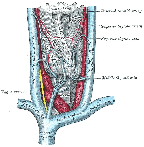
Long description
The thyroid gland and its relations. Labelled illustration from Wikimedia Commons, used under its open licence.

Figure 1: The thyroid gland and its relations.
The viewer should note the proximity of the nerve to the inferior thyroid artery. This relationship is variable but critical. The parathyroid glands are often obscured by the thyroid tissue. Careful dissection is required to locate them. The blood supply is rich and anastomotic. This ensures good healing but increases bleeding risk. The lymphatic drainage goes to deep cervical nodes. This is important in the spread of thyroid cancer. The anatomy is consistent across most individuals. Variations exist but are rare. Mastery of these relations is essential for clinical practice.
The thyroid gland consists of two lobes connected by an isthmus, typically weighing 15-25 grams. Each lobe is composed of numerous follicles, which are spherical structures lined with follicular cells. These follicles contain colloid, a substance rich in thyroglobulin, which serves as a precursor for thyroid hormone synthesis. The follicular cells are responsible for producing and secreting thyroid hormones, while parafollicular cells (C cells) are scattered throughout the gland and produce calcitonin. [1]
The parathyroid glands are typically small oval structures. They are usually located on the posterior surface of the thyroid gland, with two superior and two inferior parathyroid glands.
Histologically, the thyroid follicles consist of a single layer of cuboidal epithelial cells surrounding a central cavity filled with colloid. The follicular cells contain organelles necessary for hormone synthesis, including rough endoplasmic reticulum and Golgi apparatus. The parathyroid glands are composed primarily of chief cells, which produce parathyroid hormone, and oxyphil cells, whose function is less well understood. [1]
The blood supply to both glands is rich, with the thyroid receiving blood primarily from the superior and inferior thyroid arteries, which are branches of the external carotid and thyrocervical trunk, respectively. The parathyroid glands are supplied by branches of the inferior thyroid artery. Venous drainage occurs through corresponding thyroid veins into the brachiocephalic veins. [1] [2]
The follicles appear as small spherical structures within the thyroid tissue, filled with colloid. The superior parathyroid glands are positioned higher on the thyroid lobes, while the inferior glands are located lower.
Figure 21817 The Role of Parathyroid Hormone in Maintaining Blood Calcium Homeostasis
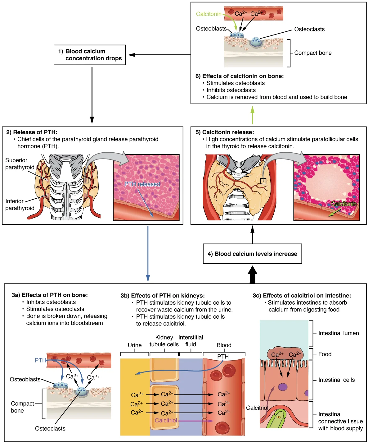
Long description
Parathyroid hormone increases blood calcium levels when they drop too low. Conversely, calcitonin, which is released from the thyroid gland, decreases blood calcium levels when they become too high. These two mechanisms constantly maintain blood calcium concentration at homeostasis.

Figure 2: Parathyroid hormone increases blood calcium levels when they drop too low.
The thyroid gland receives a rich vascular supply primarily from two pairs of arteries: the superior and inferior thyroid arteries. Understanding this arrangement is critical for surgical safety, particularly during thyroidectomy, to avoid haemorrhage and damage to adjacent structures.
The artery runs in close proximity to the external branch of the superior laryngeal nerve.
The superior thyroid artery is the first branch of the external carotid artery. It descends to the upper pole of the thyroid gland, supplying the superior portion of the lobe and the adjacent parathyroid glands. Its course is closely associated with the external branch of the superior laryngeal nerve. Surgeons must ligate this artery close to the gland capsule to preserve the nerve, which innervates the cricothyroid muscle. Damage to this nerve results in voice fatigue and inability to produce high-pitched sounds. [1]
The inferior thyroid artery arises from the thyrocervical trunk. It ascends behind the carotid sheath and then turns medially to supply the lower pole of the thyroid gland and the inferior parathyroid glands. Its relationship with the recurrent laryngeal nerve is complex and variable. The nerve may pass anterior, posterior, or between the branches of the artery. This variability necessitates careful dissection to identify and protect the nerve. The recurrent laryngeal nerve provides motor innervation to all intrinsic muscles of the larynx except the cricothyroid. Injury to this nerve causes vocal cord paralysis, leading to hoarseness or airway obstruction if bilateral.
| Structure | Origin | Relation to Nerve | Clinical Significance |
|---|---|---|---|
| Superior thyroid artery | External carotid | External branch of superior laryngeal nerve | Ligate close to capsule to spare nerve |
| Inferior thyroid artery | Thyrocervical trunk | Recurrent laryngeal nerve | Variable crossing; identify nerve before ligation |
The venous drainage of the thyroid and parathyroid glands is complex, involving multiple plexuses that ultimately empty into the internal jugular and subclavian systems. Understanding this drainage is critical for interpreting metastatic spread and surgical complications.
The superior thyroid vein accompanies the superior thyroid artery. It originates from the upper pole of the thyroid gland. The vein drains blood from the superior aspect of the gland. It typically joins the facial vein or the common facial vein. In some cases, it drains directly into the internal jugular vein. This pathway allows for rapid drainage from the upper thyroid region. [1]
The middle thyroid vein is the largest of the thyroid veins. It arises from the lateral aspect of the thyroid gland. The vein runs horizontally across the carotid sheath. It usually drains into the internal jugular vein. This direct connection to the internal jugular vein is clinically significant. It provides a route for potential spread of pathology to the neck veins. [1]
The inferior thyroid vein drains the lower pole of the thyroid gland. It originates from the inferior thyroid plexus. The vein ascends anterior to the trachea. It typically drains into the left brachiocephalic vein. This crossing of the midline is a key anatomical feature. The left inferior thyroid vein is often larger than the right. It may also receive drainage from the parathyroid glands. [1]
The parathyroid glands have a rich venous supply. They drain into the thyroid venous plexuses. Superior parathyroids usually drain via the superior thyroid vein. Inferior parathyroids often drain via the inferior thyroid vein. This integration means parathyroid pathology can affect thyroid venous patterns.
Figure 3Gray1177
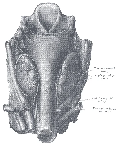
Long description
Parathyroid glands. (Halsted and Evans.)

Figure 3: Parathyroid glands (Halsted and evans.).
The middle thyroid vein crosses the carotid sheath to enter the internal jugular vein. The inferior thyroid vein ascends anterior to the trachea to join the left brachiocephalic vein.
Clinical pearlClinical Pearl
The middle thyroid vein is the most consistent vein draining into the internal jugular vein, making it a key landmark in thyroid surgery.
The lymphatic drainage of the thyroid and parathyroid glands is extensive and follows specific anatomical pathways that are critical for understanding the spread of pathology. The lymphatics originate from a rich subcapsular plexus within the glandular tissue itself. From this initial network, vessels course towards the deep cervical lymph nodes, which serve as the primary drainage basin for the neck region.
The lymphatic vessels are traced from the thyroid capsule towards the jugulodigastric nodes and the lower deep cervical nodes.
The drainage from the upper part of the thyroid gland typically flows to the pretracheal and paratracheal nodes. These nodes are situated along the trachea and the recurrent laryngeal nerves. From here, lymph may pass to the superior deep cervical nodes, also known as the jugulodigastric nodes, which lie at the angle of the mandible. The lower part of the thyroid gland drains primarily into the inferior deep cervical nodes, located along the lower segment of the internal jugular vein. Some vessels may also drain directly into the thoracic duct on the left side or the right lymphatic duct on the right side, although this is less common.
The parathyroid glands share a similar drainage pattern with the thyroid gland. Their lymphatics drain into the pretracheal, paratracheal, and deep cervical nodes. This shared drainage pathway explains why metastatic disease from thyroid carcinoma often involves these specific nodal groups. Understanding this anatomy is essential for surgical planning and staging of thyroid malignancies. The proximity of the lymphatic vessels to the recurrent laryngeal nerve is particularly important, as surgical dissection in this area carries a risk of nerve injury. The clear depiction of the vascular and neural structures alongside the lymphatic pathways provides a comprehensive overview of the region's complexity.
The innervation of the thyroid and parathyroid glands is a complex interplay of autonomic and somatic pathways, crucial for understanding both physiological regulation and surgical complications. The autonomic supply consists of sympathetic and parasympathetic fibres that primarily regulate blood flow and glandular secretion rather than direct motor control of the parenchyma. Sympathetic fibres arise from the cervical sympathetic ganglia, travelling alongside the superior and inferior thyroid arteries to reach the gland. Parasympathetic input is derived from the vagus nerve, specifically via the recurrent laryngeal and superior laryngeal nerves, which modulate vascular tone.
This anatomical arrangement explains the phenomenon of referred pain. Pain originating from the thyroid or parathyroid glands is often perceived in the anterior neck, the jaw, or the upper thoracic region, corresponding to the dermatomal levels of the cervical spinal nerves (C2–C4). This referral pattern can sometimes complicate the clinical diagnosis of thyroid pathology, as patients may present with neck stiffness or shoulder pain rather than localised thyroid tenderness.
DefinitionReferred Pain
Pain perceived at a location other than the site of the painful stimulus, caused by the convergence of sensory nerve fibres from different regions onto the same spinal cord neurons.
The relationship between the blood supply and the nerve supply is clinically significant, particularly regarding the recurrent laryngeal nerve. The recurrent laryngeal nerve ascends in the tracheoesophageal groove, running in close proximity to the inferior thyroid artery. This anatomical relationship is critical during thyroidectomy. The nerve provides motor innervation to all the intrinsic muscles of the larynx except the cricothyroid muscle. It also carries sensory fibres from the mucosa below the vocal cords.
| Branch | Target Structure | Function |
|---|---|---|
| Motor | Intrinsic laryngeal muscles | Vocal cord movement |
| Sensory | Mucosa below vocal cords | Sensation |
Damage to the recurrent laryngeal nerve during thyroidectomy can lead to vocal cord paralysis. Unilateral injury results in hoarseness, while bilateral injury can cause airway obstruction. The parathyroid glands, located on the posterior aspect of the thyroid, receive their nerve supply from the same autonomic plexus that supplies the thyroid. This shared innervation means that surgical manipulation of the thyroid can affect parathyroid function, leading to hypocalcaemia if the glands are inadvertently removed or their blood supply compromised.
Figure 41814 The Parathyroid Glands
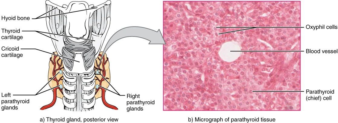
Long description
The small parathyroid glands are embedded in the posterior surface of the thyroid gland. LM × 760. (Micrograph provided by the Regents of University of Michigan Medical School © 2012)

Figure 4: The small parathyroid glands are embedded in the posterior surface of the thyroid gland (lm × 760).
To understand the functional architecture of the thyroid and parathyroid glands, one must first appreciate the distinct cellular compositions that dictate their hormonal output. The thyroid gland is composed primarily of follicles, which are spherical structures lined by a single layer of epithelial cells known as follicular cells. These cells are responsible for the synthesis and secretion of thyroxine (T4) and triiodothyronine (T3). The lumen of each follicle contains colloid, a gel-like substance rich in thyroglobulin, which serves as the precursor protein for thyroid hormone production. The follicular cells actively transport iodide from the blood into the cell, where it is organified onto tyrosine residues of thyroglobulin. This process is critical for the subsequent coupling reactions that form T3 and T4. [1]
Adjacent to the follicular cells, in the parafollicular spaces, lie the C cells, or parafollicular cells. These cells are derived from the neural crest and are responsible for secreting calcitonin. Calcitonin plays a minor role in calcium homeostasis in adults but is significant in lowering blood calcium levels by inhibiting osteoclast activity. Histologically, C cells are larger than follicular cells and have a more basophilic cytoplasm. They do not secrete into the follicular lumen but release their product directly into the surrounding capillaries.
The parathyroid glands, in contrast, lack follicles and colloid. They consist of two main cell types: chief cells and oxyphil cells. Chief cells are the predominant cell type and are responsible for the secretion of parathyroid hormone (PTH). PTH acts to increase blood calcium levels by stimulating bone resorption, increasing renal calcium reabsorption, and activating vitamin D to enhance intestinal calcium absorption. Oxyphil cells are larger, have eosinophilic granular cytoplasm, and appear in greater numbers after puberty. Their function is less clear, but they may play a role in calcium sensing or hormone secretion regulation. [2] [1]
The vascular supply to these glands is rich and highly regulated. The thyroid receives its blood supply from the superior and inferior thyroid arteries, which branch from the external carotid and subclavian arteries, respectively. The parathyroid glands typically receive their blood supply from the inferior thyroid artery. This rich vascularization ensures rapid hormone release into the systemic circulation in response to physiological stimuli.
DefinitionFollicular Cell
The primary cell type in the thyroid gland, responsible for synthesizing and secreting thyroid hormones T3 and T4.
DefinitionChief Cell
The predominant cell type in the parathyroid gland, responsible for secreting parathyroid hormone (PTH).
| Feature | Thyroid Gland | Parathyroid Gland |
|---|---|---|
| Primary Cell Type | Follicular Cells | Chief Cells |
| Secondary Cell Type | C Cells (Parafollicular) | Oxyphil Cells |
| Secretory Product | T3, T4, Calcitonin | Parathyroid Hormone (PTH) |
| Structural Unit | Follicle with Colloid | Nodules without Colloid |
| Embryonic Origin | Endoderm | Neural Crest (C cells) / Endoderm (Chief cells) |
Understanding these histological distinctions is fundamental for diagnosing pathological conditions. For instance, hyperplasia of chief cells leads to hyperparathyroidism, while neoplastic transformation of follicular cells can result in thyroid carcinoma. The presence of C cells is crucial for identifying medullary thyroid carcinoma, a rare but aggressive form of thyroid cancer.
NoteClinical Correlation
Medullary thyroid carcinoma arises from C cells and secretes calcitonin, which can be used as a tumor marker.
In summary, the histology of the thyroid and parathyroid glands reflects their distinct endocrine functions. The thyroid’s follicular structure supports the storage and release of thyroid hormones, while the parathyroid’s cellular arrangement facilitates the rapid secretion of PTH for calcium regulation. Mastery of these histological features is essential for medical students to understand the pathophysiology of endocrine disorders.
The thyroid gland originates from an endodermal outpouching of the floor of the primitive pharynx called the median thyroid diverticulum, which appears at the foramen cecum – the junction of the anterior two‑thirds and posterior one‑third of the tongue. This diverticulum elongates and descends in front of the hyoid bone and thyroid cartilage, remaining attached to the tongue by the thyroglossal duct. [1]
The gland reaches its definitive location anterior to the second and third tracheal rings, with the isthmus typically crossing the second tracheal ring. Its final position is stabilised by the pretracheal fascia, a continuation of the deep cervical fascia. The superior parathyroid glands arise from the fourth pharyngeal pouch and settle near the inferior border of the cricoid cartilage; the inferior parathyroids descend with the thymus from the third pouch, ending near the inferior pole of the thyroid or occasionally within the thymus. Because of this migration, ectopic parathyroid tissue is found in up to 20 % of re‑operative cases. [1] [2]
Figure 5Gray1175
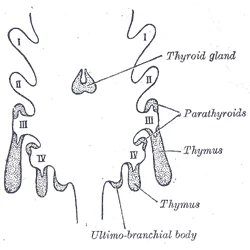
Long description
Scheme showing development of branchial epithelial bodies. (Modified from Koh.) I, II, III, IV. Branchial pouches.

Figure 5: Scheme showing development of branchial epithelial bodies.
Functional maturation begins before birth. Thyroxine (T4) synthesis is detectable during gestation, and triiodothyronine (T3) follows shortly thereafter. Parathyroid hormone (PTH) secretion starts during gestation. These hormones are essential for fetal brain development; maternal iodine deficiency can reduce fetal thyroid hormone output, leading to cretinism in low‑resource settings. [1]
Worked example – A 10‑week gestation fetus shows a thyroid primordium measuring 3 mm on ultrasound. By week 11 it begins secreting T4, measurable in cord blood at 5 µg/dL (normal > 4 µg/dL). If the thyroglossal duct fails to involute, a midline neck cyst may appear at age 5, presenting as a painless swelling that moves with tongue protrusion.
Exceptions and special populations
- Ectopic thyroid: In 1 % of the population the thyroid fails to descend, remaining at the base of the tongue (lingual thyroid). This can cause dysphagia or airway obstruction, especially in children.
- Pregnancy: Maternal TSH suppression is amplified, and the gland may increase in size by up to 30 % to meet increased hormone demand.
- Older adults: Age‑related fatty infiltration reduces gland weight from the typical 15–25 g in youth to 10–15 g, often with a nodular appearance.
- Low‑resource settings: Iodine‑deficient diets raise the prevalence of goitre to > 30 % in school‑age children, highlighting the importance of universal salt iodisation.
For a deeper dive into the step‑by‑step morphogenesis, see the dedicated [Embryology page].
The thyroid gland appears as a bilobed structure connected by the isthmus, lying anterior to the trachea. Each lobe is hypoechoic relative to the surrounding strap muscles. The isthmus crosses the midline.
During surgery, the strap muscles are retracted laterally to expose the thyroid capsule. The inferior thyroid artery is identified posterior to the gland, running in close relation to the recurrent laryngeal nerve. Preservation of the recurrent laryngeal nerve is critical to prevent vocal cord paralysis. The parathyroid glands are carefully preserved to maintain calcium homeostasis.
In cases of goitre, the thyroid gland enlarges, potentially extending into the posterior mediastinum. This substernal goitre can compress the trachea, causing respiratory distress, or the oesophagus, leading to dysphagia. The radiological anatomy of a large goitre shows significant displacement of adjacent structures. The trachea may be deviated to one side. The great vessels in the neck can be compressed or displaced laterally. Ultrasound and CT scans are used to determine the extent of the goitre and its relationship to vital structures. The parathyroid glands may be displaced or hyperplastic in association with certain types of goitre. Understanding these anatomical variations is essential for safe surgical management and accurate radiological interpretation.
The thyroid gland is a butterfly-shaped endocrine organ located in the anterior neck. It consists of two lateral lobes connected by an isthmus, lying anterior to the trachea and inferior to the larynx. The gland is enclosed within the pretracheal layer of the deep cervical fascia, which allows it to move vertically during swallowing and tongue protrusion. The posterior surface of each lobe relates closely to the parathyroid glands, the recurrent laryngeal nerve, and the inferior thyroid artery. [1]
DefinitionBerry's Ligament
A thickening of the pretracheal fascia that attaches the thyroid lobes to the cricoid cartilage and upper tracheal rings, providing structural support.
The parathyroid glands are typically four small, oval bodies located on the posterior surface of the thyroid gland. Variations in number and position are common; ectopic glands may be found within the thymus or mediastinum. [2] [1]
The thyroid isthmus crosses the tracheal rings anteriorly. The lateral lobes extend superiorly to the thyroid cartilage and inferiorly to the lower tracheal rings. The recurrent laryngeal nerves run in the tracheoesophageal groove, posterior to the thyroid lobes.
| Muscle | Origin | Insertion | Action | Nerve Supply |
|---|---|---|---|---|
| Sternohyoid | Sternum | Hyoid bone | Depresses hyoid | Ansa cervicalis |
| Sternothyroid | Sternum | Thyroid cartilage | Depresses thyroid | Ansa cervicalis |
| Thyrohyoid | Hyoid bone | Thyroid cartilage | Depresses hyoid / Elevates thyroid | C1 via hypoglossal |
Surgical exposure requires careful dissection of the strap muscles, which are innervated by the ansa cervicalis. Injury to this nerve results in loss of high-pitch voice projection. Preservation of these nerves and vessels is paramount to prevent haemorrhage and vocal cord paralysis. [2]
The thyroid gland is a butterfly-shaped endocrine organ located in the anterior neck, spanning the second to fourth tracheal rings. It consists of two lateral lobes connected by an isthmus. The gland is enclosed within the pretracheal layer of the deep cervical fascia. This fascial investment allows the thyroid to move vertically during swallowing and tongue protrusion. The superior thyroid artery, a branch of the external carotid artery, supplies the upper pole. The inferior thyroid artery, arising from the thyrocervical trunk, supplies the lower pole. Venous drainage occurs via three pairs of veins. The superior and middle thyroid veins drain into the internal jugular vein. The inferior thyroid veins drain into the brachiocephalic veins. Injury to this nerve during thyroidectomy causes vocal cord paralysis. The external branch of the superior laryngeal nerve supplies the cricothyroid muscle. Damage to this nerve results in voice fatigue and inability to hit high notes. The parathyroid glands are typically four in number, located on the posterior surface of the thyroid lobes. They regulate calcium homeostasis via parathyroid hormone secretion. [1]
The thyroid gland lies anterior to the larynx and trachea. The pretracheal fascia encloses the gland, forming a capsule. The sternothyroid and sternohyoid muscles lie anterior to the gland. The carotid sheaths, containing the common carotid artery and internal jugular vein, lie lateral to the lobes. The recurrent laryngeal nerves run posterior to the gland in the tracheoesophageal groove. This anatomical relationship is critical during ligation of the superior thyroid vessels. Surgeons must ligate the artery close to the gland t [1]
The infrahyoid muscles depress the hyoid bone and larynx. They are arranged in two layers. The superficial layer includes the sternohyoid and omohyoid muscles. The deep layer includes the sternothyroid and thyrohyoid muscles. These muscles attach to the thyroid cartilage and hyoid bone. They do not directly attach to the thyroid gland but influence its position during swallowing. [2]
| Muscle | Origin | Insertion | Action | Nerve Supply |
|---|---|---|---|---|
| Sternohyoid | Sternum | Hyoid bone | Depresses hyoid | Ansa cervicalis |
| Sternothyroid | Sternum | Thyroid cartilage | Depresses larynx | Ansa cervicalis |
| Thyrohyoid | Hyoid bone | Thyroid cartilage | Depresses hyoid | C1 via hypoglossal |
Exam trapNerve Injury
Ligation of the superior thyroid artery too far from the gland risks injury to the external branch of the superior laryngeal nerve, causing voice changes.
DefinitionRecurrent Laryngeal Nerve
A branch of the vagus nerve that loops under the subclavian artery on the right and the aortic arch on the left, ascending in the tracheoesophageal groove to innervate all intrinsic laryngeal muscles except the cricothyroid.
NoteFascial Enclosure
The thyroid is enclosed by the pretracheal fascia, which attaches to the thyroid cartilage, allowing the gland to move with the larynx during swallowing.
Clinical pearlBlood Supply
The superior thyroid artery arises from the external carotid artery. The inferior thyroid artery arises from the thyrocervical trunk. These anastomose within the gland capsule.
Exam trapParathyroid Variability
The inferior parathyroid glands are more variable in position than the superior glands, often migrating with the thymus during development.
DefinitionParathyroid Hormone
A peptide hormone secreted by the chief cells of the parathyroid glands that increases blood calcium levels by acting on bone, kidney, and intestine.
NoteVenous Drainage
The superior and middle thyroid veins drain into the internal jugular vein. The inferior thyroid veins drain into the brachiocephalic veins, providing collateral circulation.
Clinical pearlSurgical Landmark
The recurrent laryngeal nerve is the most critical structure to identify during thyroid surgery to prevent vocal cord paralysis.
Exam trapArterial Ligation
Ligating the inferior thyroid artery too far from the gland risks injury to the recurrent laryngeal nerve, which runs in close proximity.
Exam trapConfusing thyroid and parathyroid function
The thyroid gland produces hormones that regulate metabolism, while the parathyroid glands produce parathyroid hormone (PTH) that regulates calcium levels. These are distinct glands with different functions. [1]
Exam trapMisidentifying the parathyroid glands
The parathyroid glands are located on the posterior surface of the thyroid gland, embedded within its connective tissue capsule.
Exam trapIncorrect thyroid hormone synthesis
Thyroid hormones (T3 and T4) are synthesized when iodine atoms attach to thyroglobulin in the colloid, not directly by the follicle cells[1].
Exam trapConfusing recurrent laryngeal nerve branches
The recurrent laryngeal nerve (a branch of the vagus nerve) ascends in the tracheoesophageal groove and supplies all intrinsic muscles of the larynx except the cricothyroid muscle[1].
Exam trapMisunderstanding calcium regulation
Parathyroid hormone increases blood calcium levels by stimulating bone resorption, increasing renal calcium reabsorption, and activating vitamin D[1].
Exam trapIncorrect thyroid blood supply
The thyroid gland has a rich blood supply with arteries from both the superior and inferior thyroid arteries, which anastomose freely within the gland[1].
Exam trapConfusing thyroid follicle components
Thyroid follicles consist of follicle cells that secrete thyroglobulin into the colloid, where iodination occurs to form thyroid hormones[1].
At the bedside, knowledge of thyroid and parathyroid anatomy is essential for clinical examination. Palpation of the thyroid gland involves feeling for enlargement (goiter) or nodules while asking the patient to swallow, as the thyroid moves with the trachea during swallowing[1]. The parathyroid glands are not normally palpable but their enlargement can sometimes be detected in cases of primary hyperparathyroidism.
In the operating theatre, precise anatomical knowledge is critical during thyroidectomy. The surgeon must identify and preserve the recurrent laryngeal nerves to prevent vocal cord paralysis, and the parathyroid glands to avoid hypoparathyroidism[1]. The superior laryngeal nerve, another branch of the vagus, must also be identified as it supplies the cricothyroid muscle.
Thyroid Examination
- InspectionLook for visible enlargement, symmetry, and visible pulsations
- Question: Is the thyroid enlarged?
No
- PalpationUse anterior approach with patient sitting
Yes
- PalpationUse both anterior and posterior approaches
- PalpationFeel for size, consistency, tenderness, and nodules while patient swallows
- Question: Are nodules present?
No
- AuscultationListen for bruits over the thyroid
Yes
- Further investigationRefer for ultrasound and fine needle aspiration
- AuscultationListen for bruits which may suggest increased vascularity
On imaging, ultrasound is the primary modality for evaluating thyroid structure, allowing assessment of size, echotexture, and detection of nodules[1]. Nuclear medicine scans using technetium-99m or iodine-123 can evaluate thyroid function and identify nodules as hot or cold. Parathyroid imaging typically uses sestamibi scans or ultrasound to localize enlarged glands in hyperparathyroidism.
In examinations, students must demonstrate knowledge of the embryological origin of these glands. The thyroid develops from the floor of the pharynx (thyroglossal duct) while the parathyroid glands originate from the third and fourth pharyngeal pouches[1]. Understanding these origins helps explain the potential locations of ectopic tissue and the path of descent of these structures.
- The thyroid gland produces thyroid hormones (T3 and T4) while the parathyroid glands produce parathyroid hormone (PTH)[1]
- There are typically four parathyroid glands located on the posterior surface of the thyroid gland[1]
- The recurrent laryngeal nerve supplies all intrinsic laryngeal muscles except the cricothyroid[1]
- Parathyroid hormone increases blood calcium levels through bone resorption, renal reabsorption, and vitamin D activation[1]
- The thyroid has a rich blood supply from superior and inferior thyroid arteries that anastomose within the gland[1]
- Thyroid examination involves inspection, palpation during swallowing, and auscultation for bruits[1]
- The thyroid develops from the thyroglossal duct while parathyroids originate from pharyngeal pouches[1]
Answer each one in your head first, then open the answer.
Which nerve supplies all intrinsic laryngeal muscles except the cricothyroid?
Show answerHide answer
Recurrent laryngeal nerveWhat mineral is essential for thyroid hormone synthesis?
Show answerHide answer
IodineHow many parathyroid glands are typically present?
Show answerHide answer
FourWhat is the function of parathyroid hormone?
Show answerHide answer
Increase blood calcium levelsWhich embryological structure gives rise to the thyroid gland?
Show answerHide answer
Thyroglossal duct
- Betts JG, Young KA, Wise JA, et al. The Thyroid Gland. OpenStax, Anatomy and Physiology 2e (CC BY-NC-SA 4.0). Accessed .
- Betts JG, Young KA, Wise JA, et al. The Parathyroid Glands. OpenStax, Anatomy and Physiology 2e (CC BY-NC-SA 4.0). Accessed .
Figure credits
- Figure 1: Henry Vandyke Carter, via Wikimedia Commons, Public domain, Public domain.
- Figure 2: OpenStax College, via Wikimedia Commons, CC BY 3.0, CC BY 3.0.
- Figure 3: Henry Vandyke Carter, via Wikimedia Commons, Public domain, Public domain.
- Figure 4: OpenStax College, via Wikimedia Commons, CC BY 3.0, CC BY 3.0.
- Figure 5: Henry Vandyke Carter, via Wikimedia Commons, Public domain, Public domain.
Educational content for medical students and doctors. It is not a substitute for clinical judgement or local guidelines.