Cervical fascia
The cervical fascia is a system of connective tissue layers that compartmentalise the neck and can conduct infection into the mediastinum
The neck is the region between the base of the skull above and the thoracic inlet below. It carries the trachea, oesophagus, great vessels and nerves between the thorax and the head, and its fascia forms connective-tissue sheets that wrap these structures and form the walls of the compartments within the neck.
DefinitionCervical fascia
The cervical fascia is a system of connective tissue layers that divides the neck into compartments. [1]
The superficial cervical fascia lies just beneath the skin and blends with the dermis. It encloses the platysma, carries the cutaneous nerves and superficial veins, and is continuous with the superficial fascia of the face and chest. It is not a strong barrier. [1]
The deep cervical fascia lies beneath the superficial layer. Its investing layer springs from the nuchal ligament, cervical spinous processes, mastoid processes and zygomatic arches, and encircles the neck. It splits to enclose sternocleidomastoid and trapezius, attaches to the hyoid bone and mandible, and continues below into the deep fascia of the thorax. [2] [1]
The pretracheal or visceral layer invests the thyroid gland, trachea, oesophagus and pharynx. It is attached above to the hyoid bone and cricoid cartilage and descends into the superior and posterior mediastinum, where it blends with the pericardium.
The prevertebral layer covers the prevertebral muscles, namely longus colli, longus capitis and the scalenes, with the cervical nerves and sympathetic trunk. It is fixed to the base of the skull and runs down behind the oesophagus into the posterior mediastinum. [2]
| Layer | Principal contents | Continuation |
|---|---|---|
| Investing layer | Sternocleidomastoid, trapezius, submandibular and parotid glands | Deep fascia of the thorax |
| Pretracheal layer | Thyroid gland, trachea, oesophagus, pharynx | Superior and posterior mediastinum, pericardium |
| Prevertebral layer | Longus colli, longus capitis, scalenes, cervical nerves | Posterior mediastinum |
| Carotid sheath | Common carotid artery, internal jugular vein, vagus nerve | Thorax |
Between these layers lie potential spaces: the suprasternal space above the manubrium, the pretracheal space, the retropharyngeal space behind the pharynx, and the danger space between the alar and prevertebral fascia. They are collapsed in health but open when pus or blood tracks through them.
Infection from the teeth, tonsils or pharynx can pass into the retropharyngeal space, then the danger space, and so into the posterior mediastinum, causing mediastinitis. Infection in the submandibular region can spread across the floor of the mouth, as in Ludwig angina. [1]
The cervical fascia is not a single sheet but a system of connective tissue layers that compartmentalise the neck. Understanding its surface anatomy requires identifying the landmarks that mark the boundaries of these deep compartments. The most prominent superficial landmark is the sternocleidomastoid muscle. This large, strap-like muscle runs from the sternum and clavicle to the mastoid process. It divides the neck into anterior and posterior triangles. The anterior triangle is bounded by the [1]
The internal jugular vein lies deep to the sternocleidomastoid, running parallel to the common carotid artery. The carotid sinus is located at the bifurcation of the common carotid artery, just above the level of the upper border of the thyroid cartilage.
DefinitionCarotid Sheath
A tubular fascial compartment containing the common carotid artery, internal jugular vein, and vagus nerve.
The hyoid bone is a key landmark at the level of C3. It marks the division between the oral cavity and the pharynx. The thyroid cartilage, forming the laryngeal prominence or Adam's apple, is located at C4. The cricoid cartilage sits immediately below at C6. This level is critical because it marks the lower boundary of the larynx and the upper boundary of the trachea. The recurrent laryngeal nerves run in the tracheoesophageal groove at this level. Injury to these nerves during surgery can cause
| Landmark | Vertebral Level | Clinical Significance |
|---|---|---|
| Hyoid Bone | C3 | Attachment for suprahyoid and infrahyoid muscles |
| Thyroid Cartilage | C4 | Laryngeal prominence; upper airway landmark |
| Cricoid Cartilage | C6 | Complete ring; lower boundary of larynx |
| Carotid Tubercle | C6 | Anterior tubercle of transverse process |
The supraclavicular fossa is the depression above the clavicle. It contains the subclavian vessels and the lower trunk of the brachial plexus. Palpation here can reveal enlarged lymph nodes or vascular abnormalities. The posterior triangle also contains the external jugular vein, which runs superficially across the sternocleidomastoid. This vein drains blood from the scalp and face into the subclavian vein. Its visibility can indicate increased central venous pressure.
Exam trapC8 Nerve Root
The C8 nerve root emerges between C7 and T1, not C8 and T1. This is a common anatomical variation often tested in exams.
The pretracheal fascia encloses the thyroid gland and trachea. It extends laterally to form the carotid sheath. The prevertebral fascia covers the deep back muscles and vertebral column. It forms the floor of the posterior triangle. The investing layer of cervical fascia surrounds all these structures, splitting to enclose the sternocleidomastoid and trapezius muscles. This layer provides structural integrity to the neck. [1]
Neck Triangle Boundaries
- Identify SternocleidomastoidCentral landmark dividing the neck
- Question: Anterior or Posterior?
Anterior
- Bounded by SCM, midline, mandible
Posterior
- Bounded by SCM, trapezius, clavicle
The cervical fascia is a complex multilayered structure that attaches to various bony landmarks in the neck. The fascia surrounds the neck like a collar, providing support and compartmentalization for its structures. The superior attachment of the superficial layer of cervical fascia is to the nuchal ligament and the external occipital protuberance. This attachment creates a continuous sheet that extends inferiorly. [1]
Posteriorly, the fascia attaches to the mastoid process and the superior nuchal line. These attachments create a complete collar-like structure around the neck. [1]
The hyoid bone is another crucial bony landmark for cervical fascia attachments. This attachment point is important as it marks the boundary between the neck and the floor of the mouth. [1]
The clavicle and manubrium of the sternum form the inferior attachments of the cervical fascia. The fascia blends with the periosteum of the clavicle and attaches to the manubrium sterni. These inferior attachments help anchor the fascia to the thoracic skeleton. [1]
The attachment points of the cervical fascia to these structures are clearly marked, demonstrating how the fascial layers form a continuous collar around the neck.
Figure 1Gray384
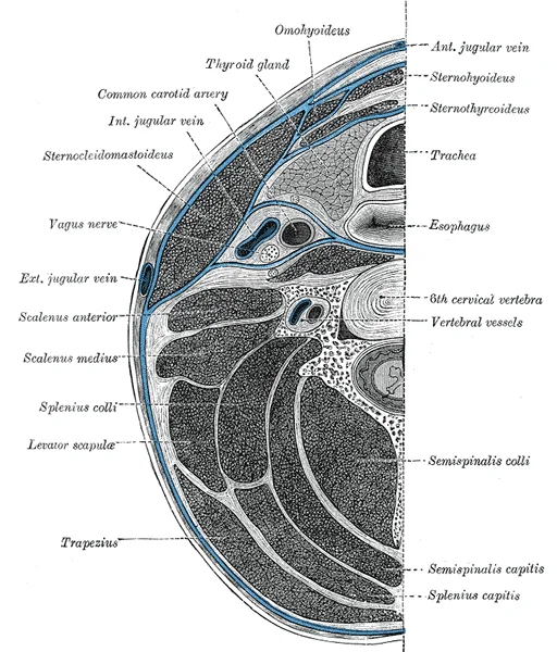
Long description
A cross-section diagram of the human neck at the level of C6 showing the fascia compartments, muscles, organs, bone, and major arteries, veins, and nerves.

Figure 1: A cross-section diagram of the human neck at the level of C6 showing the fascia compartments, muscles, organs, bone, and major arteries, veins, and nerves.
The cervical fascia is a complex multilayered connective tissue structure that divides the neck into distinct compartments. [1]
The superficial cervical fascia lies just beneath the skin. [1]
The deep cervical fascia has three main components:
- The investing layer surrounds the entire neck. [1]
- The pretracheal layer lies anterior to the vertebral column and encloses the thyroid gland, trachea, and esophagus [1]
- The prevertebral layer covers the deep muscles of the back and vertebral column
The retropharyngeal space is a common site for infection in children, particularly following upper respiratory tract infections. This space extends from the base of the skull to the level of T2.
The investing layer surrounds the sternocleidomastoid muscle, while the pretracheal layer encloses the thyroid gland and trachea. The prevertebral layer covers the vertebral column and deep neck muscles.
The cervical region contains several important muscles organized into compartments that work together to support the head, neck, and shoulder movements. These muscles can be broadly categorized into anterior, lateral, and posterior compartments based on their location and function.
| Compartment | Muscle | Origin | Insertion | Action | Nerve Supply [2] |
|---|---|---|---|---|---|
| Anterior | Sternocleidomastoid | Manubrium of sternum and medial third of clavicle | Mastoid process of temporal bone and lateral third of clavicle | Rotates head contralaterally, flexes neck | Accessory nerve (CN XI) and spinal nerves [1] |
| Scalenus anterior | Transverse processes of C3-C6 vertebrae | Upper surface of first rib | Elevates first rib during inspiration | C5-C7 spinal nerves | |
| Scalenus medius | Transverse processes of C2-C7 vertebrae | Upper surface of first rib | Elevates first rib during inspiration | C3-C8 spinal nerves [2] | |
| Scalenus posterior | Transverse processes of C5-C7 vertebrae | Outer surface of second rib | Elevates second rib during inspiration | C7-C8 spinal nerves | |
| Lateral | Levator scapulae | Transverse processes of C1-C4 vertebrae | Superior angle of scapula | Elevates scapula | [2] |
| Middle scalene | Transverse processes of C2-C7 vertebrae | Upper surface of first rib | Elevates first rib during inspiration | C3-C8 spinal nerves [2] | |
| Posterior | Splenius capitis | Ligamentum nuchae and spinous processes of C7-T3 vertebrae | Mastoid process and occipital bone | Extends and rotates head | [2] |
| Semispinalis capitis | Transverse processes of C7-T6 vertebrae | Occipital bone between superior and inferior nuchal lines | [2] | ||
| Rectus capitis posterior major | Spinous process of axis (C2) | Inferior nuchal line of occipital bone | Extends and rotates head | C1 spinal nerve (suboccipital nerve) [2] | |
| Rectus capitis posterior minor | Posterior tubercle of atlas (C1) | Inferior nuchal line of occipital bone | Extends head | [2] |
The scalene muscles are located laterally, with the anterior, middle, and posterior scalenes forming a staircase-like pattern between the transverse processes of the cervical vertebrae and the upper ribs.
The arterial supply to the cervical fascia is primarily derived from branches of the external carotid artery and the subclavian artery. The external carotid artery originates from the common carotid artery at the level of the upper border of the thyroid cartilage[1]. It ascends within the carotid triangle, lying medial to the internal carotid artery and posterior to the stylopharyngeus muscle. The external carotid artery gives off several branches that contribute to the blood supply of
The superior thyroid artery, the first branch of the external carotid artery, descends to the upper pole of the thyroid gland, supplying the infrahyoid muscles and the investing layer of deep cervical fascia[1]. The facial artery, after arising from the external carotid, curves upward over the mandible to supply the submandibular region and the platysma muscle, which is embedded in the superficial cervical fascia[1].
The posterior auricular artery, a smaller branch, ascends posterior to the ear to supply the auricle and the surrounding fascia[1].
From the subclavian artery, the thyrocervical trunk gives rise to the inferior thyroid artery, which ascends to supply the thyroid gland and the surrounding deep cervical fascia[1].
DefinitionArterial supply of cervical fascia
The arterial supply to the cervical fascia comes primarily from branches of the external carotid artery (superior thyroid, lingual, facial, occipital, posterior auricular) and the subclavian artery via the thyrocervical trunk (inferior thyroid, suprascapular).
DefinitionVenous drainage
The process by which de‑oxygenated blood from the cervical region returns to the heart through a network of veins.
Deep neck infections can present with fever, neck stiffness, dysphagia, and trismus. The clinical examination should assess for signs of airway compromise, which can develop rapidly in deep neck infections. [1]
| Vein | Origin (source territory) | Course (relationship to fascia) | Termination |
|---|---|---|---|
| Internal jugular vein | Venous plexus of the brain, orbit, and deep neck | Runs within the carotid sheath, deep to the sternocleidomastoid, parallel to the internal carotid artery | Joins the subclavian vein to form the brachiocephalic vein |
| External jugular vein | Superficial scalp and face | Lies superficial to the investing layer of cervical fascia, descends across the sternocleidomastoid | Drains into the subclavian vein |
| Vertebral vein | Deep cervical musculature and vertebral bodies | Travels within the transverse foramina of cervical vertebrae, deep to the prevertebral fascia | Empties into the brachiocephalic vein |
| Deep cervical vein | Deep cervical plexus | Courses deep to the prevertebral fascia, accompanies the deep cervical artery | Joins the vertebral or internal jugular vein |
| Thyroid veins (superior, middle, inferior) | Thyroid gland | Pass through the visceral layer of the deep cervical fascia | Drain into the internal jugular or brachiocephalic veins |
These veins follow a “complementary” pattern, often mirroring the arteries that supply the same regions. Nevertheless, considerable variability exists, and clinicians must be aware that tributaries may join the SVC through alternative routes.
Figure 6 (F‑B1‑2399‑6) illustrates the venous drainage of the cervical fascia in a sagittal view, with anterior structures to the left and posterior structures to the right. The labelled structures are:
- Internal jugular vein – located deep to the sternocleidomastoid within the carotid sheath; it receives blood from the brain and deep neck.
- External jugular vein – superficial to the investing fascia, crossing the sternocleidomastoid and descending to the subclavian vein.
- Vertebral vein – runs within the transverse foramina of C1‑C6, deep to the prevertebral fascia, and exits the foramina to join the brachiocephalic vein.
- Deep cervical vein – situated deep to the prevertebral fascia, parallel to the deep cervical artery.
- Subclavian vein – lies inferior to the clavicle, receiving the external jugular and vertebral veins.
- Brachiocephalic vein – formed by the union of the internal jugular and subclavian veins; it courses medially to join the superior vena cava.
- Superior vena cava – the final conduit that returns cervical venous blood to the right atrium. [1]
Caption: Sagittal illustration of cervical venous drainage showing the relationship of the internal and external jugular veins, vertebral and deep cervical veins, and their convergence into the brachiocephalic and superior vena cava.
Understanding this arrangement is essential for interpreting imaging, planning neck surgery, and managing venous complications such as thrombosis. [1]
The neck is supplied by both cranial and spinal nerves, classified by their connection to the central nervous system: cranial nerves connect to the brain, spinal nerves to the spinal cord. [2]
Cranial nerves carry the sensory and motor functions of the head and neck. Three are purely sensory — olfactory (CNI), optic (CNII) and vestibulocochlear (CNVIII); five strictly motor — oculomotor (CNIII), trochlear (CNIV), abducens (CNVI), accessory (CNXI) and hypoglossal (CNXII); four mixed — trigeminal (CNV), facial (CNVII), glossopharyngeal (CNIX) and vagus (CNX). Motor axons end on skeletal muscle of the head or neck. [2]
Three cranial nerves also carry autonomic fibres, and a fourth is almost purely autonomic: oculomotor fibres drive pupillary constriction, facial and glossopharyngeal fibres salivation, and the vagus targets ganglia in the thoracic and upper abdominal cavities. Terminal ganglia receiving cranial input lie in the head and neck, as well as the thoracic and upper abdominal cavities. [2]
All spinal nerves are mixed, with a sensory ganglion on the dorsal root of each. In the neck the cervical spinal nerves form the cervical plexus: cutaneous branches supply the skin, motor branches the infrahyoid muscles, and the phrenic nerve descends to the diaphragm. [2] [1]
DefinitionNerve point of the neck
The cutaneous branches of the cervical plexus emerge at the midpoint of the posterior border of sternocleidomastoid, the nerve point of the neck.
| Nerve | Muscles supplied | Skin area supplied |
|---|---|---|
| Lesser occipital | none (cutaneous) | scalp behind the auricle |
| Great auricular | none (cutaneous) | parotid region, mastoid, ear |
| Transverse cervical | none (cutaneous) | anterior neck |
| Supraclavicular | none (cutaneous) | clavicle and shoulder |
| Ansa cervicalis | infrahyoid muscles | none |
| Phrenic | diaphragm | none |
| Accessory | sternocleidomastoid, trapezius | none |
| Hypoglossal | tongue muscles | none |
At the midpoint of the posterior border of sternocleidomastoid the cutaneous branches appear: the lesser occipital ascends to the scalp behind the auricle, the great auricular to the parotid and ear, the transverse cervical forward across the neck, and the supraclavicular nerves over the clavicle.
The cervical fascia itself is largely avascular, but its layers form the boundaries of the spaces through which lymphatic vessels travel. These vessels accompany the external jugular vein and terminate in the superficial cervical lymph nodes, which lie along the external jugular vein beneath the platysma. From these nodes, efferent vessels [1]
These nodes receive afferent vessels from the superficial nodes, from the larynx, pharynx, thyroid and trachea, and from the submental and submandibular nodes. [1]
All efferent vessels from the deep cervical nodes converge to form the jugular trunk. On the right side, the jugular trunk joins the subclavian trunk to form the right lymphatic duct, which empties into the right brachiocephalic vein. On the left side, the jugular trunk drains directly into the thoracic duct, which then empties into the left brachiocephalic vein. [1]
The superficial nodes are drawn as small ovals beneath the platysma, the deep cervical chain as a vertical row of nodes along the internal jugular vein, and the jugular trunks as thick vessels descending to the venous angles on both sides.
The cervical fascia consists of several layers that define the anatomical compartments of the neck and influence the spread of infection. This layer encloses the trapezius and sternocleidomastoid muscles, forming a complete collar around the neck.
This fascial layer helps to compartmentalize the neck and can limit or direct the spread of infection from these structures. [1]
It forms a potential space between itself and the pretracheal layer known as the retropharyngeal space. This space extends superiorly to the base of the skull and inferiorly into the mediastinum, allowing infections in this region to spread along these planes. [1]
This sheath extends from the base of the skull to the root of the neck, providing a protective pathway for these vital structures. [1]
These spaces are potential routes for infection spread, with infections potentially tracking inferiorly into the mediastinum or laterally to involve the parapharyngeal space. [1]
The carotid sheath is demonstrated enclosing the carotid artery, internal jugular vein, and vagus nerve.
An enlarged retropharyngeal space on an anteroposterior view suggests fluid or infection tracking between the visceral and pre‑vertebral fasciae.
CT provides the clearest cross‑sectional view of the fascial planes. Between the investing and pre‑vertebral layers the visceral (or carotid) sheath contains the carotid artery, internal jugular vein and vagus nerve; on CT it appears as a compact, slightly hyperattenuating tube.
MRI differentiates fascial layers by signal intensity. On T1‑weighted images the superficial fascia is isointense with subcutaneous fat, while the investing fascia shows intermediate signal due to its mixed fibrous‑fat composition. Fat‑suppressed T2 sequences highlight fluid collections that dissect between these layers; for example, a retropharyngeal abscess will appear as a high‑signal pocket bounded laterally by the investing fascia and medially by the pre‑vertebral fascia.
Dynamic scanning can demonstrate the sliding of fascial layers during swallowing; loss of this glide suggests adhesions or fibrosis.
Figure 10 (F‑B1‑2399‑10) illustrates the radiological anatomy of cervical fascia on a contrast‑enhanced axial CT slice at the level of C3. Labeled structures include: Superficial cervical fascia (thin radiolucent line superficial to platysma), Investing fascia (denser band encircling sternocleidomastoid and trapezius), Carotid sheath (compact tube containing carotid artery, internal jugular vein, vagus nerve), Visceral (alar) fascia (thin line posterior to carotid sheath), Pre‑vertebral fascia (robust posterior envelope around vertebral bodies), and Danger space (potential space between alar and pre‑vertebral fascia). The image is displayed in the axial plane, with the patient’s right side on the left of the screen. Caption: Axial CT of the mid‑cervical region showing the layered cervical fascia and their relationships.
The cervical fascia plays a crucial role in the spread of deep neck infections, which can rapidly extend along fascial planes. The deep cervical fascia is divided into layers that create potential spaces where infections can spread. The pretracheal fascia surrounds the trachea and thyroid gland, while the prevertebral fascia surrounds the vertebral column and deep neck muscles. Infections in these spaces can lead to life-threatening complications such as airway obstruction or mediastinitis. [1]
Surface landmarks are essential for clinical examination and procedures.
Nerve palsies can occur due to compression or injury in the neck. Injury to this nerve can result in hoarseness due to vocal cord paralysis. [1]
The fascial planes of the neck are important in understanding the spread of infection and the potential for complications. Infections can spread along fascial planes, potentially leading to serious complications such as mediastinitis or airway obstruction. [1]
The retropharyngeal space is a common site for infection in children, particularly following upper respiratory tract infections. This space extends from the base of the skull to the level of T2.
Deep neck infections can present with fever, neck stiffness, dysphagia, and trismus. The clinical examination should assess for signs of airway compromise, which can develop rapidly in deep neck infections. [1]
Treatment of deep neck infections typically involves intravenous antibiotics and surgical drainage when indicated. In cases of airway compromise, intubation or tracheostomy may be necessary. [1]
DefinitionCervical fascia
The cervical fasciae are sheets of connective tissue beneath the skin that partition the neck; exam questions test their layers, their bony attachments and the spaces they create.
| Layer | Contents |
|---|---|
| Investing | sternocleidomastoid, trapezius, submandibular and parotid glands, suprahyoid and infrahyoid muscles |
| Pretracheal | thyroid gland, trachea, oesophagus, pharynx, larynx |
| Prevertebral | scalenes, prevertebral muscles, brachial plexus, subclavian vessels |
The cervical fascia organizes the neck into distinct surgical planes that dictate infection spread, guide operative dissection, and protect vital structures. The investing layer splits to envelop the sternocleidomastoid and trapezius, forming the roof of the posterior triangle and the floor of the submandibular region; its superior attachment to the mandible and zygomatic arch traps the facial vein and marginal mandibular branch of the facial nerve during submandibular gland excision. The pretracheal layer has a muscular portion investing the infrahyoid strap muscles and a visceral portion that suspends the thyroid and parathyroids, allowing the thyroid to move with swallowing; a breach here permits anterior visceral space infections to track into the superior mediastinum. The prevertebral layer covers the deep paraspinal muscles and forms the floor of the posterior triangle, creating the "danger space" between it and the alar fascia—a direct conduit for retropharyngeal abscesses to descend behind the pericardium. The carotid sheath, a condensation of all three layers, binds the common carotid, internal jugular, and vagus nerve; its anteromedial relation to the phrenic nerve on the anterior scalene and the sympathetic trunk posteriorly makes it the key landmark for central line placement and radical neck dissection. Lymphatic chains follow these fascial boundaries: levels I–V correspond to submental, upper jugular, mid-jugular, lower jugular, and posterior triangle groups, respectively, and their predictable drainage patterns stage head-and-neck malignancies. Mastery of these layers converts a complex anatomic region into a navigable surgical map.
The commonest error is to speak of "the cervical fascia" as if it were a single membrane. The neck is wrapped in a superficial fascia and in several deep layers, each with its own attachments and contents, and examinations test them as separate structures.
A second mistake is to confuse the investing layer with the pretracheal layer. The investing fascia encircles the neck and splits to enclose the sternocleidomastoid and trapezius; the pretracheal fascia lies deeper, enclosing the thyroid, trachea and oesophagus. Students who merge them cannot explain why a thyroid swelling moves on swallowing while a sternocleidomastoid lump does not.
A third error is to forget the attachments of the investing fascia to the mandible, the hyoid bone, the clavicle and the spine of the scapula. These attachments limit how far a superficial collection can spread, so a haematoma confined by the investing layer behaves differently from one that has breached it.
A fourth mistake is to treat the carotid sheath as just another fascial layer. It is a condensation of deep fascia around the common and internal carotid arteries, the internal jugular vein and the vagus nerve, and it is the landmark for carotid surgery and for central venous access.
A fifth error is to assume that fascial planes are barriers. They are potential spaces, and pus tracks along them. The retropharyngeal space lies between the buccopharyngeal fascia and the prevertebral fascia; behind the prevertebral fascia lies the danger space, so called because infection there descends directly into the posterior mediastinum.
A sixth mistake is to confuse the submandibular space with the submental space. The mylohyoid muscle divides them, so an infection anterior to the mylohyoid behaves differently from one posterior to it. Bilateral submandibular involvement with brawny swelling and elevation of the tongue is Ludwig's angina, and it is a airway emergency.
DefinitionCervical fascia
The cervical fasciae are layers of connective tissue that compartmentalise the neck, bind its muscles and viscera, and provide the planes along which infection, blood and air spread.
Exam trapDanger space
Do not confuse the retropharyngeal space with the danger space. The danger space lies behind the prevertebral fascia and communicates freely with the posterior mediastinum.
Clinical pearlCarotid sheath
The carotid sheath contains the common and internal carotid arteries, the internal jugular vein and the vagus nerve; it is the key landmark in carotid surgery and in central line insertion.
| Layer | Attachments and relations | Contents |
|---|---|---|
| Investing layer | Mandible, hyoid, clavicle, scapular spine; splits around sternocleidomastoid and trapezius | Roof of the anterior and posterior triangles |
| Pretracheal layer | Hyoid, thyroid cartilage, trachea, oesophagus; blends with the pericardium | Thyroid, trachea, oesophagus, recurrent laryngeal nerve |
| Prevertebral layer | Vertebral bodies and transverse processes, base of skull; extends into the posterior mediastinum | Vertebral muscles, sympathetic trunk |
| Carotid sheath | Condensation around the great vessels | Common and internal carotid arteries, internal jugular vein, vagus nerve |
| Buccopharyngeal fascia | Covers the pharynx and oesophagus | Forms the anterior wall of the retropharyngeal space |
At the bedside, a patient with sore throat, fever, trismus, drooling and neck swelling is assessed for deep neck space infection. The examiner looks for tongue elevation and floor-of-mouth swelling, and asks whether the swelling lies above or below the hyoid, because this predicts which spaces are involved. In children, a retropharyngeal abscess is a recognised cause of neck stiffness and refusal to swallow, and the airway is checked first in every case.
In the operating theatre the fascial planes guide dissection. In thyroid surgery the pretracheal fascia is opened and the gland is mobilised within its bed; in tracheostomy the pretracheal fascia and strap muscles are divided to reach the trachea; in neck dissection the investing fascia is raised with the skin flap and the carotid sheath is the deep landmark. In carotid endarterectomy the sheath is opened to expose the bifurcation, and the vagus nerve is identified and protected.
On imaging, contrast computed tomography of the neck shows which space contains a collection and whether it tracks toward the mediastinum. Magnetic resonance imaging is used when soft-tissue detail matters. The radiologist describes the collection in relation to the investing, pretracheal and prevertebral layers and notes whether the airway is narrowed.
In examinations, spotters ask for the layers of the deep cervical fascia, the contents of the carotid sheath, and the boundaries of the submandibular space. A frequent question is why a retropharyngeal abscess can cause mediastinitis, and the answer lies in the danger space behind the prevertebral fascia.
Illustrative example: a patient with a neglected dental infection develops increasing pain, trismus and a hot, tender swelling below the mandible. Because the swelling is above the hyoid and confined by the investing fascia, it points to a submandibular space infection rather than a retropharyngeal one.
These nodes receive afferent vessels from the superficial nodes, from the larynx, pharynx, thyroid and trachea, and from the submental and submandibular nodes.
- The neck is the region between the base of the skull and the thoracic inlet, carrying structures like the trachea, oesophagus, great vessels and nerves between the thorax and head.
- The cervical fascia is a system of connective tissue layers that divides the neck into compartments and can transmit infection into the mediastinum.
- The deep cervical fascia consists of three main layers: the investing layer that surrounds the neck and encloses sternocleidomastoid and trapezius muscles, the pretracheal layer that invests the thyroid gland, trachea, oesophagus and pharynx, and the prevertebral layer that covers the prevertebral muscles.
- Potential spaces between fascial layers include the suprasternal space, pretracheal space, retropharyngeal space, and danger space, which can open when pus or blood tracks through them.
- The sternocleidomastoid muscle divides the neck into anterior and posterior triangles, with the anterior triangle bounded by the sternocleidomastoid, anterior midline, and inferior border of the mandible, and the posterior triangle bounded by the sternocleidomastoid, trapezius, and clavicle.
- Key cervical landmarks include the hyoid bone at C3, thyroid cartilage at C4, and cricoid cartilage at C6, which marks the lower boundary of the larynx and upper boundary of the trachea.
Answer each one in your head first, then open the answer.
What are the three main layers of the deep cervical fascia and what structures do they contain?
Show answerHide answer
The investing layer surrounds the neck and encloses sternocleidomastoid and trapezius muscles, the pretracheal layer invests the thyroid gland, trachea, oesophagus and pharynx, and the prevertebral layer covers the prevertebral muscles.How does the sternocleidomastoid muscle divide the neck and what are the boundaries of each division?
Show answerHide answer
The sternocleidomastoid divides the neck into anterior and posterior triangles; the anterior triangle is bounded by the sternocleidomastoid, anterior midline, and inferior border of the mandible, while the posterior triangle is bounded by the sternocleidomastoid, trapezius, and clavicle.What are the key cervical landmarks at vertebral levels C3, C4, and C6?
Show answerHide answer
At C3 is the hyoid bone, at C4 is the thyroid cartilage forming the laryngeal prominence, and at C6 is the cricoid cartilage which marks the lower boundary of the larynx and upper boundary of the trachea.What potential spaces exist between the fascial layers of the neck and why are they clinically significant?
Show answerHide answer
Potential spaces include the suprasternal space, pretracheal space, retropharyngeal space, and danger space; they are clinically significant because they can open when pus or blood tracks through them, allowing infection to spread to other areas like the mediastinum.What structures are contained within the carotid sheath and how is it formed?
Show answerHide answer
The carotid sheath contains the common carotid artery, internal jugular vein, and vagus nerve, and is formed by a condensation of all three layers of cervical fascia.
- Betts JG, Young KA, Wise JA, et al. Circulatory Pathways. OpenStax, Anatomy and Physiology 2e (CC BY-NC-SA 4.0). Accessed .
- Betts JG, Young KA, Wise JA, et al. The Peripheral Nervous System. OpenStax, Anatomy and Physiology 2e (CC BY-NC-SA 4.0). Accessed .
Figure credits
Educational content for medical students and doctors. It is not a substitute for clinical judgement or local guidelines.