Carotid sheath
The carotid sheath is a fascial tube enclosing the major neurovascular bundle of the neck.
The carotid sheath is a cylindrical fascial condensation of the deep cervical fascia that encloses the major neurovascular bundle of the neck. It extends from the base of the skull superiorly to the root of the neck inferiorly, where it becomes continuous with the mediastinal fascia. This arrangement creates a
The carotid sheath lies within the carotid triangle, bounded anteriorly by the sternocleidomastoid muscle, posteriorly by the longus capitis and longus colli muscles overlying the transverse processes of the cervical vertebrae, and superiorly by the styloid process and its attached muscles. The anterolateral wall is reinforced by the investing layer of deep cervical fascia as it splits to enclose the sternocleidomastoid. The posteromedial wall relates to the prevertebral fascia covering the paraspinal muscles. The medial wall is adjacent to the pretracheal fascia investing the thyroid gland and paratracheal structures. [1]
The principal contents are the common carotid artery, internal carotid artery, internal jugular vein, and vagus nerve (cranial nerve X). The common carotid arteries have asymmetric origins: on the right side, the common carotid arises from the brachiocephalic artery, which is present only on the right and branches into the right subclavian and right common carotid arteries; on the left side, the common carotid arises directly from the aortic arch. Both common carotid arteries ascend within the s [1]
The external carotid artery gives off multiple branches that supply the face, lower jaw, neck, esophagus, and larynx; these include the lingual, facial, occipital, maxillary, and superficial temporal arteries. The internal carotid artery forms the carotid sinus, which contains baroreceptors and chemoreceptors that provide afferent input critical for maintaining cardiovascular homeostasis. [1]
The internal jugular vein lies lateral to the carotid arteries within the sheath and receives venous drainage from the brain, face, and neck. The vagus nerve runs posteriorly between the artery and vein, providing parasympathetic innervation to thoracic and abdominal viscera and sensory innervation to the larynx and pharynx.
Anatomic variations of the internal carotid artery relevant to the carotid sheath and skull base include dehiscence of the bony covering, with reported prevalence ranging from 2% to 23%, and protrusion into the sphenoid sinus, with prevalence from 5.2% to 67%. Optic nerve protrusion into the sphenoid sinus occurs in 7% to 35% of individuals and may be associated with internal carotid artery variations. These variations are important for surgical approaches to the skull base and for understanding potential pathways of infection spread from the sphenoid sinus to the cavernous sinus. [3]
The carotid sheath also contains the ansa cervicalis embedded in its anterolateral wall, sympathetic trunk fibers posteromedially, and lymphatic nodes along the internal jugular vein. The sheath's fascial investment allows it to act as a conduit for spread of infection or malignancy between the neck and mediastinum. [1]
The carotid sheath is a fascial compartment in the neck that contains the major vascular and neural structures supplying the head and neck. To locate it clinically, one must first identify the surface landmarks of the neck. The sternocleidomastoid muscle is the primary superficial landmark. Its anterior border runs obliquely from the mastoid process to the sternum. The carotid sheath lies deep to this muscle. Specifically, it is situated in the carotid triangle, which is bounded by the sternocle [1]
The sternocleidomastoid muscle is retracted laterally. Within this sheath, the common carotid artery is the most medial structure. It lies adjacent to the trachea and oesophagus. The internal jugular vein is lateral to the artery. The vagus nerve lies within the sheath.
Palpation of the carotid pulse is a key clinical skill. The pulse is felt by pressing the common carotid artery against the transverse processes of the cervical vertebrae. This is done just anterior to the sternocleidomastoid muscle. The pulse is strongest at the level of the thyroid cartilage. One must be careful not to compress both carotid arteries simultaneously, as this can reduce cerebral blood flow. The internal jugular vein is also palpable but is softer and collapses easily. It lies lateral to the artery. The vagus nerve is not palpable. [1]
The surface markings help guide invasive procedures such as central venous catheterisation. The internal jugular vein is often accessed here. The carotid artery must be avoided. Ultrasound guidance is recommended to distinguish the vein from the artery. The artery is pulsatile and round. The vein is non-pulsatile and oval. The relationship is consistent: artery medial, vein lateral. This anatomical arrangement is crucial for safe practice.
Clinical pearlClinical Pearl
Always palpate the carotid pulse on one side at a time to prevent syncope or cerebral ischaemia.
The carotid body is located at the bifurcation of the common carotid artery. It is a small, oval-shaped organ. It is not visible on the surface. It is deep to the sternocleidomastoid muscle. It is attached to the artery by connective tissue. It is involved in chemoreception. It detects changes in blood oxygen, carbon dioxide, and pH. It sends signals via the glossopharyngeal nerve. This information is vital for respiratory control.
| Structure | Position in Sheath | Relation |
|---|---|---|
| Common Carotid Artery | Medial | Adjacent to trachea |
| Internal Jugular Vein | Lateral | Superficial to artery |
| Vagus Nerve | Posterior | Between artery and vein |
The carotid sheath is a tubular fascial compartment that extends from the base of the skull to the root of the neck. It encloses three vital structures: the common carotid artery, the internal jugular vein, and the vagus nerve.
The bony landmarks defining the superior boundary are the jugular process of the occipital bone and the carotid canal. The carotid canal transmits the internal carotid artery into the cranial cavity. As noted in anatomical studies, the locations of the arteries in the brain not only provide blood flow to the brain tissue but also prevent interruption in the flow of blood. The sheath attaches superiorly to the margins of the carotid canal and the jugular foramen. [1]
Inferiorly, the sheath continues into the thorax, where the common carotid artery divides. The internal carotid artery supplies the anterior portion of the cerebrum. As a rule, branches to the anterior portion of the cerebrum are normally fed by the internal carotid arteries; the remainder of the brain receives blood flow from branches associated with the vertebral arteries. The vertebral arteries branch once they enter the cranial cavity, and some of these branches form a structure known as the arterial circle (or circle of Willis), an anastomosis that is remarkably like a traffic circle that sends off branches (in this case, arterial branches to the brain). [1]
The lateral boundary of the sheath is formed by the prevertebral fascia, while the medial boundary is adjacent to the visceral compartment of the neck. The sternocleidomastoid muscle overlies the sheath laterally. The internal jugular vein lies lateral to the common carotid artery. The vagus nerve runs between the artery and vein, posterior to both.
Clinical relevance arises when considering variations in bony anatomy. Insertion of an intersphenoid sinus septum onto the carotid canal has been reported in 4.7%. This variant is critical for sinus surgeons to recognize, as the sinus area is separated from the orbit and cranial fossa by only the thinnest of bones. Another anatomical variant is the sphenomaxillary plate, a condition where a posteriorly over-pneumatized maxillary sinus is united with the sphenoid sinus via a bony septum. This structure should be recognized on the CT images in order to avoid orbital complications during surgery. [3]
Significant associations were also found between the pneumatized pterygoid process and dehiscent or protruding internal carotid artery. [3]
The carotid sheath thus serves as a protected conduit for neurovascular structures, anchored by specific cranial base landmarks. Understanding these bony relationships prevents iatrogenic injury during approaches to the skull base and upper cervical spine.
The carotid sheath is a fascial compartment within the deep cervical fascia that encloses the major neurovascular structures of the neck. [1][2]
Within this sheath, three primary structures are identified. The common carotid artery lies medially and slightly posteriorly. The internal jugular vein is positioned laterally and anteriorly to the artery. The vagus nerve runs vertically between the artery and the vein, situated in the postero-lateral aspect of the sheath. The sheath itself is bounded by the sternocleidomastoid muscle laterally and the prevertebral fascia medially. This arrangement ensures that the neurovascular bundle is protected while remaining accessible for clinical procedures. [1]
The contents of the carotid sheath are closely related to surrounding structures. Anteriorly, the sheath is related to the thyroid gland, the strap muscles, and the sympathetic trunk, which lies on the prevertebral fascia just posterior to the sheath. Posteriorly, the sheath rests against the prevertebral muscles and the cervical plexus nerves. Laterally, the sternocleidomastoid muscle covers the sheath. The vagus nerve, contained within the sheath, gives off several branches, including the superior laryngeal nerve and the recurrent laryngeal nerve, which descend into the thorax. The carotid artery bifurcates into the internal and external carotid arteries at the level of the upper border of the thyroid cartilage. The internal jugular vein receives tributaries such as the facial and lingual veins before joining the subclavian vein to form the brachiocephalic vein. [1]
DefinitionCarotid Sheath
A fascial compartment in the neck containing the common carotid artery, internal jugular vein, and vagus nerve.
The integrity of the carotid sheath is crucial for maintaining the spatial relationships of these vital structures. Injury to the sheath can lead to compression or laceration of its contents, resulting in significant clinical consequences such as hemorrhage, neurological deficits, or airway compromise. Understanding the anatomical boundaries and contents of the carotid sheath is essential for surgeons performing neck dissections, central line placements, and other procedures in the cervical region. The sheath serves as a landmark for identifying and accessing the major vessels and nerves of the neck.
The carotid sheath is a fascial compartment within the deep cervical fascia that encloses the major neurovascular structures of the neck. It extends from the base of the skull to the root of the neck, where it blends with the mediastinal fascia. The sheath contains the common carotid artery, the internal jugular vein, and the vagus nerve. While the sheath itself is composed of connective tissue rather than contractile muscle, it is intimately associated with several key muscles that define its b [1]
The sternocleidomastoid muscle forms the lateral boundary of the sheath. The omohyoid muscle crosses the sheath anteriorly. The scalene muscles lie posteriorly.
The scalene muscles are located posteriorly.
The longus colli is an anterior vertebral muscle.
The longus colli is an anterior vertebral muscle.
The longus colli is an anterior vertebral muscle.
| Muscle | Origin | Insertion | Action | Nerve Supply |
|---|---|---|---|---|
| Sternocleidomastoid | Manubrium, medial clavicle | Mastoid process, superior nuchal line | Head flexion, rotation | Spinal accessory nerve (XI) |
| Omohyoid (Inf) | Superior scapula border | Hyoid bone (via tendon) | Depress hyoid | Ansa cervicalis [1] |
| The longus colli is an anterior vertebral muscle. | ||||
| Middle Scalene | Transverse processes C1–C6 | First rib | Lateral flexion | Anterior rami C3–C8 [1] |
| Posterior Scalene | Transverse processes C5–C7 | Second rib | Lateral flexion, rotation | Anterior rami C6–C8 [1] |
| Longus Colli | Bodies T1–T3, transverse C5–T1 | Bodies C1–C3, transverse C3–C5 | Neck flexion | Anterior rami C1–C6 |
| The scalene muscles are located posteriorly. |
These muscles provide structural support and protection to the carotid sheath. Their anatomical relationships are critical for surgical approaches and diagnostic imaging. The sternocleidomastoid is the most superficial landmark. The omohyoid crosses the sheath, dividing it into superior and inferior triangles. The scalene muscles form the posterior boundary, separating the sheath from the brachial plexus. The longus muscles form the anterior boundary, separating the sheath from the prevertebral fascia. [1]
Figure 12122 Common Carotid Artery
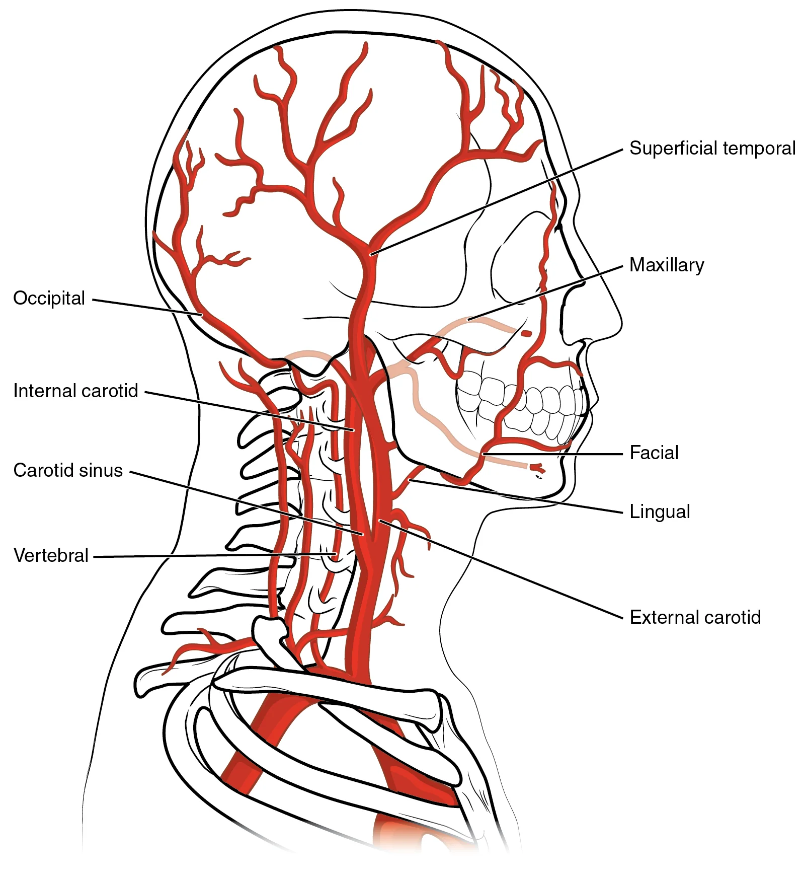
Long description
The common carotid artery gives rise to the external and internal carotid arteries. The external carotid artery remains superficial and gives rise to many arteries of the head. The internal carotid artery first forms the carotid sinus and then reaches the brain via the carotid canal and carotid fora

Figure 1: The common carotid artery gives rise to the external and internal carotid arteries.
The arterial components are derived from the aortic arch and the subclavian artery.
The common carotid artery is a major vessel that ascends in the neck within the carotid sheath. On the right side, it arises from the brachiocephalic trunk, while on the left side, it arises directly from the aortic arch. It ascends superiorly, lying deep to the sternocleidomastoid muscle. The common carotid artery terminates by dividing into the external and internal carotid arteries. This bifurcation is a cri
It ascends within the carotid sheath, initially lying posterolateral to the external carotid artery. As it passes superiorly, it enters the carotid canal in the temporal bone. The internal carotid artery initially forms an expansion known as the carotid sinus. Its primary role is to supply blood to the brain and eyes. It is often described as having no branches in the cervical region, which distinguishes it from the external carotid artery. [1]
The external carotid artery is the other terminal branch of the common carotid artery. It ascends anterior to the internal carotid artery. It gives off several branches in the neck, including the superior thyroid artery, ascending pharyngeal artery, lingual artery, facial artery, occipital artery, posterior auricular artery, and maxillary artery. These branches supply the structures of the face, neck, and scalp. The external carotid artery is distinguished from the internal carotid artery by the presence of these branches. [1]
At the level of the thyroid cartilage, it bifurcates into the internal and external carotid arteries. The internal carotid artery is positioned posterolaterally, while the external carotid artery is positioned anteromedially.
The arterial supply to the tissues surrounding the carotid sheath is provided by branches of the external carotid artery. These include the superior thyroid artery, which supplies the thyroid gland and larynx, and the lingual artery, which supplies the tongue. The facial artery supplies the face and mandible. The occipital artery supplies the posterior scalp and neck muscles. These branches arise from the external carotid artery as it ascends within the sheath.
The internal carotid artery continues its course into the skull, where it gives rise to the ophthalmic artery and the anterior and middle cerebral arteries. These vessels supply the brain and eyes. The lack of branches in the neck makes the internal carotid artery vulnerable to injury, as there are no collateral vessels in the cervical region to compensate for occlusion.
The common carotid artery is covered by the platysma, sternocleidomastoid muscle, and deep cervical fascia. It is related to the vagus nerve, which lies posteriorly within the sheath. The sympathetic trunk lies posterior to the sheath. The internal jugular vein lies lateral to the carotid artery. These relations are important for surgical approaches to the carotid sheath.
The carotid sinus is a dilation at the bifurcation of the common carotid artery. It contains baroreceptors that monitor blood pressure. The carotid body is a small structure located at the bifurcation that contains chemoreceptors. These structures are innervated by the glossopharyngeal nerve. They play a crucial role in regulating blood pressure and respiration.
The arterial supply of the carotid sheath is thus derived from the common carotid artery, which divides into the internal and external carotid arteries. The internal carotid artery supplies the brain, while the external carotid artery supplies the face and neck. The close relationship between these vessels and the internal jugular vein and vagus nerve within the sheath is a key anatomical feature.
It originates as the direct continuation of the sigmoid sinus at the jugular foramen and descends vertically through the neck within the carotid sheath, lying lateral to the common carotid artery and internal carotid artery. The internal jugular vein terminates by joining the subclavian vein to form the brachiocephalic vein. [1]
It begins at the medial angle of the eye as the angular vein and descends posterior to the mandible, where it crosses the submandibular gland. The facial vein typically drains into the internal jugular vein, though it may communicate with the pterygoid plexus and ultimately drain into the external jugular vein in some individuals.
The carotid sheath is a fascial compartment in the neck that encloses the major neurovascular structures of the head and neck. It extends from the base of the skull to the root of the neck. Within this sheath, three critical structures are closely associated: the common carotid artery, the internal jugular vein, and the vagus nerve. Understanding the precise anatomical relationships and functional supply of these nerves is essential for clinical practice and surgical safety.
The sympathetic trunk lies posterior to the sheath, resting on the prevertebral fascia.
The vagus nerve provides the primary motor and parasympathetic innervation to the thoracic and abdominal viscera, as well as motor supply to the muscles of the larynx and pharynx. It also carries sensory information from the larynx, pharynx, and viscera. The hypoglossal nerve supplies motor innervation to all the intrinsic and extrinsic muscles of the tongue, except for the palatoglossus. The glossopharyngeal nerve provides motor supply to the stylopharyngeus muscle and sensory innervation to the posterior third of the tongue, the pharynx, and the carotid body and sinus.
| Nerve | Motor Supply (Muscles) | Sensory Supply (Skin/Areas) |
|---|---|---|
| Vagus (CN X) | Muscles of pharynx, larynx, soft palate | Larynx, pharynx, external ear, tympanic membrane |
| Hypoglossal (CN XII) | Intrinsic and extrinsic tongue muscles (except palatoglossus) | None (purely motor) |
| Glossopharyngeal (CN IX) | Stylopharyngeus | Posterior 1/3 of tongue, pharynx, carotid body/sinus |
| Accessory (CN XI) | Sternocleidomastoid, trapezius | None (purely motor) |
The sympathetic trunk runs vertically along the posterior aspect of the carotid sheath. It provides sympathetic innervation to the head and neck, including vasoconstriction of blood vessels and sweat gland activity. The superior cervical ganglion, the largest of the cervical sympathetic ganglia, is located at the level of the second and third cervical vertebrae, posterior to the internal carotid artery.
Lesions of these nerves can result in significant clinical deficits. Damage to the vagus nerve may cause hoarseness due to vocal cord paralysis, dysphagia, and loss of gag reflex. Hypoglossal nerve injury leads to tongue deviation towards the side of the lesion on protrusion. Glossopharyngeal nerve damage can result in loss of taste and sensation from the posterior tongue and impaired swallowing.
Exam trapNerve Identification
Do not confuse the hypoglossal nerve with the vagus nerve. The hypoglossal nerve is purely motor to the tongue, while the vagus nerve has mixed motor, sensory, and parasympathetic functions.
The lymphatic vessels from the thyroid gland and larynx follow the carotid sheath inferiorly, draining into the deep cervical lymph nodes. These lymph nodes are strategically located along the internal jugular vein, which is a key component of the carotid sheath. This anatomical relationship creates a potential pathway for the spread of infections or malignancies from the thyroid and laryngeal regions. [1]
The deep cervical lymph nodes can be divided into upper and lower groups based on their anatomical position. In clinical practice, palpation of these nodes is an important part of the head and neck examination, as enlargement may indicate
Lymph from these nodes ultimately drains into the jugular lymph trunk, which typically empties into the venous angle at the junction of the internal jugular and subclavian veins on the left side, and into the right subclavian vein on the right side. This drainage pattern explains why infections or malignancies in the carotid sheath region can lead to palpable lymphadenopathy in the neck. The jugular lymph trunk may vary in its termination point, with approximately 80% draining into the venous an [1]
For example, thyroid carcinomas commonly metastasize to the deep cervical lymph nodes along the carotid sheath, while laryngeal cancers may spread through these same pathways. Understanding these drainage patterns is essential for surgical planning, as lymph node dissection often follows these natural drainage routes. In elderly patients, lymphatic [1]
The sheath extends from the base of the skull down to the thorax, where it fuses with the endothoracic fascia. Understanding the spatial arrangement within this sheath is fundamental for clinical procedures such as central line insertion or neck dissection.
These structures are not randomly placed; they maintain specific relationships with one another.
The common carotid artery lies medially within the sheath. It ascends through the neck, supplying blood to the head and brain. As it approaches the level of the upper border of the thyroid cartilage, it bifurcates into the internal and external carotid arteries. This bifurcation is a critical landmark. The internal carotid artery continues superiorly to supply the brain, while the external carotid artery supplies the face and neck structures. The artery is relatively deep and protected by the surrounding fascia and muscles. [1]
The vein begins at the jugular foramen as the continuation of the sigmoid sinus. It descends through the neck, receiving various tributaries along its course. Because it is lateral and superficial to the artery, it is the preferred site for central venous access. The vein empties into the subclavian vein to form the brachiocephalic vein. [1]
The vagus nerve (cranial nerve X) lies posterior and between the artery and the vein. It runs in the neurovascular bundle, providing parasympathetic innervation to the thoracic and abdominal viscera. The nerve also supplies motor and sensory fibers to the larynx and pharynx. Its position between the artery and vein protects it from direct compression but makes it vulnerable during surgical dissection if the sheath is opened incorrectly.
The carotid sheath is surrounded by other important structures. Anteriorly, it is related to the sternocleidomastoid muscle, which covers the sheath laterally. The sternocleidomastoid muscle originates from the sternum and clavicle and inserts into the mastoid process. It is innervated by the accessory nerve. Posteriorly, the sheath is related to the prevertebral fascia and the deep neck muscles. Medially, the sheath is adjacent to the pharynx and larynx. Laterally, it is covered by the sternocleidomastoid muscle and the platysma. [1]
The sympathetic trunk lies posterior to the sheath, lying on the prevertebral fascia. It is not contained within the sheath but is in close proximity. Injury to the sympathetic trunk can result in Horner’s syndrome, characterized by ptosis, miosis, and anhidrosis.
The internal carotid artery and internal jugular vein are key components of the circulatory system in the head and neck. The internal jugular vein drains blood from the brain and superficial facial regions. Blood from the more superficial portions of the head, scalp, and cranial regions, including the temporal vein and maxillary vein, flow into each external jugular vein. Although the external and internal jugular veins are separate vessels, there are anastomoses between them close to the thoracic region. [1]
The common carotid artery supplies blood to the head and neck. It is a continuation of the brachiocephalic trunk on the right and the aortic arch on the left. The artery ascends without branches until it bifurcates. The bifurcation is marked by the carotid sinus and carotid body, which monitor blood pressure and chemical composition.
The vagus nerve provides critical autonomic and somatic functions. It descends through the neck within the carotid sheath, giving off branches such as the superior laryngeal nerve and the recurrent laryngeal nerve. The superior laryngeal nerve innervates the cricothyroid muscle and provides sensory innervation to the larynx above the vocal cords. The recurrent laryngeal nerve innervates all intrinsic muscles of the larynx except the cricothyroid.
On cross-sectional imaging, the internal jugular vein appears as a large, compressible, low-density structure lateral to the artery. The common carotid artery is circular and maintains a consistent diameter. The vagus nerve lies between the artery and vein, often appearing as a small, rounded structure with intermediate signal intensity. Understanding these relations is critical for surgical planning and interventional procedures. [1]
DefinitionCarotid Sheath
A fascial compartment in the neck enclosing the common carotid artery, internal jugular vein, and vagus nerve.
Computed tomography (CT) provides high-resolution anatomical detail of the carotid sheath structures. It allows for clear differentiation between the vascular lumen and surrounding soft tissues. Contrast enhancement highlights the arterial and venous systems, aiding in the identification of thrombosis or stenosis. Magnetic resonance imaging (MRI) offers superior soft tissue contrast, particularly for evaluating the vagus nerve and adjacent lymph nodes. MRI sequences such as T1-weighted and T2-weighted images help distinguish between solid masses and cystic lesions. Ultrasound is the primary modality for dynamic assessment of blood flow and vessel compressibility. It is widely used for guided interventions and real-time evaluation of vascular patency.
| Structure | CT Appearance | MRI Appearance | Ultrasound Appearance |
|---|---|---|---|
| Internal Jugular Vein | Low density, compressible | Low signal on T1, high on T2 | Anechoic, compressible |
| Common Carotid Artery | Circular, contrast-filled | Intermediate signal | Pulsatile, echogenic walls |
| Vagus Nerve | Intermediate density | Intermediate signal | Hypoechoic, honeycomb pattern |
The anatomical variations of the carotid sheath are significant for radiological interpretation. Variations in the branching pattern of the carotid artery can alter the expected landmarks. The relationship between the sheath and surrounding muscles, such as the sternocleidomastoid, provides additional context for localization. Radiologists must be aware of these variations to avoid misdiagnosis. The clarity and accuracy of CT examinations are paramount for identifying abnormalities. Preoperative imaging serves as a roadmap for surgical approaches, ensuring safe navigation through complex anatomical regions.
NoteClinical Pearl
CT provides an anatomical “road map” to identify significant abnormalities with increased clarity and accuracy prior to surgical approaches. [3]
The carotid sheath is a fibrous connective tissue tube that encloses the major neurovascular structures of the neck. It extends from the base of the skull to the root of the neck, where it blends with the mediastinal fascia. Understanding its contents is vital for surgical safety and diagnostic imaging.
The internal jugular vein lies lateral and slightly anterior within the sheath. The common carotid artery (or internal carotid artery superiorly) is positioned medial and posterior to the vein. Surrounding the sheath are the sternocleidomastoid muscle laterally and the prevertebral muscles posteriorly. Recognising this spatial arrangement allows clinicians to identify structures on ultrasound or CT scans accurately.
DefinitionCarotid Sheath Contents
The carotid sheath contains the common carotid artery, internal jugular vein, and vagus nerve.
Trauma to the neck can damage structures within the carotid sheath. Penetrating injuries may lacerate the internal jugular vein, leading to significant haemorrhage or air embolism. Arterial injury to the carotid artery can cause stroke or pseudoaneurysm formation. Compression of the vagus nerve results in vocal cord paralysis, presenting as hoarseness. This occurs because the recurrent laryngeal nerve, a branch of the vagus, innervates the intrinsic muscles of the larynx.
| Structure | Location in Sheath | Clinical Consequence of Injury |
|---|---|---|
| Internal Jugular Vein | Lateral | Haemorrhage, air embolism |
| Carotid Artery | Medial | Stroke, pseudoaneurysm |
| Vagus Nerve | Posterior | Vocal cord paralysis, hoarseness |
Clinical procedures often utilise the carotid sheath anatomy. Carotid endarterectomy involves exposing the common carotid artery within the sheath to remove atherosclerotic plaque. Surgeons must identify the vagus nerve to avoid iatrogenic injury. Palpation of the carotid pulse is performed at the level of the upper border of the thyroid cartilage, where the common carotid artery is superficial. This landmark guides needle insertion for central venous catheterisation of the internal jugular vein, which lies just lateral to the artery. Ultrasound guidance is standard to distinguish the compressible vein from the pulsatile artery, reducing the risk of arterial puncture.
Clinical pearlSurgical Safety
Always identify the vagus nerve before dividing any tissue in the carotid triangle to prevent vocal cord paralysis.
The carotid bifurcation, where the common carotid artery divides into internal and external branches, is a critical landmark. It typically occurs at the level of the upper border of the thyroid cartilage, corresponding to the fourth cervical vertebra. This area is rich in sensory innervation from the carotid sinus nerve, a branch of the glossopharyngeal nerve, which monitors blood pressure. Awareness of these relationships prevents complications during neck dissections and vascular interventions.
DefinitionCarotid Sheath Definition
The carotid sheath is a fascial tube enclosing the major neurovascular structures of the neck, formed by contributions from the investing, pretracheal, and prevertebral layers of the deep cervical fascia. [1]
Understanding this spatial relationship is critical for surgical approaches and ultrasound imaging. The common carotid artery lies most medially, adjacent to the trachea and oesophagus. The internal jugular vein is positioned laterally and is often the largest vein in the neck. The vagus nerve runs posteriorly within the sheath, between the artery and vein.
Clinical pearlBifurcation Level
The common carotid artery typically bifurcates into the internal and external carotid arteries. [1]
The level of bifurcation is a key anatomical landmark. This variation has significant implications for carotid endarterectomy and central venous catheterization procedures. The internal carotid artery ascends vertically within the carotid sheath and enters the skull through the carotid canal. It does not give off branches in the neck, unlike the exte [1]
Exam trapNerve Position
The vagus nerve lies posterior to the common carotid artery and within the carotid sheath, giving off branches to the pharynx, larynx, and heart. [1]
The vagus nerve provides parasympathetic innervation to the thoracic and abdominal viscera. Within the carotid sheath, it gives off the superior laryngeal nerve and the pharyngeal branches. It is crucial not to confuse the carotid sheath with the carotid sinus, which is a baroreceptor located at the bifurcation of the common carotid artery. The carotid sinus monitors blood pressure, while the carotid body monitors blood oxygen and carbon dioxide levels. [1]
Clinical pearlPalpation Landmark
NoteVenous Drainage
The internal jugular vein drains blood from the brain, face, and neck, and is the largest vein in the neck. [1]
It receives the facial vein, lingual vein, and superior thyroid vein. It joins the subclavian vein to form the brachiocephalic vein.
Exam trapAortic Course
The aorta is the largest artery in the body. It arises from the left ventricle and eventually descends to the abdominal region, where it bifurcates at the level of the fourth lumbar vertebra into the two common iliac arteries. [1]
The aorta consists of the ascending aorta, the aortic arch, and the descending aorta. After exiting the heart, the ascending aorta moves in a superior direction for approximately 5 cm and ends at the sternal angle. Following this ascent, it reverses direction, forming a graceful arc to the left, called the aortic arch. The aortic arch descends toward the inferior portions of the body and ends at the level of the intervertebral disk between the fourth and fifth thoracic vertebrae. Beyond this point, the descending aorta continues close to the bodies of the vertebrae and passes through an opening in the diaphragm known as the aortic hiatus.
Clinical pearlAbdominal Aorta
After crossing through the diaphragm at the aortic hiatus, the thoracic aorta is called the abdominal aorta. This vessel remains to the left of the vertebral column and is embedded in adipose tissue behind the peritoneal cavity. It formally ends at approximately the level of vertebra L4, where it bifurcates to form the common iliac arteries. [1]
The abdominal aorta gives rise to several important branches before this division. The inferior mesenteric artery supplies blood to the distal segment of the large intestine, including the rectum. It arises approximately 5 cm superior to the common iliac arteries. [1]
Students often confuse the carotid sheath with the investing layer of deep cervical fascia. The carotid sheath is a distinct tubular condensation of fascia that surrounds the major neurovascular bundle of the neck, whereas the investing layer splits to enclose the sternocleidomastoid and trapezius muscles. The sheath is formed by contributions from the prevertebral, pretracheal, and investing layers, but it is a separate fascial compartment.
A frequent error is to list the internal jugular vein as lying medial to the common carotid artery. In the carotid sheath, the internal jugular vein lies lateral to the common carotid artery at the base of the neck, though the two vessels overlap and the vein may appear posterior or anterolateral depending on the level. The vagus nerve runs in a groove between the artery and vein, posterior to both, and is often omitted from diagrams.
Another mistake is to assume the carotid sheath extends uninterrupted from the base of the skull to the mediastinum. The sheath is continuous with the prevertebral fascia superiorly and blends with the mediastinal fascia inferiorly, but it ends at the level where the internal carotid artery enters the carotid canal and the internal jugular vein passes through the jugular foramen. The external carotid artery leaves the sheath at the level of the greater horn of the hyoid bone.
Students sometimes think the ansa cervicalis lies within the carotid sheath. The ansa cervicalis is embedded in the anterior wall of the sheath or lies on its surface, not within the neurovascular compartment. The sympathetic trunk runs posterior to the sheath, embedded in the prevertebral fascia, and is not a content of the sheath.
A common oversight is forgetting that the carotid sheath contains deep cervical lymph nodes. These nodes lie alongside the internal jugular vein and are surgically relevant in neck dissections. They are often omitted from simplified lists of sheath contents.
Another pitfall is misidentifying the plane of the carotid sheath during surgery. The sheath lies deep to the sternocleidomastoid muscle and the investing fascia. Blunt dissection through the investing layer reveals the sheath as a glistening white tube; incising it exposes the common carotid artery medially, the internal jugular vein laterally, and the vagus nerve posteriorly between them.
Identifying carotid sheath contents at the bedside
- Palpate common carotid pulseMedial landmark for sheath
- Question: Is the vessel pulsatile?
Yes
- Common carotid arteryMedial structure in sheath
No
- Internal jugular veinLateral, compressible, non-pulsatile
- Vagus nervePosterior between artery and vein; not palpable
- Deep cervical nodesAlong internal jugular vein; may be palpable if enlarged
At the bedside, knowledge of the carotid sheath guides central venous catheter placement. The internal jugular vein is accessed lateral to the carotid pulse; ultrasound confirmation shows the vein lateral and superficial to the artery within the sheath. Misidentification risks arterial puncture, haematoma formation, or vagal stimulation causing bradycardia. The sheath's fascial boundaries also explain why a carotid artery injury can be compressed effectively against the transverse processes of the cervical vertebrae.
In the operating theatre, the carotid sheath is the key anatomical reference for neck dissections. In a selective neck dissection (levels II–IV), the surgeon preserves the internal jugular vein, vagus nerve, and carotid artery within the sheath while removing lymph nodes from the lateral compartment. In a radical neck dissection, the sheath is opened and its contents may be sacrificed if involved by tumour. The ansa cervicalis, lying on the anterior sheath wall, is identified and preserved when possible to maintain infrahyoid muscle function.
On imaging, the carotid sheath appears as a distinct fascial envelope on contrast-enhanced CT and MRI. The "carotid space" corresponds to the sheath and contains the carotid artery, internal jugular vein, vagus nerve, and deep cervical nodes. Pathology such as carotid artery dissection, jugular vein thrombosis, vagal schwannoma, or nodal metastasis is localised to this space. The sheath's continuity with the mediastinum explains the spread of infection from the neck to the thorax (descending necrotising mediastinitis).
In examinations, students are tested on the sheath's formation, contents, relations, and surgical relevance. Typical questions ask for the fascial layers contributing to the sheath, the arrangement of structures within it (artery medial, vein lateral, nerve posterior), the level at which the external carotid artery exits, and the clinical significance of the ansa cervicalis and sympathetic trunk lying outside the sheath. Diagrams often require labelling of the sheath in cross-section at the level of the thyroid cartilage.
- The carotid sheath is a fascial tube enclosing the common carotid artery, internal jugular vein, vagus nerve, and deep cervical lymph nodes.
- It is formed by contributions from the investing, pretracheal, and prevertebral layers of deep cervical fascia.
- The internal jugular vein lies lateral to the common carotid artery; the vagus nerve runs posteriorly between them.
- The external carotid artery leaves the sheath at the level of the greater horn of the hyoid bone.
- The ansa cervicalis lies on the anterior wall of the sheath; the sympathetic trunk lies posterior to it in the prevertebral fascia.
- The sheath extends from the skull base to the mediastinum, providing a pathway for infection spread.
- Ultrasound-guided internal jugular vein access uses the carotid artery as a medial landmark within the sheath.
- Neck dissection classification depends on whether the sheath contents are preserved or removed.
Answer each one in your head first, then open the answer.
Which three fascial layers contribute to the formation of the carotid sheath?
Show answerHide answer
The investing layer, the pretracheal layer, and the prevertebral layer of deep cervical fascia.What is the typical arrangement of the three main structures within the carotid sheath at the level of the thyroid cartilage?
Show answerHide answer
Common carotid artery medially, internal jugular vein laterally, vagus nerve posteriorly between them.At what anatomical level does the external carotid artery exit the carotid sheath?
Show answerHide answer
At the level of the greater horn of the hyoid bone.Where does the ansa cervicalis lie in relation to the carotid sheath?
Show answerHide answer
Embedded in or on the anterior wall of the sheath, not within the neurovascular compartment.Why is the carotid sheath clinically important in the spread of infection?
Show answerHide answer
It is continuous superiorly with the prevertebral fascia and inferiorly with the mediastinal fascia, allowing infection to track from the neck into the thorax.
- Betts JG, Young KA, Wise JA, et al. Circulatory Pathways. OpenStax, Anatomy and Physiology 2e (CC BY-NC-SA 4.0). Accessed .
- Fernandez R, Nardocci G, Navarro C, Reyes EP, Acuña-Castillo C, Cortes PP. Neural reflex regulation of systemic inflammation: potential new targets for sepsis therapy. Frontiers in physiology 2014. Accessed .
- Papadopoulou AM, Chrysikos D, Samolis A, Tsakotos G, Troupis T. Anatomical Variations of the Nasal Cavities and Paranasal Sinuses: A Systematic Review. Cureus 2021. Accessed .
Figure credits
Educational content for medical students and doctors. It is not a substitute for clinical judgement or local guidelines.