Cervical triangles
Anatomical regions of the neck defined by muscle boundaries, crucial for localising masses and understanding neurovascular pathways.
The neck is a complex anatomical region that serves as a conduit between the head and the thorax. It is subdivided into two large lateral compartments known as the anterior and posterior cervical triangles. These triangles are defined by the sternocleidomastoid muscle and the trapezius muscle, which act as primary landmarks for clinical examination and surgical access. Understanding these boundaries is essential for the anatomical diagnosis of neck masses, as the location of a swelling often dictates its likely origin, whether it be lymphatic, thyroid, or vascular in nature.
The posterior triangle is bounded anteriorly by the posterior border of the sternocleidomastoid, posteriorly by the anterior border of the trapezius, and inferiorly by the middle third of the clavicle. Its apex is located at the superior nuchal line of the occipital bone. This region contains the spinal accessory nerve, which is responsible for controlling the muscles of the neck, along with cervical spinal nerves. The floor of this triangle is formed by several muscles, including the splenius capitis, levator scapulae, and the scalene muscles. [2] [3]
The anterior triangle is bounded anteriorly by the midline of the neck, posteriorly by the anterior border of the sternocleidomastoid, and superiorly by the inferior border of the mandible. It is further subdivided into four smaller triangles: the submental, submandibular, carotid, and muscular triangles. The carotid triangle contains the common carotid artery, internal jugular vein, and the vagus nerve. The vagus nerve is responsible for contributing to homeostatic control of the organs of the thoracic and upper abdominal cavities. The hypoglossal nerve is responsible for controlling the muscles of the lower throat and tongue. [2]
The vertebral column forms the posterior wall of the neck. In the neck, there are seven cervical vertebrae, each designated with the letter “C” followed by its number. Typical cervical vertebrae, such as C4 or C5, have several characteristic features that differentiate them from thoracic or lumbar vertebrae. Cervical vertebrae have a small body, reflecting the fact that they carry the least amount of body weight. The transverse processes of the cervical vertebrae are sharply curved (U-shaped) to allow for passage of the cervical spinal nerves. [3] [2]
Clinical pearlClinical Landmark
The sternocleidomastoid muscle divides the neck into anterior and posterior triangles, providing a reliable guide for locating deep structures during physical examination.
| Muscle | Action | Nerve Supply |
|---|---|---|
| Sternocleidomastoid | Rotates head, flexes neck | Spinal accessory nerve |
| Trapezius | Elevates scapula, extends neck | Spinal accessory nerve |
| Scalene | Elevates ribs, flexes neck | Cervical spinal nerves |
The surface anatomy of the neck is characterized by several important palpable landmarks that help clinicians identify underlying structures. The sternocleidomastoid muscle is a prominent surface landmark that divides the neck into anterior and posterior triangles. This muscle originates from the manubrium sterni and medial clavicle, and inserts on the mastoid process of the temporal bone. When contracted, it can be easily palpated and serves as an important guide for locating deeper structures. [3]
The anterior triangle of the neck is bounded by the inferior border of the mandible, the anterior midline of the neck, and the sternocleidomastoid muscle. Within this triangle, the hyoid bone is a crucial palpable landmark that lies at the level of the C3 vertebra. The thyroid cartilage, which forms the Adam's apple, is another important structure that can be palpated in the midline at the level of C4-C5 vertebrae. [3]
Figure 1Gray384
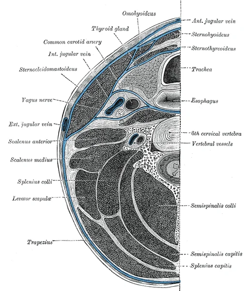
Long description
Section of the neck at about the level of the sixth cervical vertebra. Showing the arrangement of the fascia coli.

Figure 1: Section of the neck at about the level of the sixth cervical vertebra.
The posterior triangle is bounded by the sternocleidomastoid muscle, the trapezius muscle, and the clavicle. The greater auricular nerve, which is a branch of the cervical plexus, can be palpated as it crosses the posterior border of the sternocleidomastoid muscle about two-thirds of the way from the angle of the jaw to the clavicle.
The external jugular vein is another important surface landmark that can be seen in individuals with low subcutaneous fat. It begins behind the angle of the mandible and crosses the sternocleidomastoid muscle to empty into the subclavian vein. This vein is often used as an access point for venous cannulation. [1]
The anterior triangle contains the infrahyoid muscles, which include the sternohyoid, omohyoid, sternothyroid, and thyrohyoid muscles. These muscles are located inferior to the hyoid bone and help in depressing the hyoid and larynx during swallowing. The posterior triangle contains the scalene muscles, which are important for elevating the first rib during respiration. [3]
It can be palpated along its entire length and serves as an attachment point for several muscles including the trapezius, pectoralis major, and deltoid. The suprasternal notch, located at the superior border of the manubrium, is another palpable landmark that lies between the medial ends of the clavicles. [3]
Figure 2Gray387
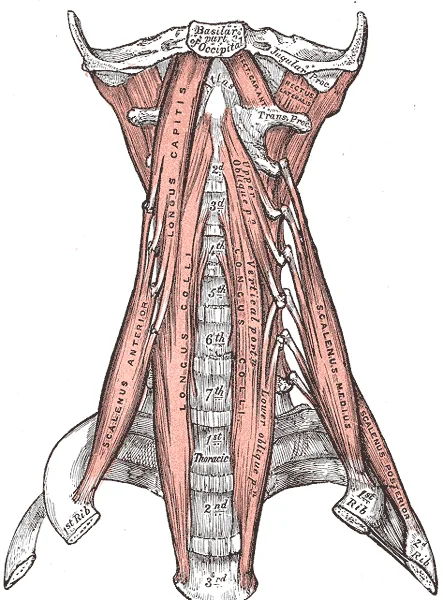
Long description
The anterior vertebral muscles. Labelled illustration from Wikimedia Commons, used under its open licence.

Figure 2: The anterior vertebral muscles.
The cervical vertebrae form the posterior boundary. A typical cervical vertebra is characterised by a small body, a bifid spinous process, and U-shaped transverse processes containing a transverse foramen. The atlas (C1) lacks a body, consisting instead of a bony ring formed by anterior and posterior arches, and articulates with the dens of the axis (C2). The axis features the dens projecting upward from its vertebral body. These vertebrae are smaller than lumbar vertebrae because they support less body weight. The intervertebral discs between them are thin in the cervical region, accounting for approximately 25 percent of body height between the pelvis and skull. [3]
The clavicle forms the anterior and inferior boundary of the posterior triangle. It is a long bone that articulates medially with the sternum and laterally with the acromion of the scapula. The sternocleidomastoid muscle attaches to the mastoid process of the temporal bone and the manubrium of the sternum, dividing the neck into anterior and posterior triangles. The hyoid bone, located in the anterior triangle, serves as an attachment point for suprahyoid and infrahyoid muscles but does not articulate with other bones.
The scapula provides the posterior anchor for the shoulder girdle. Its spine and acromion process are palpable landmarks. The subclavian artery, which supplies the arms, chest, shoulders, back, and central nervous system, passes behind the clavicle. It gives rise to the internal thoracic artery, the vertebral artery, and the thyrocervical artery. The vertebral artery passes through the vertebral foramen in the cervical vertebrae and then through the foramen magnum into the cranial cavity. [1]
This knowledge is critical at the bedside for assessing neurological status. Given the central role and vital importance of the brain to life, it is critical that blood supply to this organ remains uninterrupted. Clinicians must recognise that loss of blood flow for longer periods, typically between 3 and 4 minutes, will likely produce irreversible brain damage or a stroke, also called a cerebrovascular accident (CVA). This threshold guides emergency interventions. In the operating theatre, surg [1]
The cervical region is organised into distinct anatomical spaces defined by layers of deep cervical fascia. These fascial planes create compartments that contain muscles, nerves, and vessels, providing both structural support and pathways for the spread of infection or fluid. Understanding these boundaries is essential for interpreting clinical imaging and performing surgical procedures safely.
Deep to this lies the pretracheal fascia, which encloses the thyroid gland and trachea. The prevertebral fascia covers the vertebral column and associated muscles, extending laterally to form the floor of the posterior triangle.
The cervical triangles are bounded by specific muscular and skeletal landmarks. The anterior triangle is bounded anteriorly by the midline of the neck, posteriorly by the anterior border of the sternocleidomastoid, and superiorly by the inferior border of the mandible. This triangle is further subdivided into four smaller triangles: the submental, submandibular, carotid, and muscular triangles. The submental triangle is bounded by the anterior bellies of the digastric muscles and the hyoid bone. The submandibular triangle is bounded by the anterior and posterior bellies of the digastric muscle and the inferior border of the mandible. The carotid triangle is bounded by the posterior belly of the digastric, the sternocleidomastoid, and the superior belly of the omohyoid. The muscular triangle is bounded by the superior belly of the omohyoid, the sternohyoid, and the midline.
Figure 3Gray512
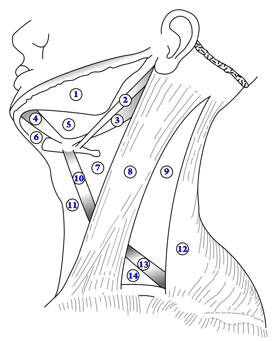
Long description
The triangles of the neck. Labelled illustration from Wikimedia Commons, used under its open licence.

Figure 3: The triangles of the neck.
The posterior triangle is bounded anteriorly by the posterior border of the sternocleidomastoid, posteriorly by the anterior border of the trapezius, and inferiorly by the middle third of the clavicle. The apex of the posterior triangle is at the superior nuchal line. This triangle is subdivided by the inferior belly of the omohyoid muscle into the occipital triangle above and the supraclavicular triangle below.
DefinitionCarotid Sheath
A tubular fascial compartment containing the common carotid artery, internal jugular vein, and vagus nerve.
| Muscle | Origin | Insertion | Action | Nerve Supply |
|---|---|---|---|---|
| Sternocleidomastoid | Manubrium and clavicle | Mastoid process | Neck flexion and rotation | Accessory nerve |
| Trapezius | Occipital bone and spine | Clavicle and scapula | Shoulder elevation and retraction | Accessory nerve |
| Omohyoid | Scapula | Hyoid bone | Depresses hyoid bone | Ansa cervicalis |
The fascial compartments also influence the spread of pathology. Infections can track along these potential spaces, leading to deep neck space infections that may compromise the airway. The pretracheal space allows communication between the neck and the superior mediastinum. The retropharyngeal space lies between the buccopharyngeal fascia and the prevertebral fascia, extending from the base of the skull to the posterior mediastinum. Awareness of these anatomical relationships is critical for diagnosis and management.
Figure 4Gray794
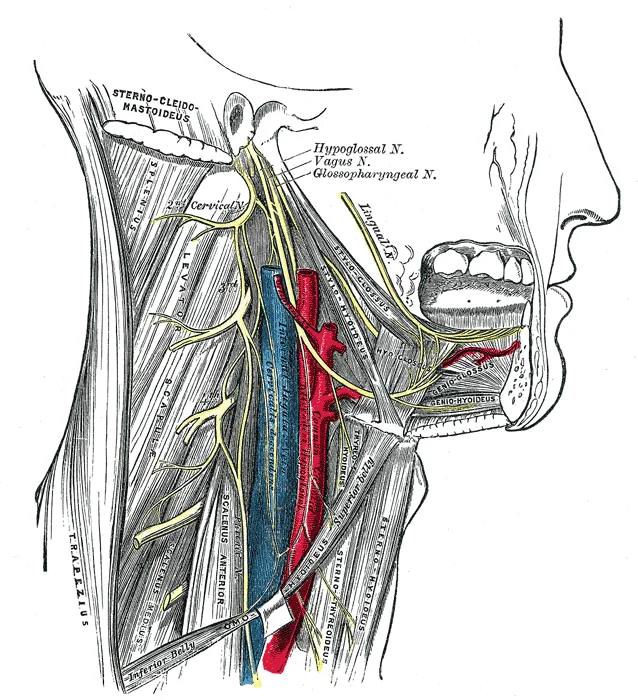
Long description
Hypoglossal nerve, cervical plexus, and their branches.

Figure 4: Hypoglossal nerve, cervical plexus, and their branches.
The cervical region is anatomically divided into distinct triangles, primarily the anterior and posterior triangles, which are bounded by key muscular landmarks. Understanding the muscles within these compartments is fundamental to locating neurovascular structures.
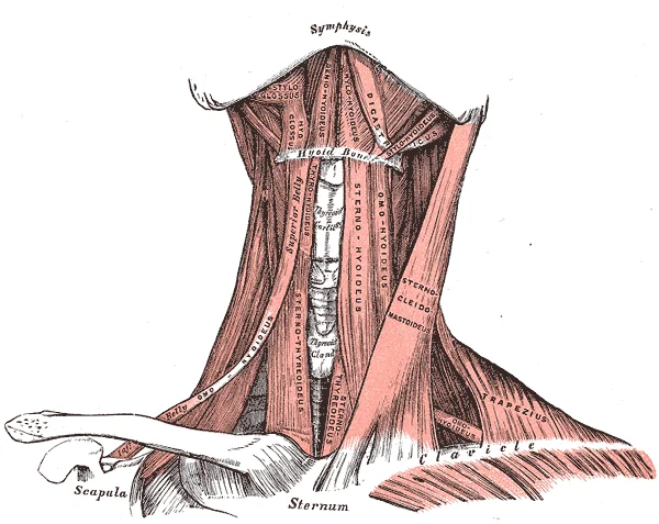
The anterior triangle is bounded by the sternocleidomastoid posteriorly, the midline of the neck anteriorly, and the inferior border of the mandible superiorly. Within this triangle lie the strap muscles, also known as the infrahyoid muscles. These muscles depress the hyoid bone and larynx during swallowing and speech. They include the sternohyoid, sternothyroid, thyrohyoid, and omohyoid muscles. The sternohyoid originates from the posterior surface of the manubrium and inserts onto the inferior border of the hyoid bone. The sternothyroid originates from the posterior surface of the manubrium and inserts onto the oblique line of the thyroid cartilage. The thyrohyoid originates from the thyroid cartilage and inserts onto the hyoid bone. The omohyoid has two bellies; the inferior belly originates from the superior border of the scapula and the superior belly inserts onto the hyoid bone. All strap muscles are innervated by the ansa cervicalis, derived from the cervical plexus (C1-C3), except for the thyrohyoid, which is innervated directly by C1 fibres travelling with the hypoglossal nerve.
Figure 5Gray386
Long description
Muscles of the neck. Anterior view. Labelled illustration from Wikimedia Commons, used under its open licence.

Figure 5: Muscles of the neck (anterior view).
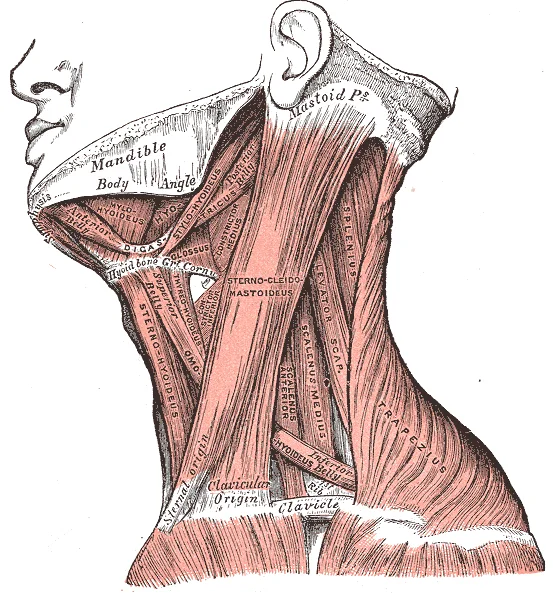
The posterior triangle is bounded by the sternocleidomastoid anteriorly, the trapezius posteriorly, and the clavicle inferiorly. It contains the trapezius muscle, which originates from the external occipital protuberance, the nuchal ligament, and the spinous processes of C7-T12. It inserts onto the lateral third of the clavicle, the acromion, and the spine of the scapula. Its actions include elevation, retraction, and rotation of the scapula. It is innervated by the spinal accessory nerve (CN XI). The cervical plexus also contributes to the innervation of the neck, with its cutaneous branches emerging from the posterior border of the sternocleidomastoid. These branches provide sensory innervation to the skin of the neck and shoulder. The relationship between the sternocleidomastoid, the accessory nerve, and the cervical plexus is critical for surgical approaches to the neck. [3]
Figure 6Gray385
Long description
Muscles of the neck. Lateral view. Labelled illustration from Wikimedia Commons, used under its open licence.

Figure 6: Muscles of the neck (lateral view).
| Muscle | Origin | Insertion | Action | Nerve Supply |
|---|---|---|---|---|
| Sternocleidomastoid | Manubrium, medial clavicle | Mastoid process, superior nuchal line | Head rotation, neck flexion | Spinal accessory nerve (CN XI), C2-C3 |
| Sternohyoid | Posterior manubrium | Inferior hyoid border | Depresses hyoid | Ansa cervicalis (C1-C3) |
| Sternothyroid | Posterior manubrium | Thyroid cartilage | Depresses larynx | Ansa cervicalis (C1-C3) |
| Thyrohyoid | Thyroid cartilage | Hyoid bone | Depresses hyoid, elevates larynx | C1 via hypoglossal nerve |
| Omohyoid | Scapula superior border | Hyoid bone | Depresses hyoid | Ansa cervicalis (C1-C3) |
| Trapezius | Occipital bone, nuchal ligament, spinous processes | Lateral clavicle, acromion, scapular spine | Scapular movement | Spinal accessory nerve (CN XI) |
The arterial supply to the cervical region primarily originates from branches of the aortic arch and the subclavian arteries. The aortic arch gives rise to three major branches: the brachiocephalic trunk, the left common carotid artery, and the left subclavian artery[1]. The brachiocephalic trunk further divides into the right common carotid artery and the right subclavian artery[1].
The common carotid arteries ascend through the neck and bifurcate at the level of the fourth cervical vertebra (C4) into the internal and external carotid arteries[1]. The internal carotid artery enters the cranial cavity through the carotid canal and contributes to the formation of the arterial circle (circle of Willis)[1]. The external carotid artery supplies structures of the face, scalp, and neck with its branches including the superior thyroid, lingual, facial, occipital, and maxillary arteries[1].
The subclavian arteries pass posterior to the clavicles and give rise to several important branches. The vertebral arteries arise from the subclavian arteries and ascend through the transverse foramina of the cervical vertebrae before entering the cranial cavity to join in forming the basilar artery[1]. The thyrocervical trunk branches from the subclavian artery and gives rise to the inferior thyroid, suprascapular, and transverse cervical arteries[1].
The costocervical trunk, another branch of the subclavian artery, provides blood to the neck and thoracic wall through its superior intercostal and deep cervical branches[1]. The internal thoracic artery (also known as the internal mammary artery) originates from the subclavian artery and descends along the anterior chest wall, giving off anterior intercostal branches and contributing to the blood supply of the diaphragm[1].
DefinitionVenous drainage of the neck
The network of veins that collects de‑oxygenated blood from the cervical region and empties it into the systemic venous system.
The cervical region is drained by a superficial system that runs just beneath the skin and a deep system that follows the arteries within the carotid sheath. Both systems ultimately converge on the internal jugular vein (IJV), which descends within the carotid sheath and joins the subclavian vein to form the brachio‑cephalic vein. The brachio‑cephalic veins on each side merge to become the superior vena cava (SVC), delivering blood to the right atrium.
The external jugular vein (EJV) originates from the confluence of the posterior auricular vein and the retromandibular vein. It runs superficially across the sternocleidomastoid (SCM) muscle, lying lateral to the SCM and deep to the platysma. It receives tributaries from the scalp and face before piercing the investing layer of the deep cervical fascia to empty into the subclavian vein just posterior to the clavicle. [1]
The facial vein begins at the angular vein near the medial canthus, descends across the face, and joins the EJV at the level of the angle of the mandible.
The posterior auricular vein drains the posterior scalp and ear, joining the EJV near its origin.
The internal jugular vein begins at the jugular foramen as a continuation of the sigmoid sinus. It runs deep to the SCM, medial to the carotid artery, and receives the following branches:
| Branch | Origin (drained area) | Course & relations | Termination |
|---|---|---|---|
| Superior thyroid vein | Thyroid gland | Descends anterior to the carotid sheath, crosses the carotid artery | Joins IJV near its origin |
| Middle thyroid vein | Thyroid isthmus | Runs laterally across the carotid sheath | Empties into IJV mid‑level |
| Inferior thyroid vein | Lower thyroid | Passes posterior to the carotid sheath | Drains into IJV near its lower third |
| Vertebral vein | Cervical vertebral venous plexus | Ascends within the transverse foramina of C1‑C6, lies posterior to the carotid artery | Enters IJV at the base of the skull |
| Deep cervical vein | Deep cervical musculature | Runs alongside the deep cervical artery, deep to the SCM | Joins IJV near its lower third |
The IJV then unites with the subclavian vein to form the brachio‑cephalic vein; the right and left brachio‑cephalic veins create the SVC, which returns blood to the right atrium. [1]
Figure 6 (F‑B1‑2401‑6) illustrates the venous drainage of the anterior and posterior cervical triangles. The diagram is a frontal view with the patient’s head at the top. Labeled structures include:
- External jugular vein (EJV) – runs superficially over the SCM, shown in blue‑green, draining into the subclavian vein (labelled “Subclavian”).
- Internal jugular vein (IJV) – depicted deep to the SCM, coursing vertically within the carotid sheath, receiving the superior, middle, and inferior thyroid veins (small branches labelled “Sup. Thyroid”, “Mid. Thyroid”, “Inf. Thyroid”).
- Vertebral vein – shown ascending through the transverse foramina of C1‑C6, posterior to the carotid artery (labelled “Carotid”).
- Facial vein – entering the EJV at the mandibular angle.
The caption reads: “Figure 6. Venous drainage of the cervical triangles, frontal view, anterior‑to‑posterior orientation.”
These veins collectively ensure that low‑oxygen blood from the head, neck, and upper limbs reaches the SVC and ultimately the right atrium, completing the systemic venous circuit. [1]
Figure 71321 Spinal Nerve Plexuses
Long description
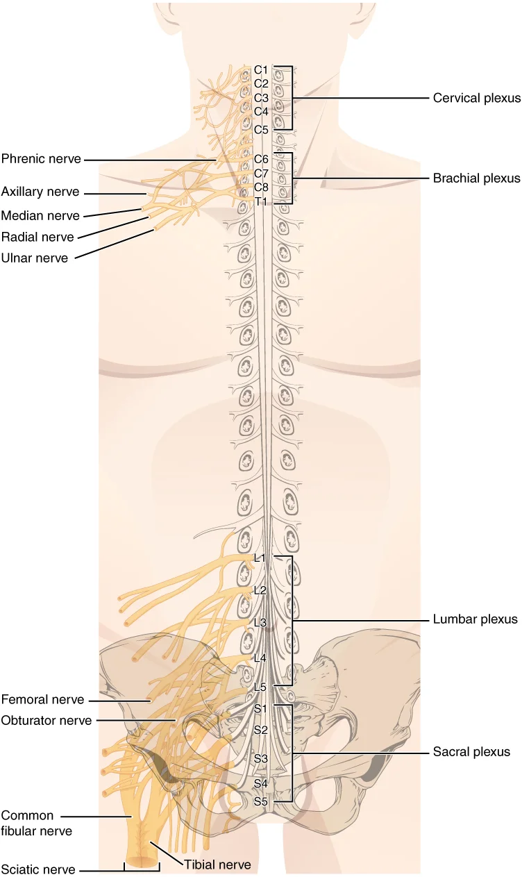
There are four main nerve plexuses in the human body. The cervical plexus supplies nerves to the posterior head and neck, as well as to the diaphragm. The brachial plexus supplies nerves to the arm. The lumbar plexus supplies nerves to the anterior leg. The sacral plexus supplies nerves to the poste

Figure 7: There are four main nerve plexuses in the human body.
The cervical triangles are traversed by a mixture of cranial and spinal nerves that provide both motor and cutaneous innervation to the neck. Motor fibres arise from motor nuclei in the brain‑stem or from the anterior horn of the spinal cord and terminate on skeletal muscles of the head and neck. Sensory fibres arise from dorsal root ganglia or cranial sensory ganglia and end in cutaneous receptors of the overlying skin. All spinal nerves are mixed, containing both sensory and motor fibres. < [2]
DefinitionCranial nerve
A cranial nerve is a bundle of axons that connects the brain to peripheral structures; twelve are numbered I–XII. [2]
Figure 7 illustrates the nerves that cross the anterior and posterior cervical triangles. In the anterior view the labelled structures are: the spinal accessory nerve (CN XI) running deep to the sternocleidomastoid, the vagus nerve (CN X) descending within the carotid sheath, the cervical plexus (C2–C4) emerging at the posterior border of the sternocleidomastoid, and the ansa cervicalis looping around the internal jugular vein. In the posterior view the labelled structures are: the greater auricular nerve (C2–C3) emerging superior to the sternocleidomastoid, the transverse cervical nerve (C2–C3) crossing the posterior triangle, and the supraclavicular nerves (C3–C4) descending toward the clavicle. The figure is a lateral view of the neck, with anterior structures shown on the left side of the image and posterior structures on the right side. Caption: “Lateral view of the cervical triangles showing cranial and cervical plexus nerves; anterior structures on the left, posterior on the right.”
The motor and cutaneous supply of the cervical triangles can be summarised in Table 1. The spinal accessory nerve (CN XI) is strictly motor and supplies the sternocleidomastoid and trapezius muscles. The vagus nerve (CN X) is mixed, providing parasympathetic fibres to thoracic and abdominal viscera and a small cutaneous branch to the ear. The cervical plexus (C2–C4) is mixed, with motor branches to the infrahyoid muscles and cutaneous branches to the skin over the neck. The trigeminal nerve (CN V) is mixed, contributing sensory fibres to the skin of the lower face and motor fibres to the muscles of mastication, but its branches do not enter the cervical triangles. [2]
| Nerve | Motor target (muscle) | Cutaneous target (skin) |
|---|---|---|
| Spinal accessory (CN XI) | Sternocleidomastoid, trapezius | – |
| Vagus (CN X) | Visceral smooth muscle (parasympathetic) | Small auricular area |
| Cervical plexus (C2‑C4) | Infrahyoid muscles | Skin over the anterolateral neck |
| Trigeminal (CN V) | Muscles of mastication | Skin of lower face (outside triangle) |
| Facial (CN VII) | Muscles of facial expression | Posterior auricular skin |
The three purely sensory cranial nerves (I, II, VIII) do not traverse the cervical triangles, whereas five cranial nerves are strictly motor (III, IV, VI, XI, XII) and four are mixed (V, VII, IX, X). Understanding the origin, course and relationships of these nerves is essential for safe surgical dissection of the cervical triangles. [2]
Figure 8Cervical lymph nodes and levels
Long description
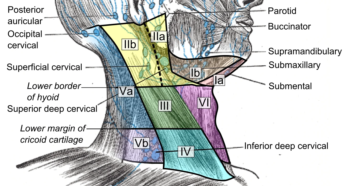
Superficial cervical lymph nodes and lymph node levels, initially described by the Memorial Sloan-Kettering Cancer Center (New York, NY) and further specified by the Committee for Head and Neck Surgery and Oncology of th

Figure 8: Superficial cervical lymph nodes and lymph node levels, initially described by the Memorial Sloan-Kettering Cancer Center (New York, NY) and further specified by the Committee for Head and Neck Surgery and Oncology of th.
The lymphatic drainage of the cervical region is organized into several groups of lymph nodes that receive lymph from different areas of the head and neck. These nodes are arranged in a hierarchical fashion, with superficial nodes receiving lymph from more superficial structures and deeper nodes receiving lymph from deeper structures as well as from the superficial nodes.
The deep cervical lymph nodes form a chain along the internal jugular vein and are divided into superior, middle, and inferior groups. These nodes receive lymph from the superficial nodes as well as from deeper structures such as the larynx, pharynx, and thyroid gland. The deep cervical nodes ultimately drain into the jugular lymphatic trunk, which empties into the venous angle at the junction of the internal jugular and subclavian veins. [1]
The submandibular nodes are located beneath the mandible, collecting lymph from the medial face.
The cervical region is divided into anterior and posterior triangles, each with distinct boundaries and contents. The anterior triangle is bounded anteriorly by the midline of the neck, posteriorly by the sternocleidomastoid muscle, and superiorly by the mandible. This triangle contains important structures including the carotid artery, internal jugular vein, and vagus nerve. The carotid triangle, a subdivision of the anterior triangle, is bounded by the sternocleidomastoid posteriorly, the posterior belly of the digastric muscle superiorly, and the superior belly of the omohyoid muscle inferiorly. Within this triangle lie the common carotid artery, which bifurcates into the internal and external carotid arteries, and the internal jugular vein.
The cervical region is a complex anatomical zone where soft tissue planes and bony landmarks create distinct compartments. Understanding these triangles requires correlating surface anatomy with deeper structures visible on imaging. This image shows a coronal CT reconstruction of the neck, oriented with the patient’s right side on the viewer’s left. The caption states: "Coronal CT view of cervical triangles showing key muscular and vascular landmarks."
In this view, the sternocleidomastoid muscle is the most prominent lateral structure. It inserts onto the mastoid process of the temporal bone and the superior nuchal line. The muscle divides the neck into two major triangles: the anterior and posterior triangles. The anterior triangle is bounded by the midline of the neck, the inferior border of the mandible, and the anterior border of the sternocleidomastoid. The posterior triangle is bounded by the posterior border of the sternocleidomastoid, the anterior border of the trapezius, and the middle third of the clavicle. [3]
On CT, the carotid sheath is visible as a fascial plane containing the common carotid artery, internal jugular vein, and vagus nerve. The common carotid artery bifurcates at the level of the upper border of the thyroid cartilage. This bifurcation is a critical landmark for identifying the internal and external carotid arteries. The internal jugular vein lies lateral to the carotid artery within the sheath. The vagus nerve runs posteriorly between the artery and vein. [1]
Ultrasound is particularly useful for evaluating the thyroid gland and lymph nodes within the anterior triangle. The thyroid gland is located anterior to the trachea, spanning from the fifth cervical to the first thoracic vertebrae. It consists of two lobes connected by an isthmus. The recurrent laryngeal nerves run in the tracheoesophageal groove, posterior to the thyroid lobes. This relationship is crucial during thyroid surgery to avoid nerve injury.
MRI provides superior soft tissue contrast for evaluating the brachial plexus and cervical spine. The brachial plexus emerges from the intervertebral foramina between C5 and T1. The roots of the plexus are visible as hypoechoic structures on ultrasound and hypointense on T1-weighted MRI.
The vertebral column provides the posterior boundary for these triangles. The transverse processes of the cervical vertebrae serve as attachment sites for the scalene muscles. The spinous processes are palpable midline landmarks. The articular processes form the zygapophyseal joints, which allow for movement and stability. Understanding these relationships allows for accurate interpretation of radiological images and safe clinical procedures.
The cervical region is clinically divided into two major triangles by the sternocleidomastoid muscle. This anatomical division is critical for localising pathology and guiding surgical access. The sternocleidomastoid itself originates from the manubrium of the sternum and the medial third of the clavicle. Its primary actions include ipsilateral lateral flexion and contralateral rotation of the neck. It is innervated by the spinal accessory nerve (cranial nerve XI) and cervical spinal nerves C2 and C3.
| Muscle | Origin | Insertion | Action | Nerve Supply |
|---|---|---|---|---|
| Sternocleidomastoid | Manubrium, medial clavicle | Mastoid process, superior nuchal line | Ipsilateral flexion, contralateral rotation | Spinal accessory nerve (CN XI), C2-C3 |
| Omohyoid | Superior scapular notch | Hyoid bone | Depresses hyoid bone | Ansa cervicalis (C1-C3) |
| Sternohyoid | Manubrium, medial clavicle | Hyoid bone | Depresses hyoid bone | Ansa cervicalis (C1-C3) |
The anterior triangle is bounded by the anterior border of the sternocleidomastoid, the midline of the neck, and the inferior border of the mandible. It contains the carotid sheath, which houses the common carotid artery, internal jugular vein, and vagus nerve. Clinically, this area is the site for carotid endarterectomy and lymph node biopsy. Injury to the vagus nerve here can cause hoarseness due to vocal cord paralysis. The hypoglossal nerve crosses the internal and external carotid arteries in this region. Damage during surgery can lead to tongue deviation towards the side of the lesion.
The posterior triangle is bounded by the posterior border of the sternocleidomastoid, the anterior border of the trapezius, and the middle third of the clavicle. It contains the brachial plexus roots and trunks, the subclavian artery, and the external jugular vein. This is a common site for iatrogenic injury to the spinal accessory nerve during lymph node excision. Such injury results in shoulder drop and weakness in arm abduction. The phrenic nerve also courses through this triangle, lying on the anterior surface of the scalenus anterior muscle.
The sternocleidomastoid muscle is retracted laterally to expose the underlying structures. The internal jugular vein lies lateral to the artery. The vagus nerve is positioned posteriorly within the sheath.
Clinical pearlClinical Pearl
Always identify the spinal accessory nerve before dissecting the posterior triangle to prevent iatrogenic shoulder dysfunction.
The cervical region is defined by two major triangular spaces: the anterior and posterior triangles. These are bounded by the sternocleidomastoid muscle, the trapezius muscle, and the clavicle. Understanding their boundaries is critical for surgical access and clinical examination.
The anterior triangle is bounded anteriorly by the midline of the neck, posteriorly by the sternocleidomastoid, and superiorly by the inferior border of the mandible. It contains vital structures including the carotid arteries, jugular veins, and cranial nerves.
The carotid sheath encloses the common carotid artery, internal jugular vein, and vagus nerve. The common carotid artery arises from the brachiocephalic trunk on the right and directly from the aortic arch on the left. It bifurcates at the level of the upper border of the thyroid cartilage into the internal and external carotid arteries. This landmark is crucial for locating the pulse.
The internal jugular vein descends within the carotid sheath, lateral to the internal carotid artery. It joins the subclavian vein to form the brachiocephalic vein. The vagus nerve runs between the artery and vein within the sheath.
The submental and submandibular triangles subdivide the anterior triangle. The submandibular triangle is bounded by the anterior and posterior bellies of the digastric muscle and the inferior border of the mandible. It contains the submandibular gland and facial artery.
The posterior triangle is bounded anteriorly by the sternocleidomastoid, posteriorly by the trapezius, and inferiorly by the middle third of the clavicle. The floor is formed by the prevertebral fascia covering deep neck muscles.
The spinal accessory nerve (CN XI) crosses the posterior triangle, supplying the sternocleidomastoid and trapezius muscles. Injury to this nerve results in shoulder droop and weakness in shrugging.
The external jugular vein drains into the subclavian vein, crossing the sternocleidomastoid superficially.
Lymph nodes in the anterior triangle drain the face and neck. Enlargement may indicate infection or malignancy. The carotid sinus, located at the bifurcation, contains baroreceptors regulating blood pressure.
The posterior triangle contains lymph nodes that drain the scalp and neck. A mass here may suggest lymphadenopathy. The supraclavicular lymph nodes are clinically significant; enlargement may indicate thoracic or abdominal pathology.
Clinical pearlCarotid Bifurcation
The common carotid artery divides into internal and external carotid arteries. [1]
DefinitionCervical Triangles
The neck is divided into anterior and posterior triangles by the sternocleidomastoid muscle. These spaces contain vital neurovascular structures. [1]
NoteLymphatic Drainage
Anterior triangle nodes drain the face; posterior triangle nodes drain the scalp. Supraclavicular nodes indicate distant pathology. [1]
Clinical pearlVagus Nerve
The vagus nerve runs within the carotid sheath, between the internal jugular vein and the common carotid artery.
DefinitionCarotid Sheath
The carotid sheath encloses the common carotid artery, internal jugular vein, and vagus nerve.
NoteDigastric Muscle
The digastric muscle divides the anterior triangle into submental and submandibular triangles. [1]
Exam trapExternal Jugular Vein
The external jugular vein crosses the sternocleidomastoid superficially. It is often used for central venous access assessment. [1]
NoteThyroid Cartilage
The upper border of the thyroid cartilage marks the level of the carotid bifurcation and the C4 vertebra. [1]
Students frequently confuse the anatomical boundaries of the neck with the vascular territories of the brain, leading to errors in localising pathology. A primary error is assuming that the internal carotid artery is the sole supplier of blood to the human brain. This is incorrect; the internal carotid arteries along with the vertebral arteries are the two primary suppliers of blood to the human brain. Failing to recognise the vertebral contribution can lead to misinterpretation of posterior circulation strokes. Another frequent mistake involves the timing of ischaemic events. Students often underestimate the speed of neuronal damage. When blood flow is interrupted, even for just a few seconds, a transient ischemic attack (TIA), or mini-stroke, may occur, resulting in loss of consciousness or temporary loss of neurological function. This highlights that neurological deficits can be transient yet indicative of severe underlying vascular compromise. [1]
A second pitfall relates to the structural integrity of the spine, which underpins the neck’s anatomy. Students may overlook the role of the intervertebral discs in weight bearing and flexibility. Each intervertebral disc consists of two parts. The anulus fibrosus is the tough, fibrous outer layer of the disc. It forms a circle (anulus = “ring” or “circle”) and is firmly anchored to the outer margins of the adjacent vertebral bodies. Inside is the nucleus pulposus, consisting of a softer, more gel-like material. With increasing age, the water content of the nucleus pulposus gradually declines. This causes the disc to become thinner, decreasing total body height somewhat, and reduces the flexibility and range of motion of the disc, making bending more difficult. In older adults, this degeneration increases the risk of herniation if the anulus fibrosus is weakened or damaged, the nucleus pulposus can protrude outward, resulting in a herniated disc. [3]
A third error is neglecting the venous drainage patterns of the abdomen and lower thorax, which can confuse students regarding systemic return. Other than the small amount of blood drained by the azygos and hemiazygos veins, most of the blood inferior to the diaphragm drains into the inferior vena cava before it is returned to the heart. The lumbar portions of the abdominal wall and spinal cord are drained by a series of lumbar veins, usually four on each side. The ascending lumbar veins drain into either the azygos vein on the right or the hemiazygos vein on the left, and return to the superior vena cava. The remaining lumbar veins drain directly into the inferior vena cava. Confusing these pathways can lead to errors in understanding collateral circulation. [1]
Finally, students often ignore individual variations in anatomical structures. Also, please keep in mind that individual variations in circulation patterns are not uncommon. Assuming a rigid, universal anatomy can lead to diagnostic errors in patients with congenital anomalies. [1]
Exam trapVascular Supply
Do not assume the internal carotid is the only brain supplier; the vertebral arteries are also primary suppliers.
DefinitionDisc Structure
The anulus fibrosus is the tough, fibrous outer layer of the disc.
This knowledge is critical at the bedside for assessing neurological status. Given the central role and vital importance of the brain to life, it is critical that blood supply to this organ remains uninterrupted. Clinicians must recognise that loss of blood flow for longer periods, typically between 3 and 4 minutes, will likely produce irreversible brain damage or a stroke, also called a cerebrovascular accident (CVA). This threshold guides emergency interventions. In the operating theatre, surg
On imaging, radiologists identify the coccyx, or tailbone, is derived from the fusion of four very small coccygeal vertebrae. It articulates with the inferior tip of the sacrum. It is not weight bearing in the standing position, but may receive some body weight when sitting. This distinction is vital when interpreting pelvic X-rays. In examinations, students must describe the ligaments supporting the spine. Additional ligaments are located inside the vertebral canal, next to the spinal cord, along the length of the vertebral column. The posterior longitudinal ligament is found anterior to the spinal cord, where it is attached to the posterior sides of the vertebral bodies. Posterior to the spinal cord is the ligamentum flavum (“yellow ligament”). This consists of a series of short, paired ligaments, each of which interconnects the lamina regions of adjacent vertebrae. The ligamentum flavum has large numbers of elastic fibers, which have a yellowish color, allowing it to stretch and then pull back. Both of these ligaments provide important support for the vertebral column when bending forward. [3]
This concept is tested in questions regarding drug metabolism and liver disease. This system of vessels is referred to as the pulmonary circuit. Understanding this flow is essential for interpreting echocardiograms. [1]
The coronary arteries encircle the heart, forming a ring-like structure that divides into the next level of branches that supplies blood to the heart tissues. (Seek additional content for more detail on cardiac circulation.) This ring-like structure is a key landmark in cardiac surgery. The median sacral crest is formed by the fused vertebral spinous processes and the lateral sacral crest is derived from the fused transverse processes. Anterior (ventral) and posterior (dorsal) sacral foramina allow branches of the sacral spinal nerves to exit the sacrum. The auricular surfaces are articulation sites on the lateral sacrum that anchor the sacrum to the hipbones to form the pelvis. These landmarks are crucial for pelvic anatomy exams. [3] [1]
Ischaemic Time Course
- Blood Flow Interrupted...
- Question: Duration < few seconds?
Yes
- TIA: Transient Ischaemic AttackLoss of consciousness or temporary loss of neurological function
No
- Question: Duration 3-4 minutes?
Yes
- Stroke (CVA)Irreversible brain damage
No
- Continue Monitoring...
The internal carotid and vertebral arteries are the two primary suppliers of blood to the human brain. Loss of blood flow for longer periods, typically between 3 and 4 minutes, will likely produce irreversible brain damage or a stroke. The anulus fibrosus is the tough, fibrous outer layer of the disc. With increasing age, the water content of the nucleus pulposus gradually declines. The ligamentum flavum has large numbers of elastic fibers, which have a yellowish color. The coccyx is derived from the fusion of four very small coccygeal vertebrae. The auricular surfaces are articulation sites on the lateral sacrum that anchor the sacrum to the hipbones. Individual variations in circulation patterns are not uncommon.
Answer each one in your head first, then open the answer.
What are the two primary suppliers of blood to the human brain?
Show answerHide answer
The internal carotid arteries along with the vertebral arteriesHow long can blood flow be interrupted before irreversible brain damage occurs?
Show answerHide answer
Typically between 3 and 4 minutesWhat is the tough, fibrous outer layer of the intervertebral disc called?
Show answerHide answer
The anulus fibrosusWhich ligament has large numbers of elastic fibers and a yellowish color?
Show answerHide answer
The ligamentum flavumFrom how many vertebrae is the coccyx derived?
Show answerHide answer
Four very small coccygeal vertebrae
- Betts JG, Young KA, Wise JA, et al. Circulatory Pathways. OpenStax, Anatomy and Physiology 2e (CC BY-NC-SA 4.0). Accessed .
- Betts JG, Young KA, Wise JA, et al. The Peripheral Nervous System. OpenStax, Anatomy and Physiology 2e (CC BY-NC-SA 4.0). Accessed .
- Betts JG, Young KA, Wise JA, et al. The Vertebral Column. OpenStax, Anatomy and Physiology 2e (CC BY-NC-SA 4.0). Accessed .
Figure credits
- Figure 1: Henry Vandyke Carter, via Wikimedia Commons, Public domain, Public domain.
- Figure 2: Henry Vandyke Carter, via Wikimedia Commons, Public domain, Public domain.
- Figure 3: Henry Vandyke Carter, via Wikimedia Commons, Public domain, Public domain.
- Figure 4: Henry Vandyke Carter, via Wikimedia Commons, Public domain, Public domain.
- Figure 5: Henry Vandyke Carter, via Wikimedia Commons, Public domain, Public domain.
- Figure 6: Henry Vandyke Carter, via Wikimedia Commons, Public domain, Public domain.
- Figure 7: OpenStax, via Wikimedia Commons, CC BY 4.0, CC BY 4.0.
- Figure 8: Mikael Häggström, M.D., via Wikimedia Commons, CC0, CC0.
Educational content for medical students and doctors. It is not a substitute for clinical judgement or local guidelines.