Cranial nerves
Twelve pairs of nerves controlling head and neck functions.
The twelve cranial nerves serve the head and neck, controlling motor and sensory functions[1]. They can be categorized by their functions: three nerves are strictly responsible for special senses (smell, vision, equilibrium, and hearing), while four others contain fibers for both special and general senses[1]. Three nerves control the extraocular muscles, governing gaze, and four connect to muscles of the face, oral cavity, and pharynx, controlling facial expressions, mastication, swallowing, and speech[1]. Four nerves form the cranial component of the parasympathetic nervous system, responsible for pupillary constriction, salivation, and regulation of thoracic and upper abdominal organs[1]. The final nerve controls neck muscles, assisting with spinal control of head and neck movement[1]. [2]
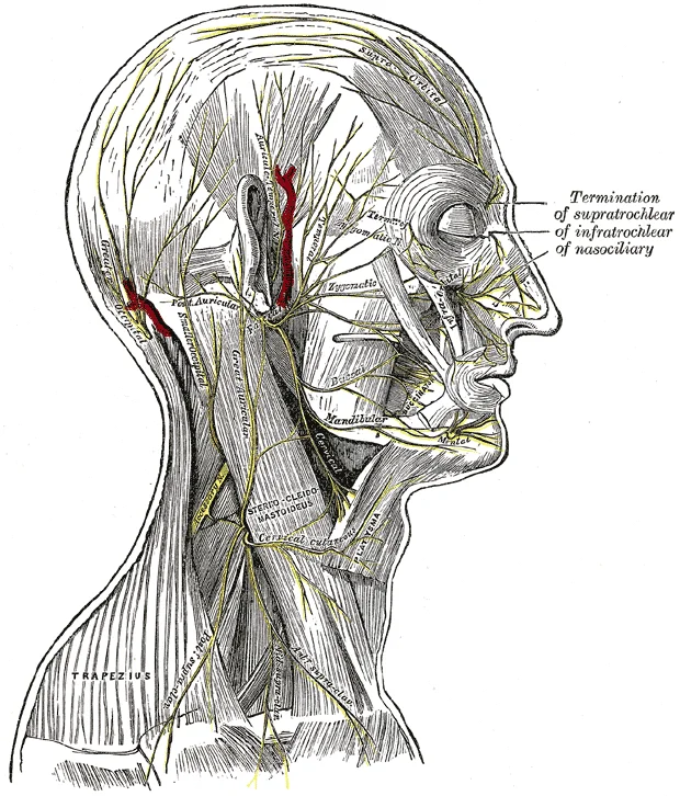
Figure 1Gray790
Long description
The nerves of the scalp, face, and side of neck.

Figure 1: The nerves of the scalp, face, and side of neck.
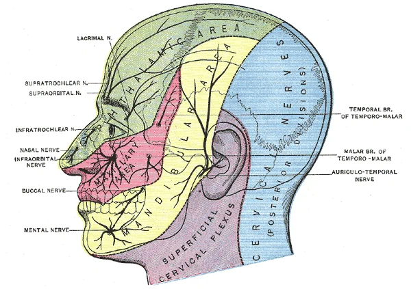
Figure 2Gray784
Long description
Sensory areas of the head, showing the general distribution of the three divisions of the fifth nerve. (Modified from Testut.)

Figure 2: Sensory areas of the head, showing the general distribution of the three divisions of the fifth nerve (Modified from Testut.).
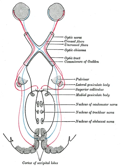
Figure 3Gray774
Long description
Scheme showing central connections of the optic nerves and optic tracts.

Figure 3: Scheme showing central connections of the optic nerves and optic tracts.
The cranial nerves exit the skull through various foramina, with MRI considered the gold standard for studying their anatomy[1]. CT can be useful for evaluating intraosseous segments, skull base foramina, and bony traumatic lesions[1]. The nuclei of cranial nerves (except I and II) are located in the brainstem, and from their origin, nerves are divided into cisternal, intracranial, and extracranial segments[1]. [2]
DefinitionCranial Nerve Classification
The twelve cranial nerves can be separated into four major groups: sensory nerves, nerves controlling eye movement, nerves of the oral cavity and superior pharynx, and the nerve controlling neck movements[1].
The corneal reflex involves the trigeminal nerve (CN V) for sensory input and the facial nerve (CN VII) for motor response. The pupillary reflex is mediated by the optic nerve (CN II) for afferent input and the oculomotor nerve (CN III) for efferent output, causing pupillary constriction. The jaw jerk reflex is a monosynaptic reflex arc involving the trigeminal nerve (CN V) as the afferent limb and the masseter muscle as the effector. The gag reflex involves the glossopharyngeal nerve (CN IX) for sensory input and the vagus nerve (CN X) for motor output. The cough reflex similarly involves the vagus nerve (CN X) as the primary nerve controlling the motor response of coughing. [2]
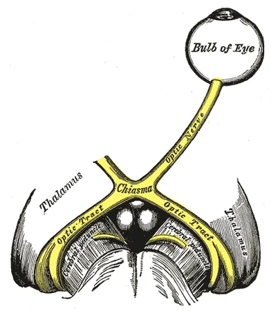
Figure 4Gray773
Long description
The left optic nerve and the optic tracts.

Figure 4: The left optic nerve and the optic tracts.
Lacrimation is controlled by parasympathetic fibers from the facial nerve (CN VII), which synapse in the pterygopalatine ganglion before postganglionic fibers innervate the lacrimal gland[1]. [2]
| Cranial Nerve | Foramen of Exit |
|---|---|
| I (Olfactory) | Cribriform plate |
| II (Optic) | Optic canal |
| III (Oculomotor) | Superior orbital fissure |
| IV (Trochlear) | Superior orbital fissure |
| V (Trigeminal) | Foramen ovale (V3), Superior orbital fissure (V1,V2) |
| VI (Abducens) | Superior orbital fissure |
| VII (Facial) | Internal acoustic meatus, Stylomastoid foramen |
| VIII (Vestibulocochlear) | Internal acoustic meatus |
| IX (Glossopharyngeal) | Jugular foramen |
| X (Vagus) | Jugular foramen |
| XI (Spinal Accessory) | Jugular foramen |
| XII (Hypoglossal) | Hypoglossal canal |
Figure 5Gray697
Long description
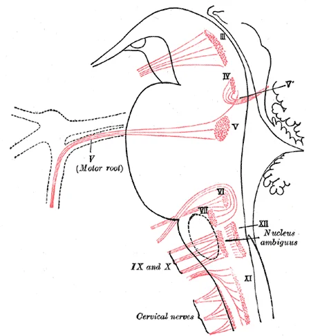
Nuclei of origin of cranial motor nerves schematically represented; lateral view.

Figure 5: Nuclei of origin of cranial motor nerves schematically represented; lateral view.
The cranial nerves originate from specific nuclei within the brainstem, with their roots emerging at distinct levels to innervate structures in the head, neck, and thoracoabdominal viscera. Understanding their formation requires tracing the path from nucleus to target, noting the unique anatomical relationships they maintain with vessels and other nerves.
The accessory nerve has both cranial and spinal roots. Cranial roots emerge into the lateral cerebellomedullary cistern, while spinal roots emerge from upper cervical segment of the spinal cord (from C0 to C5) and pass superiorly through the foramen magnum into the cisterna magna. Successively, conjoined cranial and spinal roots enter the jugular foramen, posterior to the glossopharyngeal and vagus nerve. Within the jugular foramen, cranial roots mix with vagus nerve at the level of the superior [2]
The trigeminal nerve is the largest cranial nerve, composed of sensory and motor roots. Four trigeminal nuclei extend from the midbrain down to the upper cervical medulla. The nerve emerges from the lateral pons, passes through Meckel’s cave where the trigeminal ganglion is located, and splits into three subdivisions. The ophthalmic (V1) and maxillary (V2) divisions pass in the lateral wall of the cavernous sinus, inferior to the sixth cranial nerve. V1 exits the skull via the superior orbital f [2]
Figure 6Gray792
Long description
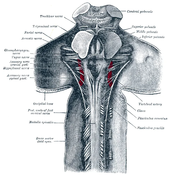
Upper part of medulla spinalis and hind- and mid-brains; posterior aspect, exposed in situ.

Figure 6: Upper part of medulla spinalis and hind- and mid-brains; posterior aspect, exposed in situ.
Figure 7Gray782
Long description
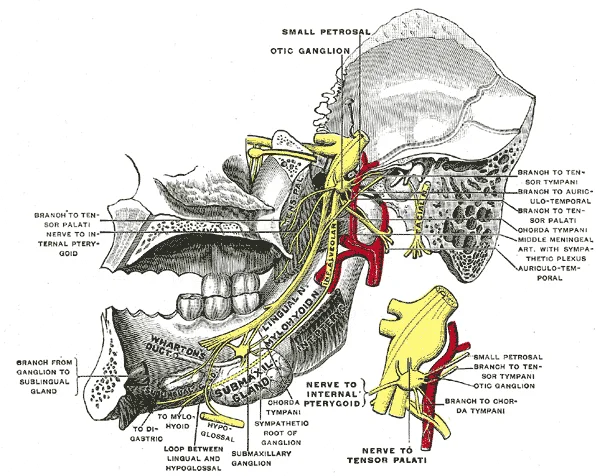
Mandibular division of trifacial nerve, seen from the middle line. The small figure is an enlarged view of the otic ganglion. (Testut.)

Figure 7: Mandibular division of trifacial nerve, seen from the middle line.
Figure 8Gray781
Long description
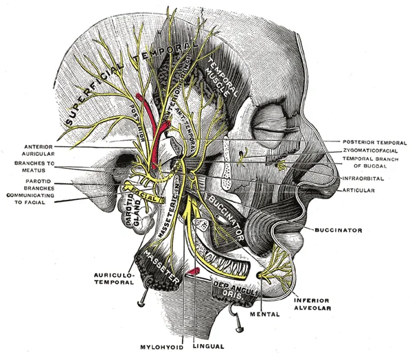
Mandibular division of the trifacial nerve. (Testut.)

Figure 8: Mandibular division of the trifacial nerve (Testut.).
Figure 9Gray780
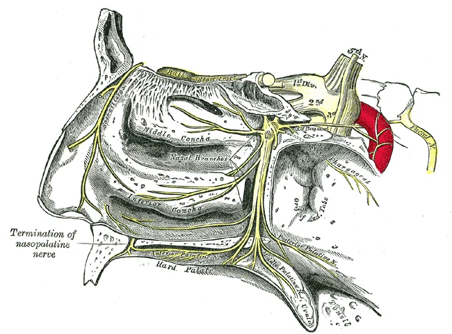
Long description
The sphenopalatine ganglion and its branches.

Figure 9: The sphenopalatine ganglion and its branches.
Figure 10Gray779
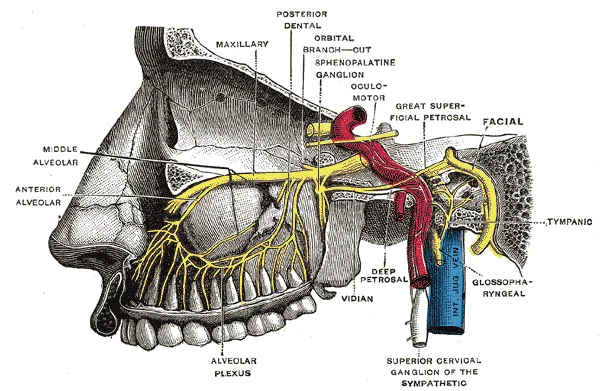
Long description
Alveolar branches of superior maxillary nerve and sphenopalatine ganglion. (Testut.)

Figure 10: Alveolar branches of superior maxillary nerve and sphenopalatine ganglion (Testut.).
The trochlear nerve runs along the free margin of the tentorium before entering the cavernous sinus, inferior to the third nerve and superior to the ophthalmic division of the fifth nerve. It enters the orbit through the superior orbital fissure to innervate only the superior oblique muscle. The abducens nerve descends near the clivus, passing through Dorello’s canal, and enters the cavernous sinus where it is the unique cranial nerve to course through the central venous portion. It enters the orbit via the superior orbital fissure to innervate the lateral rectus muscle. [2]
Figure 11Gray787
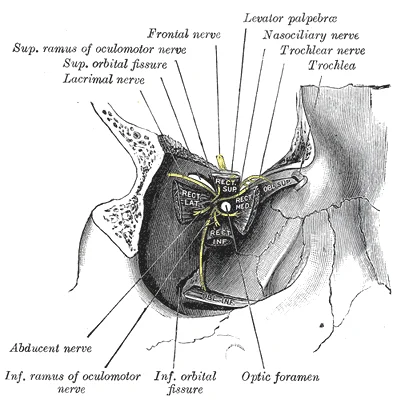
Long description
Dissection showing origins of right ocular muscles, and nerves entering by the superior orbital fissure.

Figure 11: Dissection showing origins of right ocular muscles, and nerves entering by the superior orbital fissure.
The vestibulocochlear nerve traverses the internal acoustic meatus with the facial nerve. In the meatus, the facial nerve is anterosuperior, the superior vestibular nerve is posterosuperior, the cochlear nerve is anteroinferior, and the inferior vestibular nerve is posteroinferior. The vagus nerve exits the skull through the jugular foramen (pars vascularis) between the glossopharyngeal and accessory nerves. The accessory nerve, also referred to as the spinal accessory nerve, innervates the sternocleidomastoid and trapezius muscles. It descends vertically in the retrostyloid space with the carotid artery, giving off the [1][2]
The cavernous sinus contains critical neurovascular structures. The ophthalmic and maxillary divisions of the trigeminal nerve pass in the lateral wall, inferior to the sixth cranial nerve. The trochlear nerve enters the cavernous sinus inferior to the third nerve and superior to the ophthalmic division of the fifth nerve. Neurovascular compression syndromes often involve vascular structures contacting the cisternal segment of these nerves, particularly at the transition zone between central and peripheral myelin. [2]
Figure 12Gray786
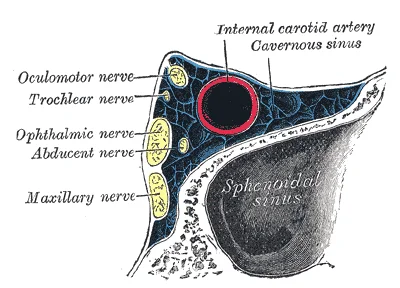
Long description
Oblique section through the right cavernous sinus.

Figure 12: Oblique section through the right cavernous sinus.
NoteClinical Relevance
Cranial nerve dysfunctions may result from pathological processes of the nerve itself, tumours, inflammation, infection, or trauma. Knowledge of anatomy is crucial for detecting the site of pathological alterations. [2]
The anatomy of cranial nerves is complex and its knowledge is crucial to detect pathological alterations in case of nervous disorders. Magnetic resonance imaging (MRI) is the gold standard technique in the study of the cranial nerves. Steady-state free procession (SSFP) images are the best sequences for the visualization of the cisternal segments showing dark cranial nerves against a background of bright cerebrospinal fluid (CSF). [2]
The cranial nerves appear as dark linear structures against the bright cerebrospinal fluid. This contrast allows for clear identification of each nerve as it exits the brainstem. The facial nerve runs alongside it. The glossopharyngeal and vagus nerves emerge from the medulla. The spinal accessory nerve exits laterally. The hypoglossal nerve emerges from the medullary pyramids. The oculomotor nerve emerges from the midbrain. The trochlear nerve is the only cranial nerve to exit from the dorsal aspect of the brainstem. The abducens nerve emerges from the pontomedullary junction. The trigeminal nerve has a large root entering the pons. Understanding this course is essential for localizing lesions. [2]
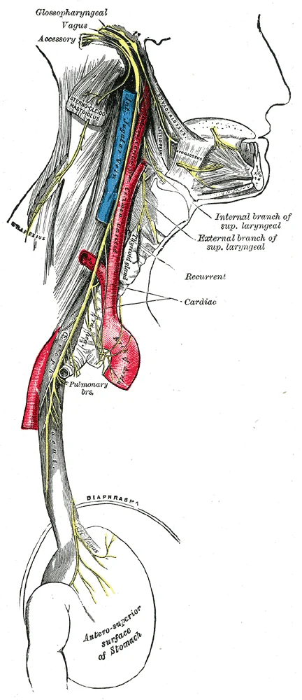
Figure 13Gray793
Long description
Course and distribution of the glossopharyngeal, vagus, and accessory nerves.

Figure 13: Course and distribution of the glossopharyngeal, vagus, and accessory nerves.
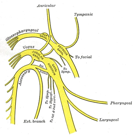
Figure 14Gray791
Long description
Plan of upper portions of glossopharyngeal, vagus, and accessory nerves.

Figure 14: Plan of upper portions of glossopharyngeal, vagus, and accessory nerves.
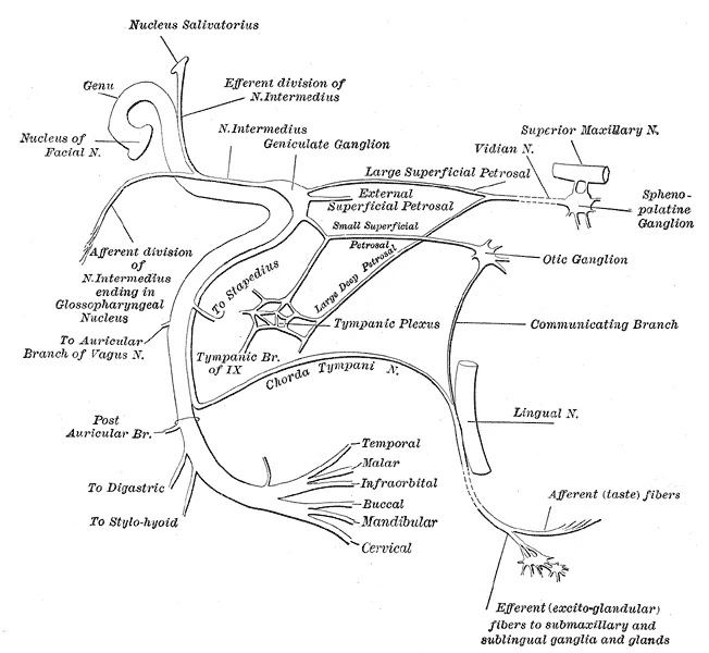
Figure 15Gray788
Long description
Plan of the facial and intermediate nerves and their communication with other nerves.

Figure 15: Plan of the facial and intermediate nerves and their communication with other nerves.
The three nerves that control the extraocular muscles are the oculomotor, trochlear, and abducens nerves, which are the third, fourth, and sixth cranial nerves. As the name suggests, the abducens nerve is responsible for abducting the eye, which it controls through contraction of the lateral rectus muscle. The trochlear nerve controls the superior oblique muscle to rotate the eye along its axis in the orbit medially, which is called intorsion, and is a component of focusing the eyes on an object close to the face. The oculomotor nerve controls all the other extraocular muscles, as well as a muscle of the upper eyelid. Movements of the two eyes need to be coordinated to locate and track visual stimuli accurately. [1]
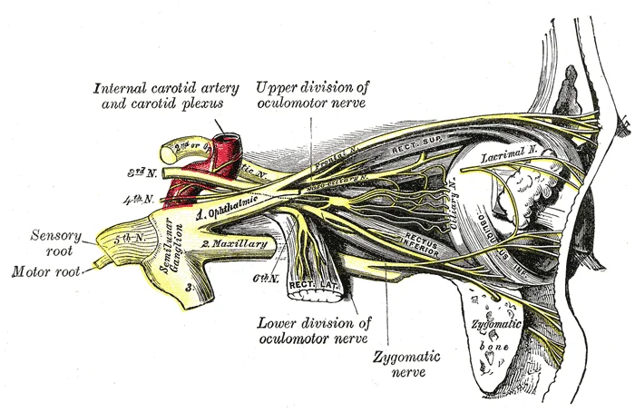
Figure 16Gray777
Long description
Nerves of the orbit, and the ciliary ganglion. Side view.

Figure 16: Nerves of the orbit, and the ciliary ganglion (side view).
Figure 17Gray776
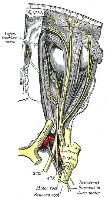
Long description
Nerves of the orbit. Seen from above. Labelled illustration from Wikimedia Commons, used under its open licence.

Figure 17: Nerves of the orbit (seen from above).
Figure 18Gray775
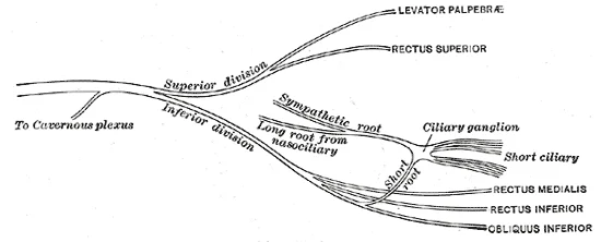
Long description
Plan of oculomotor nerve. Labelled illustration from Wikimedia Commons, used under its open licence.

Figure 18: Plan of oculomotor nerve.
Figure 19Gray785
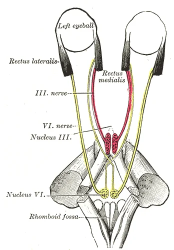
Long description
Figure showing the mode of innervation of the Recti medialis and lateralis of the eye (after Duval and Laborde).

Figure 19: Figure showing the mode of innervation of the Recti medialis and lateralis of the eye (after Duval and Laborde).
The olfactory, optic, and vestibulocochlear nerves are strictly sensory nerves for smell, sight, and balance and hearing, whereas the trigeminal, facial, and glossopharyngeal nerves carry somatosensation of the face, and taste—separated between the anterior two-thirds of the tongue and the posterior one-third. Special senses are tested by presenting the particular stimuli to each receptive organ. General senses can be tested through sensory discrimination of touch versus painful stimuli. [1] [2]
Microscopically, cranial nerves are surrounded by a series of a connective tissue sheaths: endoneurium, perineurium, and epineurium. Axons of each nerve, except I and II, are myelinated by Schwann cells. This structural organization supports the transmission of neural signals. The sheaths also provide protection and structural integrity. Damage to these sheaths can lead to nerve dysfunction. [2]
Cranial nerve dysfunctions may be the result of pathological processes of the cranial nerve itself or may be associated with tumor, inflammation, infectious processes, or traumatic injuries of adjacent structures. Pathology of cranial nerves involves a wide spectrum of etiologies that includes primary or secondary neoplastic involvement, inflammation, infections, traumatic injuries, ischemic lesions, compression by adjacent structures, and congenital pathologies. [2]
Tests of equilibrium are important for coordination and gait and are related to other aspects of the neurological exam. The vestibulo-ocular reflex involves the cranial nerves for gaze control. Balance and equilibrium, as tested by the Romberg test, are part of spinal and cerebellar processes and involved in those components of the neurological exam, as discussed later. [1]
The facial and glossopharyngeal nerves are also responsible for the initiation of salivation. Neurons in the salivary nuclei of the medulla project through these two nerves as preganglionic fibers, and synapse in ganglia located in the head. The parasympathetic fibers of the glossopharyngeal nerve synapse in the otic ganglion, which projects to the parotid gland. Salivation in response to food in the oral cavity is based on a visceral reflex arc within the facial or glossopharyngeal nerves. Other stimuli that stimulate salivation are coordinated through the hypothalamus, such as the smell and sight of food. [1]
Figure 20Gray783
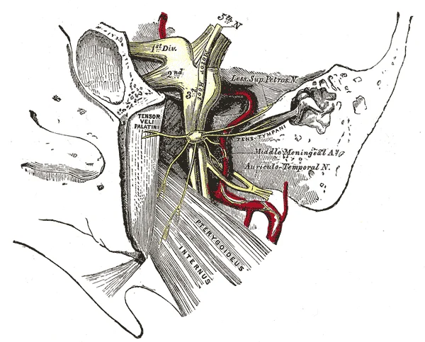
Long description
The otic ganglion and its branches. Labelled illustration from Wikimedia Commons, used under its open licence.

Figure 20: The otic ganglion and its branches.
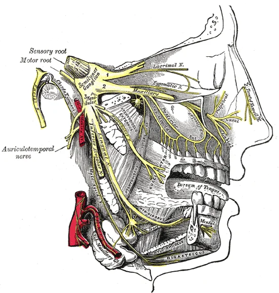
Figure 21Gray778
Long description
Distribution of the maxillary and mandibular nerves, and the submaxillary ganglion.

Figure 21: Distribution of the maxillary and mandibular nerves, and the submaxillary ganglion.
DefinitionCisternal Segment
The portion of the cranial nerve between the brainstem and its entry into a skull foramen, visible on MRI as dark against bright CSF.
NoteMyelination Exception
Cranial nerves I and II are not myelinated by Schwann cells; they are central nervous system tracts.
| Nerve | Motor Branches | Sensory Branches |
|---|---|---|
| Vestibulocochlear (VIII) | None | Equilibrium from vestibule; Auditory from cochlea |
| Facial (VII) | Nerve of stapedius; Muscles of facial expression | Intermediate nerve fibers |
| Glossopharyngeal (IX) | Stylopharyngeus muscle | Lingual branch (posterior tongue); Pharyngeal nerve; Sinus nerve; Tympanic branch |
| Accessory (XI) | Sternocleidomastoid; Trapezius | None (primarily motor) |
The trigeminal system's sensory component is tested using a cotton-tipped applicator, where the cotton end provides a touch stimulus and the pointed end provides a painful or sharp stimulus. The primary sensory subtest is sensory discrimination, requiring the patient to identify whether the stimulus is sharp or dull while their eyes are closed. These stimuli are processed by the trigeminal system separately. Perineural invasion (PNI) is a microscopic process affecting small nerves, while perineural spread of tumor (PNS) extends along the sheaths covering larger central nerves. The incidence of perineural spread is about 2.5–5% and may occur with any head and neck malignancy. It is typically associated with carcinoma arising from minor or major salivary glands, mucosal or cutaneous squamous cell carcinoma, basal cell carcinoma, melanoma, lymphoma, and sarcoma. In most cases, the peripheral branches of the second (V2) and third (V3) divisions of the trigeminal nerve, and the descending branch of the facial nerve serve as conduits for perineural spread. Also, the ophthalmic nerve and the hypoglossal nerve may be involved. [1] [2]
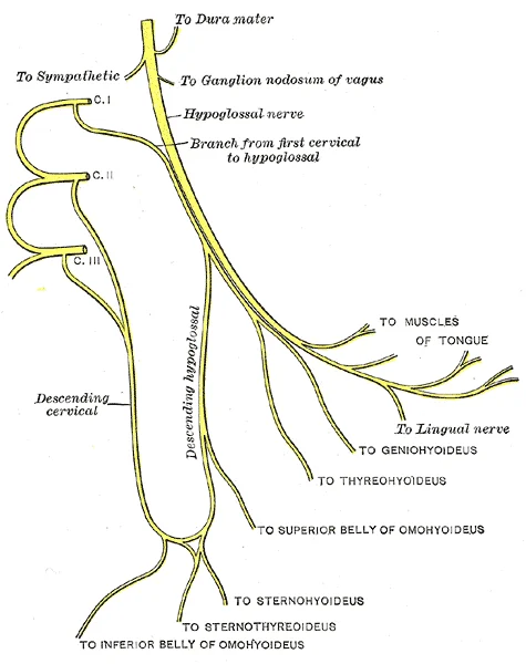
Figure 22Gray795
Long description
Plan of hypoglossal nerve. Labelled illustration from Wikimedia Commons, used under its open licence.

Figure 22: Plan of hypoglossal nerve.
Lesions of the cranial nerves are classified by their anatomical location, which dictates the specific clinical deficit. Understanding the origin, course, relations, branches, and termination of each nerve allows for precise localisation of the pathology.
The olfactory nerve consists of short axons that originate from neurons in the olfactory epithelium. These neurons have a limited life span and are replaced regularly. The axons grow back into the central nervous system by following existing pathways. This regeneration represents one of the few examples of such growth in the mature nervous system. Loss of smell, known as anosmia, can result from blunt head trauma or aging. If the brain moves within the cranium during a motor vehicle accident, al [1]
The frontal lobes lie superior to the bulbs. Damage here shears the delicate axons.
Hearing is tested using a tuning fork to distinguish between conductive and sensorineural hearing. Conductive hearing relies on vibrations conducted through the ossicles of the middle ear. Sensorineural hearing involves transmission through neural components of the inner ear and cranial nerve. In the Rinne test, a vibrating fork is placed on the mastoid process until sound is no longer heard, then moved next to the ear canal. If sound is conducted better through the temporal bone than via the air, a conductive hearing deficit is present. The vestibulo-ocular reflex stabilises gaze during head movements by coordinating equilibrium sensations with eye movement systems. [2] [1]
The facial nerve conveys gustatory stimulation to the brain. Afferent sensory fibers from the first two-thirds of the tongue arrive at the geniculate ganglion via the chorda tympani. These fibers run with the intermediate nerve in the internal acoustic meatus and cerebellopontine cistern to terminate in the nucleus of the tractus solitarius. Parasympathetic fibers arise from the superior salivatory nucleus. At the geniculate ganglion, the great petrosal nerve provides parasympathetic fibers for the lacrimal gland and mucosa of the mouth, nose, and pharynx. [2]
The glossopharyngeal nerve relays general sensations from the pharyngeal walls and certain taste stimuli. These sensations stimulate the gag reflex. Inspection of the oral cavity and pharynx, including the fauces, tests these functions. Moving a tongue depressor to contact the lateral wall of the fauces should elicit the gag reflex. Stimulation of either side should elicit an equivalent response. [1]
The vagus nerve controls the palatoglossus muscle, unlike other tongue muscles. It also contributes to the gag reflex efferent limb.
The hypoglossal nerve emerges from the medulla oblongata as rootlets from the ventrolateral sulcus. It enters the lateral cerebellomedullary cistern near the vertebral artery and posterior inferior cerebellar artery (PICA). The nerve exits via the hypoglossal canal and runs medial to the glossopharyngeal, vagus, and accessory nerves. It innervates most extrinsic and intrinsic tongue muscles. The genioglossus muscle is responsible for tongue protrusion. If both nerves work, the tongue sticks straight out. If one side is damaged, the tongue points to the side of the lesion. [2] [1]
| Nerve | Key Deficit | Common Cause |
|---|---|---|
| CN I | Anosmia | Blunt trauma, aging |
| CN VIII | Conductive/Sensorineural loss | Ossicle damage, neural damage |
| CN VII | Taste loss, dry eye | Geniculate ganglion injury |
| CN IX | Absent gag reflex | Pharyngeal wall trauma |
| CN XII | Tongue deviation to lesion | Hypoglossal canal injury |
The olfactory nerve conveys olfactory stimuli from the nasal cavity to the brain. It consists of white matter tracts not surrounded by Schwann cells. Olfactory receptors are located in the mucosa of nasal cavity and olfactory filiae represent their axons. They enter the anterior cranial fossa through the cribriform plate and terminate in the olfactory bulb. Successively, the nerve courses between the gyrus rectus and the medial orbital gyrus. The secondary axons terminate in the inferomedial temporal lobe, uncus, and entorhinal cortex. If the nerve is not completely severed, the anosmia may be temporary as new neurons can eventually reconnect. Clinical testing involves assessing the patient's ability to identify distinct odours, such as coffee or vanilla, while one nostril is occluded. [2] [1]
The optic nerve represents an extension of central nervous system and is myelinated by oligodendrocytes. Optic nerve emerges from the posterior pole of the ocular globe. It is approximately 50 mm in length, divided in four segments: intraocular (1 mm), intraorbital (25 mm), intracanalicular (9 mm), and prechiasmatic (16 mm). The nerve joins the contralateral to form the optic chiasma, where the nasal fibers from each nerve decussate, while the temporal fibers not. From the optic chiasm, optic tract courses posteriorly along the cerebral peduncles and synapses at the lateral geniculate nuclei. From the lateral geniculate body, optic radiation reaches the primary visual cortex in the occipital lobe (Brodmann area 17). Examination includes visual acuity, visual fields, and fundoscopy to assess the optic disc for swelling or pallor. [2]
These nerves control extraocular movements. To elevate the eyes, the oculomotor nerve on either side stimulates the contraction of both superior rectus muscles. To depress the eyes, the oculomotor nerve on either side stimulates the contraction of both inferior rectus muscles. Clinical examination involves observing eye alignment at rest and during gaze in all nine positions. Pupillary light reflex and accommodation are tested to assess parasympathetic function of the oculomotor nerve. Ptosis and diplopia are key signs of dysfunction. [1]
The trigeminal nerve controls the muscles of chewing, which are tested for stretch reflexes. Sensory examination tests the three divisions: ophthalmic, maxillary, and mandibular. Motor function is assessed by palpating the temporalis and masseter muscles during clenching. Corneal reflex testing evaluates both the afferent limb (trigeminal) and efferent limb (facial) pathways. [1]
Motor functions of the facial nerve are usually obvious if facial expressions are compromised, but can be tested by having the patient raise their eyebrows, smile, and frown. Bell’s palsy (idiopathic facial palsy) is the most common cause of unilateral peripheral nerve neuropathy. Clinical presentation is variable, with dysgeusia, mastoid pain, impaired salivation, and lacrimation. A Herpes-Simplex-Virus reactivation seems to be the most probable etiology. Prognosis is often good and the treatment is based on corticosteroid therapy, often with the addition of acyclovir. Typical Bell’s palsy does not require imaging studies. However, MRI may be useful to exclude other lesions responsible of atypical Bell’s palsy (gradual-onset palsy, failure to improve with time) or recurrent palsy. On MRI, the facial nerve shows increased enhancement on post-contrast sequences that may involve one or more segments, without nodularity. Enhancement of distal intrameatal and labyrinthine segments is typical. On T2-weighted sequences, the nerve may be hyperintense. [1] [2]
Superficial siderosis is a rare pathological condition that results from chronic deposition of hemosiderin in the subpial layers of the brain and spinal cord. Classically, it is bilateral and it may present with the typical triad: ataxia, sensorineural hearing loss, and myelopathy. Other clinical presentations include dysarthria, nystagmus, myelopathy, bladder dysfunction, sensory and pyramidal signs. Hemosiderin deposition has predilection for the superior cerebellar vermis, cerebellar folia, frontal lobe, temporal cortex, brainstem, spinal cord, nerve roots and cranial nerve, mainly the VIII. Rarely, extraocular nerve palsies, optic and trigeminal neuropathy have been reported in literature. MRI is useful to identify hemosiderin deposition, based on magnetic susceptibility, better demonstrated as hypointensity on T2*-weighted sequences (blooming effect). Hearing tests (Rinne, Weber) and balance assessments are standard. [2]
Movements of the tongue, soft palate, or superior pharynx can be observed directly while the patient swallows, while the gag reflex is elicited, or while the patient says repetitive consonant sounds. The motor response, through contraction of the muscles of the pharynx, is mediated through the vagus nerve. Normally, the vagus nerve is considered autonomic in nature. The vagus nerve directly stimulates the contraction of skeletal muscles in the pharynx and larynx to contribute to the swallowing and speech functions. Further testing of vagus motor function has the patient repeating consonant sounds that require movement of the muscles around the fauces. The patient is asked to say “lah-kah-pah” or a similar set of alternating sounds while the examiner observes the movements of the soft palate and arches between the palate and tongue. The motor control of the gag reflex is largely controlled by fibers in the vagus nerve and constitutes a test of that nerve because the parasympathetic functions of that nerve are involved in visceral regulation, such as regulating the heartbeat and digestion. [1]
To test these muscles, the patient is asked to flex and extend the neck or shrug the shoulders against resistance, testing the strength of the muscles. Lateral flexion of the neck toward the shoulder tests both at the same time. Any difference on one side versus the other would suggest damage on the weaker side. These strength tests are common for the skeletal muscles controlled by spinal nerves and are a significant component of the motor exam. Deficits associated with the accessory nerve may have an effect on orienting the head, as described with the VOR. Movement of the head and neck using the sternocleidomastoid and trapezius muscles is controlled by the accessory nerve. Flexing of the neck and strength testing of those muscles reviews the function of that nerve. [1]
Examination involves inspecting the tongue for atrophy or fasciculations at rest and asking the patient to protrude it. Deviation towards the side of the lesion indicates hypoglossal nerve palsy.
The optic nerves enter the cranium via the optic canals and converge at the optic chiasm, where medial retinal fibres decussate to process the peripheral visual field. [1]
The vestibular and cochlear nuclei are anatomical neighbours, meaning a single lesion in the cerebellopontine angle can impair both balance and hearing. [1]
Horizontal gaze requires coordinated action of the lateral rectus (CN VI) and medial rectus (CN III) muscles. The abducens nucleus in the superior medulla controls the lateral rectus, while the oculomotor nucleus in the midbrain controls the medial rectus. Testing involves tracking a pen through the visual field to assess conjugate eye movements. [1]
INO is characterised by failure of one eye to abduct while the other adducts during horizontal gaze. This results from damage to the medial longitudinal fasciculus (MLF) and causes diplopia. Conjugate movement failure indicates dysfunction of the related nuclei or the MLF. [1]
Convergence involves bilateral adduction of the eyes to focus on near objects. Accommodation, the change in lens shape for near focus, is controlled by parasympathetic fibres of the oculomotor nerve. These functions are tested by moving a pen towards the patient’s face. [1]
Esthesioneuroblastoma arises from the olfactory epithelium, typically presenting in the second and sixth decades with nasal obstruction or epistaxis. Optic nerve gliomas are rare, often occurring in children, and are frequently associated with Neurofibromatosis type 1 (NF1). In NF1 patients, the optic nerve is the most common site, whereas chiasmal involvement is more frequent in non-NF1 cases. Meningiomas are the commonest extra-axial CNS tumours, often showing a “cleft sign” on MRI due to a ri [2]
Exam trapVisual Fields vs Eye Movement
Do not confuse visual field testing (peripheral stimulus fixation) with eye movement testing (tracking a moving target). The former assesses the optic nerve pathway; the latter assesses cranial nerves III, IV, and VI and the MLF. [2]
Clinical pearlChiasm Anatomy
Remember that the optic chiasm processes the peripheral visual field because the fibres carrying this information originate from the medial retinae and decussate.
| Nerve | Lesion Sign | Key Anatomical Relation |
|---|---|---|
| CN II | Bitemporal hemianopia | Optic chiasm (pituitary inferior) |
| CN VI | Failure to abduct eye | Lateral rectus muscle |
| CN III | Failure to adduct/converge | Medial rectus muscle |
| MLF | Internuclear ophthalmoplegia | Connects CN III and CN VI nuclei |
DefinitionMénière’s Disease
A disorder of the inner ear characterised by vertigo, tinnitus, and hearing loss, caused by fluid accumulation in the vestibule or cochlea. [1]
NoteImaging Features
Meningiomas appear as lobular extra-axial masses with a broad-based dural attachment and a high T2 signal “cleft sign” separating them from the brain.
Students frequently confuse the sensory modalities carried by the three divisions of the trigeminal nerve. A common error is assuming that pain and temperature are processed in the same brainstem nucleus as light touch. In reality, the chief nucleus receives information about light touch as well as proprioceptive information about the mandible, which are both relayed to the thalamus and, ultimately, to the postcentral gyrus of the parietal lobe. Conversely, contact with the pointed end of the applicator is a painful stimulus relayed by the spinal trigeminal nucleus. Failure to discriminate these stimuli can localize problems within the brain stem. If a patient cannot recognize a painful stimulus, that might indicate damage to the spinal trigeminal nucleus in the medulla. [1]
Another frequent mistake involves the interpretation of the Weber test. Students often assume that sound lateralising to the affected ear indicates sensorineural loss. However, with unilateral conductive hearing loss, however, the tuning fork sounds louder in the ear with hearing loss. This is because the sound of the tuning fork has to compete with background noise coming from the outer ear, but in conductive hearing loss, the background noise is blocked in the damaged ear, allowing the tuning fork to sound relatively louder in that ear. With unilateral sensorineural hearing loss, however, damage to the cochlea or associated nervous tissue means that the tuning fork sounds quieter in that ear. [1]
Visual field testing is often misunderstood as merely checking for blindness rather than mapping boundaries. Testing the extent of the visual field means that the examiner can establish the boundaries of peripheral vision as simply as holding their hands out to either side and asking the patient when the fingers are no longer visible without moving the eyes to track them. Furthermore, students may overlook the impact of anosmia on patient quality of life. Olfaction is not the pre-eminent sense, but its loss can be quite detrimental. Anosmia means that food will not seem to have the same taste, though the gustatory sense is intact, and food will often be described as being bland. [1]
Eye movement errors also arise from ignoring the geometry of extraocular muscles. Purely vertical movements of the eyes are not very common. The superior and inferior rectus muscles are not perfectly oriented with the line of sight. The origin for both muscles is medial to their insertions, so elevation and depression may require the lateral rectus muscles to compensate for the slight adduction inherent in the contraction of those muscles, requiring MLF activity as well. [1]
Finally, localising tongue lesions requires awareness of adjacent structures. Loss of function of the tongue can interfere with speech and swallowing. Additionally, because the location of the hypoglossal nerve and nucleus is near the cardiovascular center, inspiratory and expiratory areas for respiration, and the vagus nuclei that regulate digestive functions, a tongue that protrudes incorrectly can suggest damage in adjacent structures that have nothing to do with controlling the tongue. [1]
At the bedside, cranial nerve assessment is fundamental for localising neurological deficits. Vision testing relies on the tests that are common in an optometry office. The Snellen chart demonstrates visual acuity by presenting standard Roman letters in a variety of sizes. The result of this test is a rough generalization of the acuity of a person based on the normal accepted acuity. To have reduced vision, for example, means that the smallest letters that a person can see at a 20-foot distance could b [1]
In the operating theatre and emergency settings, understanding the trigeminal system is critical. The trigeminal system of the head and neck is the equivalent of the ascending spinal cord systems of the dorsal column and the spinothalamic pathways. Somatosensation of the face is conveyed along the nerve to enter the brain stem at the level of the pons. Synapses of those axons, however, are distributed across nuclei found throughout the brain stem. The mesencephalic nucleus processes proprioceptive information of the face, which is the movement and position of facial muscles. It is the sensory component of the jaw-jerk reflex, a stretch reflex of the masseter muscle. [1] [2]
Imaging plays a pivotal role in diagnosing complex cranial nerve pathologies. MR imaging findings include iso-hypointensity on T1-weighted sequences, high signal on T2-weighted sequences, and marked enhancement on post-contrast images, eventually with unenhanced cystic spaces. For conditions like Tolosa-Hunt, imaging techniques can show an infiltrative soft tissue mass within the cavernous sinus that appears enlarged. The tissue is hypo-isointense on T2-weighted sequences, with avid contrast enhancement on post-gadolinium images [2]
In examinations, students must distinguish between conductive and sensorineural hearing loss using the Weber test. The Weber test also uses a tuning fork to differentiate between conductive versus sensorineural hearing loss. In this test, the tuning fork is placed at the top of the skull, and the sound of the tuning fork reaches both inner ears by travelling through bone. In a healthy patient, the sound would appear equally loud in both ears. [1]
Weber Test Interpretation
- Place tuning fork on skull vertexAsk patient where sound is heard
- Question: Sound equal in both ears?
Yes
- Normal hearing or symmetric loss
No
- Question: Lateralises to affected ear?
Yes
- Conductive hearing loss in that ear
No
- Sensorineural hearing loss in that ear
- Anosmia causes food to taste bland despite intact gustatory function.
- The Snellen chart measures acuity based on a 5-minute arc at 20 feet.
- The chief nucleus processes light touch and proprioception.
- The spinal trigeminal nucleus processes painful stimuli.
- Weber test lateralises to the ear with conductive loss.
- Weber test lateralises away from the ear with sensorineural loss.
- Superior and inferior rectus muscles cause slight adduction during vertical movement.
- Hypoglossal lesions may indicate adjacent brainstem damage.
Answer each one in your head first, then open the answer.
What does anosmia primarily affect regarding food perception?
Show answerHide answer
The enjoyment of food, making it seem bland despite intact tasteWhere does the trigeminal nerve enter the brainstem?
Show answerHide answer
At the level of the ponsWhich nucleus processes proprioceptive information of the face?
Show answerHide answer
The mesencephalic nucleusIn Weber test, where does sound localise in conductive loss?
Show answerHide answer
In the ear with hearing lossWhat imaging feature is typical for Tolosa-Hunt syndrome?
Show answerHide answer
Infiltrative soft tissue mass within the cavernous sinus
- Betts JG, Young KA, Wise JA, et al. The Cranial Nerve Exam. OpenStax, Anatomy and Physiology 2e (CC BY-NC-SA 4.0). Accessed .
- Romano N, Federici M, Castaldi A. Imaging of cranial nerves: a pictorial overview. Insights into imaging 2019;10(1):33. doi:10.1186/s13244-019-0719-5. Accessed .
Figure credits
- Figure 1: Henry Vandyke Carter, via Wikimedia Commons, Public domain, Public domain.
- Figure 2: Henry Vandyke Carter, via Wikimedia Commons, Public domain, Public domain.
- Figure 3: Henry Vandyke Carter, via Wikimedia Commons, Public domain, Public domain.
- Figure 4: Henry Vandyke Carter, via Wikimedia Commons, Public domain, Public domain.
- Figure 5: Henry Vandyke Carter, via Wikimedia Commons, Public domain, Public domain.
- Figure 6: Henry Vandyke Carter, via Wikimedia Commons, Public domain, Public domain.
- Figure 7: Henry Vandyke Carter, via Wikimedia Commons, Public domain, Public domain.
- Figure 8: Henry Vandyke Carter, via Wikimedia Commons, Public domain, Public domain.
- Figure 9: Henry Vandyke Carter, via Wikimedia Commons, Public domain, Public domain.
- Figure 10: Henry Vandyke Carter, via Wikimedia Commons, Public domain, Public domain.
- Figure 11: Henry Vandyke Carter, via Wikimedia Commons, Public domain, Public domain.
- Figure 12: Henry Vandyke Carter, via Wikimedia Commons, Public domain, Public domain.
- Figure 13: Henry Vandyke Carter, via Wikimedia Commons, Public domain, Public domain.
- Figure 14: Henry Vandyke Carter, via Wikimedia Commons, Public domain, Public domain.
- Figure 15: Henry Vandyke Carter, via Wikimedia Commons, Public domain, Public domain.
- Figure 16: Henry Vandyke Carter, via Wikimedia Commons, Public domain, Public domain.
- Figure 17: Henry Vandyke Carter, via Wikimedia Commons, Public domain, Public domain.
- Figure 18: Henry Vandyke Carter, via Wikimedia Commons, Public domain, Public domain.
- Figure 19: Henry Vandyke Carter, via Wikimedia Commons, Public domain, Public domain.
- Figure 20: Henry Vandyke Carter, via Wikimedia Commons, Public domain, Public domain.
- Figure 21: Henry Vandyke Carter, via Wikimedia Commons, Public domain, Public domain.
- Figure 22: Henry Vandyke Carter, via Wikimedia Commons, Public domain, Public domain.
Educational content for medical students and doctors. It is not a substitute for clinical judgement or local guidelines.