Cervical plexus
The cervical plexus is a network of nerves formed by spinal nerves C1-C5.
The cervical plexus is a network of fibers formed by the anterior rami of spinal nerves C1 through C5. This plexus represents one of four major nerve plexuses in the human body, with the other three being the brachial plexus, lumbar plexus, and sacral plexus. The cervical plexus is anatomically significant as it gives rise to both sensory and motor branches that innervate structures of the posterior neck, head, and the diaphragm. [1] [3]
Its terminal cutaneous branches emerge at the midpoint of the posterior border of the sternocleidomastoid muscle, a location clinically known as "Erb's point". From this point, the cervical plexus gives rise to four main cutaneous branches: the lesser occipital, great auricular, transverse cervical, and supraclavicular nerves. [3]
The cervical plexus serves important clinical functions, particularly in surgical anesthesia. A superficial cervical plexus block (SCPB) can be performed at Erb's point to anesthetize the sensory branches of C2-C4. This block achieves postoperative analgesia by interrupting nociceptive input from the posterior scalp and upper neck. The SCPB is technically straightforward, employing a single superficial subcutaneous injection along the posterior border of the sternocleidomastoid muscle. [3]
The cervical plexus has both sensory and motor components. Its sensory branches, particularly the lesser occipital (C2-C3) and great auricular (C2-C3) nerves, innervate the upper and posterior aspect of the scalp, auricle, and mastoid region. These areas are directly involved in occipital craniotomy incisions, making the cervical plexus block valuable in neurosurgical procedures. [3]
DefinitionErb's Point
The midpoint of the posterior border of the sternocleidomastoid muscle where the cutaneous branches of the cervical plexus emerge.
| Branch | Nerve Roots | Distribution |
|---|---|---|
| Lesser occipital | C2-C3 | Upper and posterior scalp |
| Great auricular | C2-C3 | Auricle and mastoid region |
| Transverse cervical | C2-C3 | Anterior neck |
| Supraclavicular | C3-C4 | Superior shoulder and chest wall |
The cervical plexus block can be performed reliably using either a landmark-guided or ultrasound-assisted approach. With the patient supine and the head turned contralateral to the side of the block, the SCPB can be performed within five minutes, making it a simple addition to routine neurosurgical preparation. This technique contrasts with deep cervical plexus blocks, which require needle advancement to the transverse processes and deeper fascial layers, increasing procedural complexity and risk. [3]
These roots emerge from the spinal cord and exit the vertebral column via the intervertebral foramina. Unlike cranial nerves, which have ganglia outside the skull, spinal nerve roots are associated with dorsal root ganglia located just outside the spinal cord. The anterior rami of C1–C4 loop around to form the cervical plexus, situated deep to the sternocleidomastoid muscle and anterior to the scalene muscles. This arrangement allows for the reorganization of axons from different spinal levels into systemic nerves that follow distinct courses. [1]
Systemic opioids are often undertreated due to risks such as respiratory depression. [3]
The formation branches of the cervical plexus are divided into two main groups: the ansa cervicalis and the cutaneous nerves. It loops around the internal jugular vein and descends to innervate the infrahyoid muscles. The cutaneous nerves arise directly from the plexus and pierce the investing fascia to supply the skin of the neck. These include the lesser occipital nerve (C2), great auricular nerve (C2–C3), transverse cervical nerve (C2–C3), and supraclavicular nerves (C3–C4).
It descends through the neck anterior to the anterior scalene muscle. It provides motor innervation to the diaphragm, which is essential for respiration. The phrenic nerve also carries sensory fibres from the pericardium, pleura, and peritoneum.
The block is administered subcutaneously along the posterior sternocleidomastoid border, which bathes these superficial branches after they pierce the investing fascia. This technique targets the cutaneous nerves to provide anaesthesia to the neck region. Understanding the formation and branching of the cervical plexus is crucial for both anatomical knowledge and clinical practice.
These nerves emerge from the spinal cord and pass through the intervertebral foramina. Within the neck, they are situated deep to the sternocleidomastoid muscle and lie anterior to the scalene muscles. The plexus is divided into two main components: the deep motor branches and the superficial sensory branches. [1]
The deep branches arise from the anterior rami and primarily supply the muscles of the neck and the diaphragm. The phrenic nerve, a key deep branch, descends through the neck anterior to the anterior scalene muscle. It provides motor innervation to the diaphragm, which is essential for respiration. Injury to this nerve can result in diaphragmatic paralysis, a significant clinical concern during neck surgeries. The most commonly reported complication when this occurs is paralysis of the ipsilateral phrenic nerve and, less commonly, recurrent laryngeal nerve paresis. [3]
The superficial branches emerge from the posterior border of the sternocleidomastoid muscle. They ascend and descend to supply the skin of the neck, the scalp, and the shoulder. These branches pierce the deep cervical fascia to reach the subcutaneous tissue. The lesser occipital nerve supplies the skin behind the ear. The transverse cervical nerve crosses the sternocleidomastoid to supply the anterior neck. The supraclavicular nerves descend to supply the skin over the clavicle and upper chest.
Figure 1Gray805
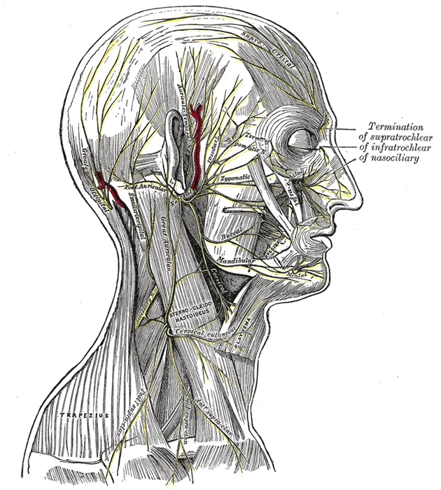
Long description
The nerves of the scalp, face, and side of neck.

Figure 1: The nerves of the scalp, face, and side of neck.
DefinitionCervical Plexus
A network of nerves formed by the anterior rami of C1–C4, providing motor and sensory innervation to the neck and shoulder region.
The anatomical relations of the cervical plexus are critical for safe regional anaesthesia. The plexus lies in close proximity to the carotid sheath, which contains the common carotid artery, internal jugular vein, and vagus nerve. The sympathetic trunk also lies posterior to the carotid sheath. Understanding these relations is vital to avoid complications such as vascular puncture or sympathetic blockade. [2]
| Branch | Origin | Course | Area of Supply |
|---|---|---|---|
| Lesser Occipital | C2 | Ascends along posterior border of SCM | Skin behind ear |
| Great Auricular | C2, C3 | Ascends over SCM | Skin over parotid, ear, mastoid |
| Transverse Cervical | C2, C3 | Crosses SCM anteriorly | Skin of anterior neck |
| Supraclavicular | C3, C4 | Descends over SCM | Skin over clavicle, upper chest |
The deep branches include the muscular branches to the prevertebral and infrahyoid muscles, and the phrenic nerve. The phrenic nerve descends on the anterior surface of the anterior scalene muscle, deep to the prevertebral fascia. It passes behind the subclavian artery and vein to enter the thorax. The motor supply to the diaphragm is crucial, and blockade of this nerve can lead to respiratory compromise.
Exam trapPhrenic Nerve Risk
The phrenic nerve lies on the anterior scalene muscle. Deep cervical plexus blocks carry a risk of phrenic nerve blockade, leading to hemidiaphragmatic paralysis. [3]
The cervical plexus is located in the posterior triangle of the neck, bounded by the sternocleidomastoid, trapezius, and clavicle. The omohyoid muscle often crosses this triangle. The plexus is protected by the prevertebral fascia. Superficial cervical plexus blocks target the nerves as they emerge from the posterior border of the sternocleidomastoid, avoiding the deep structures. This technique is safer as it avoids the vertebral artery and phrenic nerve. [3]
Clinical pearlSuperficial Block Safety
Superficial cervical plexus blocks are safe because injection remains superficial, avoiding the vertebral artery and phrenic nerve, unlike deep techniques. [3]
The blood supply to the region is provided by branches of the subclavian artery. The thyrocervical trunk supplies the thyroid and cervical region. The vertebral artery passes through the vertebral foramina of the cervical vertebrae. The internal thoracic artery supplies the anterior chest wall. Venous drainage occurs via the internal jugular and vertebral veins, which flow into the brachiocephalic vein. [2]
| Structure | Relation to Plexus | Clinical Significance |
|---|---|---|
| Sternocleidomastoid | Superficial | Landmark for nerve emergence |
| Carotid Sheath | Medial | Avoid during deep blocks |
| Phrenic Nerve | Deep/Anterior Scalene | Risk of respiratory compromise |
| Vertebral Artery | Deep/Posterior | Risk of intravascular injection |
The cervical plexus plays a vital role in neck sensation and motor control. Its anatomical position makes it accessible for regional anaesthesia. Superficial blocks provide effective analgesia for neck and scalp surgeries with minimal risk. Understanding the course and relations of the plexus is essential for safe and effective clinical practice.
Figure 2Gray794A
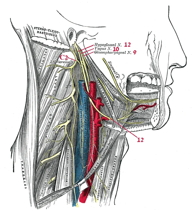
Long description
Hypoglossal nerve, cervical plexus, and their branches.

Figure 2: Hypoglossal nerve, cervical plexus, and their branches.
Figure 31321 Spinal Nerve Plexuses
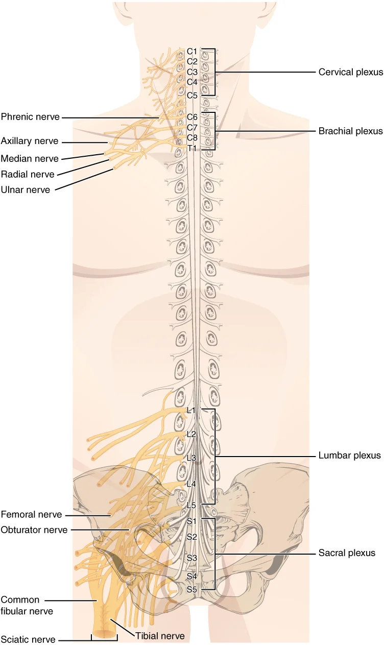
Long description
There are four main nerve plexuses in the human body. The cervical plexus supplies nerves to the posterior head and neck, as well as to the diaphragm. The brachial plexus supplies nerves to the arm. The lumbar plexus supplies nerves to the anterior leg. The sacral plexus supplies nerves to the poste

Figure 3: There are four main nerve plexuses in the human body.
Figure 4Gray794
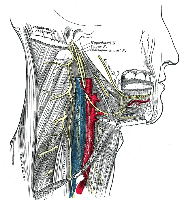
Long description
Hypoglossal nerve, cervical plexus, and their branches.

Figure 4: Hypoglossal nerve, cervical plexus, and their branches.
Figure 5Gray804
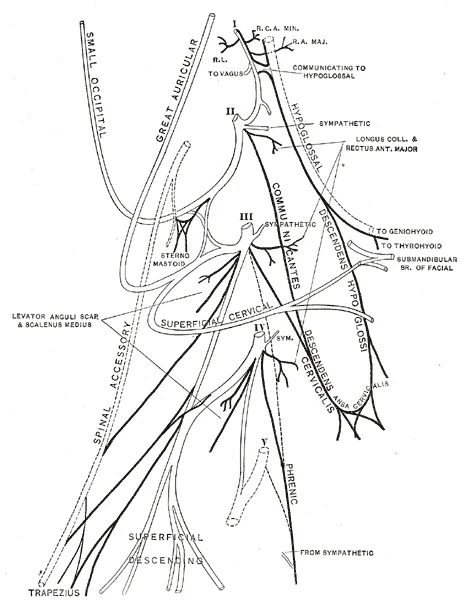
Long description
Plan of the cervical plexus. (Gerrish.). Labelled illustration from Wikimedia Commons, used under its open licence.

Figure 5: Plan of the cervical plexus (Gerrish.).
It is a network of nerves located in the posterior triangle of the neck, deep to the sternocleidomastoid muscle. The plexus is divided into superficial and deep branches. The superficial branches are primarily sensory, while the deep branches are primarily motor. Understanding this distinction is crucial for clinical anatomy, as injury to the plexus can result in specific deficits depending on which branch is affected. [1]
The superficial branches of the cervical plexus emerge from the posterior border of the sternocleidomastoid muscle and pierce the deep cervical fascia. They provide cutaneous sensation to the neck, ear, and part of the scalp.
| Branch | Origin | Distribution |
|---|---|---|
| Lesser occipital nerve | C2, C3 | Skin of the posterior scalp behind the ear |
| Great auricular nerve | C2, C3 | Skin of the external ear and angle of the jaw |
| Transverse cervical nerve | C2, C3 | Skin of the anterior neck |
| Supraclavicular nerves | C3, C4 | Skin over the clavicle, shoulder, and upper chest |
The deep branches of the cervical plexus remain deep to the sternocleidomastoid and supply muscles of the neck and larynx. These branches are motor in function and innervate specific extrinsic muscles.
| Branch | Origin | Muscles Supplied |
|---|---|---|
| Nerve to thyrohyoid | C1 (via hypoglossal) | Thyrohyoid muscle |
| Descending branch of hypoglossal | C1 (via hypoglossal) | Sternohyoid, sternothyroid, omohyoid (inferior belly) |
| Phrenic nerve | C3, C4, C5 | Diaphragm |
It descends through the neck, passing anterior to the subclavian artery, and enters the thorax to supply the diaphragm. The mnemonic "C3, 4, 5 keeps the diaphragm alive" highlights its importance. Injury to the phrenic nerve results in paralysis of the diaphragm, leading to respiratory compromise. The other deep branches supply the infrahyoid muscles, which depress the hyoid bone and larynx.
DefinitionCervical Plexus
The cervical plexus is a network of nerves formed by the ventral rami of C1–C4, located in the posterior triangle of the neck, providing sensory and motor innervation to the head, neck, and diaphragm.
Exam trapPhrenic Nerve Injury
Damage to the phrenic nerve (C3–C5) causes diaphragmatic paralysis. Clinically, this presents with elevated hemidiaphragm on chest X-ray and respiratory distress. Always assess diaphragmatic function in patients with neck trauma or surgery near the scalene muscles.
It lies deep to the sternocleidomastoid muscle, resting on the prevertebral fascia. Understanding lesions requires mapping the specific nerve roots to their functional deficits.
Lesions affecting the first and second cervical nerves are rare in isolation but significant when part of a broader cervical plexus injury. The C1 nerve root typically travels with the hypoglossal nerve without contributing to it directly, though it provides motor fibres to the geniohyoid and thyrohyoid muscles. Damage here may result in subtle weakness of tongue protrusion or hyoid depression. The C2 nerve root gives rise to the greater occipital nerve. A lesion at this level, such as from a posterior fossa tumour or trauma, causes loss of sensation over the occipital scalp. This is a key clinical sign distinguishing cervical plexus issues from cranial nerve pathologies. [3]
The C3 and C4 nerves contribute to the formation of the phrenic nerve, which descends through the thorax to innervate the diaphragm. A lesion at the C3–C4 level, particularly if bilateral, results in diaphragmatic paralysis. This is a life-threatening deficit requiring immediate mechanical ventilation. Unilateral lesions cause partial diaphragmatic weakness, leading to dyspnoea on exertion. Additionally, C3 and C4 contribute to the supraclavicular nerves, which supply sensation to the skin over the clavicle and upper shoulder. Loss of sensation in this region is a localising sign for cervical plexus pathology.
While C5 is not part of the cervical plexus proper (it joins the brachial plexus), high cervical plexus lesions often extend to involve the C5 root due to anatomical proximity. Generalised cervical plexus lesions, such as those from deep neck infections or surgical trauma, present with a combination of motor and sensory deficits. Motor involvement includes weakness of the sternocleidomastoid and trapezius muscles (via the accessory nerve, which receives fibres from C1–C3). Sensory deficits include anaesthesia of the neck, ear, and shoulder regions.
| Nerve Level | Key Branches Affected | Resulting Deficit |
|---|---|---|
| C1–C2 | Greater Occipital Nerve | Loss of sensation over occipital scalp |
| C3–C4 | Phrenic Nerve | Diaphragmatic paralysis (bilateral) |
| C3–C4 | Supraclavicular Nerves | Anaesthesia over clavicle and shoulder |
| General | Sternocleidomastoid/ Trapezius | Weakness in head rotation and shoulder shrug |
Clinical pearlPhrenic Nerve
Remember the mnemonic C3, 4, 5 keep the diaphragm alive. A lesion at C3–C4 is a surgical emergency due to respiratory compromise.
These nerves emerge from the spinal cord and pass through the intervertebral foramina. In the peripheral nervous system, bundles of axons are referred to as nerves. These structures differ from central counterparts called tracts. Nerves contain connective tissue investments and blood vessels. The outer surface is covered by the epineurium. Inside, axons are bundled into fascicles surrounded by the perineurium. Finally, individual axons are surrounded by loose connective tissue called the endoneurium. This organisation supports the nerve’ [1]
The great auricular nerve arises from C2–C3, ascending vertically across the SCM. [3]
Motor examination focuses on the ansa cervicalis, which supplies the infrahyoid muscles. These muscles depress the hyoid bone and larynx. Weakness may indicate damage to the C1–C3 contributions. It supplies the diaphragm. Injury to the phrenic nerve causes diaphragmatic paralysis. The cervical plexus also provides sensory innervation to the anterior neck. This is relevant in surgical procedures such as craniotomy. [1]
DefinitionCervical Plexus
A network of nerves formed by the anterior rami of C1–C4, providing cutaneous innervation to the neck and motor supply to the infrahyoid muscles.
The clinical significance of the cervical plexus is highlighted in post-craniotomy pain management. Post-craniotomy pain is prevalent, with up to two-thirds of patients reporting moderate to severe discomfort in the first postoperative days. Systemic opioids are often undertreated due to risks such as respiratory depression. The superficial cervical plexus block (SCPB) targets cutaneous branches to render the posterior scalp and upper neck insensible. This approach avoids phrenic nerve injury. Cl [3]
| Nerve | Origin | Distribution |
|---|---|---|
| Lesser occipital | C2 (occasionally C3) | Postauricular scalp |
| Great auricular | C2–C3 | Parotid region, auricle, mastoid |
| Transverse cervical | C2–C3 | Anterior neck |
| Supraclavicular | C3–C4 | Skin over clavicle and shoulder |
The superficial technique eliminates concerns about phrenic nerve involvement. Diaphragmatic motion and pulmonary function are preserved. This ma [3]
It lies deep to the sternocleidomastoid muscle and superficial to the prevertebral fascia. A key anatomical landmark is Erb’s point, located approximately 1 cm posterior to the posterior border of the sternocleidomastoid muscle at the level of the cricoid cartilage. This is the site where superficial branches emerge and where cervical plexus blocks are commonly performed.
It innervates the skin over the parotid region, auricle, and mastoid area. [3]
The deep branches arise from the cervical plexus loops and include the ansa cervicalis, the phrenic nerve, and direct muscular branches. The ansa cervicalis is formed by a superior root from C1 and an inferior root from C2 and C3. Injury to the ansa cervicalis results in weakness of these muscles but does not affect swallowing significantly because the stylopharyngeus and other pharyngeal muscles remain intact.
The phrenic nerve is the most clinically significant branch, arising primarily from C4 with contributions from C3 and C5. It descends through the neck anterior to the anterior scalene muscle, passing deep to the sternocleidomastoid and internal jugular vein. It enters the thorax and innervates the diaphragm, providing both motor and sensory supply. Damage to the phrenic nerve leads to paralysis of the hemidiaphragm on the affected side, causing respiratory distress.
A common trap in examinations is confusing the cervical plexus with the brachial plexus. Another frequent error is misidentifying the nerve supply to the diaphragm; it is the phrenic nerve (C3–C5), not the intercostal nerves. Additionally, students often forget that the C1 nerve fibres travel with the hypoglossal nerve (CN XII) for a short distance before branching off to join the ansa cervicalis. [1]
Clinical relevance includes the risk of injury during neck surgeries, such as lymph node biopsies or carotid endarterectomy. Compression of the cervical plexus can occur in trauma or tumours. Understanding the anatomical relations is crucial for performing cervical plexus blocks for anaesthesia during neck operations. The block targets the superficial branches as they emerge from the posterior border of the sternocleidomastoid.
Exam trapAnsa Cervicalis Roots
The ansa cervicalis has a superior root from C1 and an inferior root from C2 and C3. It does not supply the thyrohyoid or geniohyoid muscles, which are supplied directly by C1 fibres travelling with the hypoglossal nerve.
| Nerve | Origin | Area of Supply |
|---|---|---|
| Lesser Occipital | C2 | Scalp behind the ear |
| Great Auricular | C2–C3 | Skin over parotid, ear, angle of mandible |
| Transverse Cervical | C2–C3 | Anterior neck skin |
| Supraclavicular | C3–C4 | Clavicle and upper shoulder skin |
Students frequently confuse the cervical plexus with the brachial plexus, leading to errors in local anaesthetic planning. The cervical plexus is formed by the anterior rami of C1 to C4, whereas the brachial plexus arises from C5 to T1. Confusing these origins results in incorrect block placement and failed anaesthesia for neck procedures.
A second common error involves the phrenic nerve. Students often forget that the phrenic nerve arises from C3, C4, and C5. This is critical because local anaesthetic spread to the cervical plexus can inadvertently block the phrenic nerve, causing hemidiaphragmatic paralysis. This risk is heightened in patients with pre-existing respiratory compromise.
The ansa cervicalis is another structure often misidentified. It is formed by fibres from C1 and C2, which travel with the hypoglossal nerve before descending to join C3. It supplies the infrahyoid muscles. Missing this anatomical relationship can lead to unexpected weakness in neck flexion or swallowing difficulties post-block.
Students also frequently overlook the cutaneous branches of the cervical plexus. The lesser occipital, great auricular, transverse cervical, and supraclavicular nerves provide sensory innervation to the posterior and anterior neck. Failing to map these branches leads to incomplete sensory blockade and patient discomfort during surgery.
Another pitfall is the assumption that the cervical plexus is deep and inaccessible. In reality, the superficial branches emerge at the posterior border of the sternocleidomastoid muscle. This "nerve point" is a key landmark for superficial cervical plexus blocks. Ignoring this surface anatomy makes the block technically difficult and painful.
Finally, students may underestimate the proximity of the carotid sheath. The cervical plexus lies deep to the sternocleidomastoid and anterior to the scalene muscles. Accidental puncture of the carotid artery or injection into the vertebral artery are serious complications. Understanding the spatial relationship between the plexus and major vessels is essential for safety.
Exam trapPhrenic Nerve Risk
The phrenic nerve arises from C3, C4, and C5. Blockade can cause hemidiaphragmatic paralysis, which is dangerous in patients with poor respiratory reserve.
In the operating theatre, the cervical plexus is targeted for surgeries involving the anterior neck. Thyroidectomy and parathyroidectomy are common indications. The block provides analgesia to the skin and subcutaneous tissues of the neck. It reduces the need for systemic opioids and improves postoperative comfort.
At the bedside, knowledge of the cervical plexus aids in diagnosing neck pain. Patients with referred pain from the cervical spine may present with symptoms in the distribution of the supraclavicular nerves. Recognising this pattern helps differentiate cervical radiculopathy from other causes of shoulder pain.
On imaging, the cervical plexus is visualised in the prevertebral fascia. Ultrasound guidance is now standard for performing cervical plexus blocks. The sonographer identifies the sternocleidomastoid muscle and the carotid artery. The plexus appears as a cluster of hypoechoic structures deep to the sternocleidomastoid. This visual confirmation reduces the risk of vascular injury.
In examinations, students are often asked to identify the branches of the cervical plexus. They must distinguish between the motor and sensory components. The motor branches include the ansa cervicalis and the phrenic nerve. The sensory branches are the cutaneous nerves listed above. Memorising this distinction is crucial for passing anatomy exams.
The cervical plexus is also relevant in trauma surgery. Patients with neck lacerations may have injury to the cervical plexus branches. Understanding the anatomy helps surgeons repair damaged nerves and predict sensory deficits. This knowledge is vital for comprehensive patient care in emergency settings.
Cervical Plexus Block Planning
- Identify IndicationNeck surgery or pain
- Question: Patient Respiratory Status?
Compromised
- Avoid Phrenic Nerve BlockConsider alternative analgesia
Normal
- Proceed with BlockUse ultrasound guidance
Clinical pearlNerve Point
The cutaneous branches emerge at the posterior border of the sternocleidomastoid muscle. This is the key landmark for superficial cervical plexus blocks.
| Branch | Type | Origin | Distribution |
|---|---|---|---|
| Phrenic | Motor/Sensory | C3-C5 | Diaphragm |
| Ansa Cervicalis | Motor | C1-C3 | Infrahyoid muscles |
| Lesser Occipital | Sensory | C2 | Scalp behind ear |
| Great Auricular | Sensory | C2-C3 | Ear and parotid region |
| Transverse Cervical | Sensory | C2-C3 | Anterior neck |
| Supraclavicular | Sensory | C3-C4 | Shoulder and chest wall |
The cervical plexus is formed by C1 to C4 anterior rami. The phrenic nerve arises from C3, C4, and C5. The ansa cervicalis supplies the infrahyoid muscles. Cutaneous branches emerge at the posterior border of the sternocleidomastoid. Ultrasound guidance improves safety and efficacy of blocks. Phrenic nerve blockade can cause hemidiaphragmatic paralysis. The cervical plexus is targeted for thyroid and parathyroid surgery. Accidental vascular puncture is a serious complication.
Answer each one in your head first, then open the answer.
Which spinal nerves form the cervical plexus?
Show answerHide answer
C1 to C4What is the origin of the phrenic nerve?
Show answerHide answer
C3, C4, and C5Which muscle group is supplied by the ansa cervicalis?
Show answerHide answer
Infrahyoid musclesWhere do the cutaneous branches of the cervical plexus emerge?
Show answerHide answer
Posterior border of the sternocleidomastoid muscleWhat is a major risk of cervical plexus blockade?
Show answerHide answer
Hemidiaphragmatic paralysis
- Betts JG, Young KA, Wise JA, et al. The Peripheral Nervous System. OpenStax, Anatomy and Physiology 2e (CC BY-NC-SA 4.0). Accessed .
- Betts JG, Young KA, Wise JA, et al. Circulatory Pathways. OpenStax, Anatomy and Physiology 2e (CC BY-NC-SA 4.0). Accessed .
- Ahmadzadeh S, Ford BM, Hollander AV, et al. Superficial Cervical Plexus Block for Postoperative Pain Management in Occipital Craniotomies: A Narrative Review. Medical sciences (Basel, Switzerland) 2025;13(3):101. doi:10.3390/medsci13030101. Accessed .
Figure credits
- Figure 1: Henry Vandyke Carter, via Wikimedia Commons, Public domain, Public domain.
- Figure 2: Henry Vandyke Carter, via Wikimedia Commons, Public domain, Public domain.
- Figure 3: OpenStax, via Wikimedia Commons, CC BY 4.0, CC BY 4.0.
- Figure 4: Henry Vandyke Carter, via Wikimedia Commons, Public domain, Public domain.
- Figure 5: Henry Vandyke Carter, via Wikimedia Commons, Public domain, Public domain.
Educational content for medical students and doctors. It is not a substitute for clinical judgement or local guidelines.