Blood supply of the head and neck
Blood supply to head and neck via carotid and vertebral systems
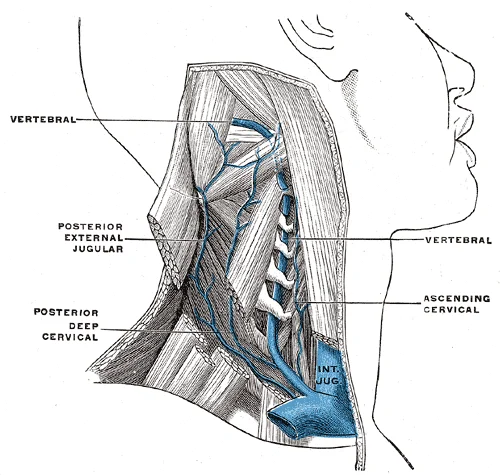
The blood supply to the head and neck is primarily derived from the subclavian and vertebral arteries. The subclavian arteries give rise to the vertebral arteries, which ascend through the transverse foramina of the cervical vertebrae to enter the cranial cavity through the foramen magnum. These arteries merge to form the basilar artery, which contributes to the circle of Willis, the anastomotic ring at the base of the brain that ensures adequate collateral circulation. [1]
Figure 1Gray563
Long description
The vertebral vein. (Poirier and Charpy.)

Figure 1: The vertebral vein (Poirier and Charpy.).
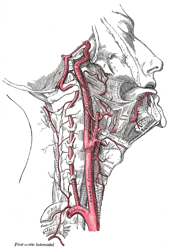
The external carotid artery provides the main arterial supply to the face and scalp. The carotid sinus, a dilated area at the bifurcation, contains baroreceptors that help regulate blood pressure. The internal carotid artery ascends through the neck without branching and enters the skull through the carotid canal, supplying the brain and eyes. The lingual artery may arise from the common carotid artery or the carotid bifurcation. [2]
Figure 2Gray513
Long description
The internal carotid and vertebral arteries. Right side.

Figure 2: The internal carotid and vertebral arteries (right side).
Figure 3Gray508
Long description
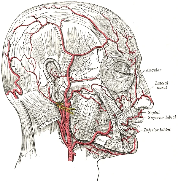
The arteries of the face and scalp. 98. Labelled illustration from Wikimedia Commons, used under its open licence.

Figure 3: The arteries of the face and scalp (98).
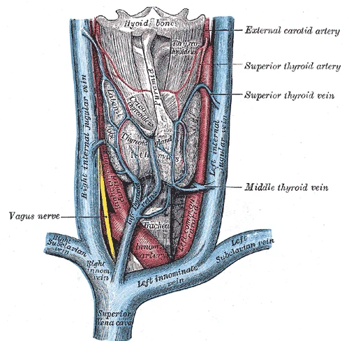
Venous drainage occurs primarily through the jugular veins. The internal jugular vein, the largest vein in the neck, receives blood from the brain, face, and neck via the dural venous sinuses. It descends vertically in the neck to join the subclavian vein and form the brachiocephalic vein[1]. The external jugular vein drains blood from the scalp and face, emptying into the subclavian vein[1]. [3]
Figure 4Gray560
Long description
The veins of the thyroid gland. Labelled illustration from Wikimedia Commons, used under its open licence.

Figure 4: The veins of the thyroid gland.
The dural venous sinuses are specialized venous channels located between the layers of the dura mater. They receive blood from the brain and drain into the internal jugular vein. Major sinuses include the superior sagittal sinus, which runs along the midline of the skull, and the transverse sinuses, which course horizontally at the base of the skull[1].
Angiographic anatomy reveals the complex three-dimensional relationships of these vessels. The lingual artery, for example, emerges anteromedially from the external carotid artery above the superior thyroid artery and below the facial artery. Its variations in origin, course, and branches are important considerations for surgical planning and interventional procedures. Side branches may theoretically be utilized for lead placement, however these branches are generally quite apical and hence not suitable for lead placement [3]
DefinitionCircle of Willis
An anastomotic ring at the base of the brain formed by the anterior and posterior communicating arteries, linking the internal carotid and vertebral arterial systems.
The blood supply of the head and neck is complex and variable, with several important arteries that follow specific patterns of origin, course, and termination. The lingual artery (LA) typically originates from the external carotid artery (ECA) at the level of the greater horn of the hyoid bone. However, anatomical variations are common, with atypical origins reported from the common carotid artery, carotid bifurcation, or shared origins with the facial artery (FA) or superficial temporal artery [2]
Figure 5Gray507
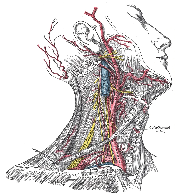
Long description
Superficial dissection of the right side of the neck, showing the carotid and subclavian arteries.

Figure 5: Superficial dissection of the right side of the neck, showing the carotid and subclavian arteries.
The three-dimensional configuration of the LA's origin and course makes it a challenging anatomical structure to identify during angiographic procedures or while interpreting imaging. The posterior loop of its origin, with its interchanging course with the FA artery, can make differentiating these arteries difficult near their origin from the ECA. [2]
Figure 6Gray509
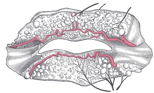
Long description
The labial coronary arteries, the glands of the lips, and the nerves of the right side seen from the posterior surface after removal of the mucous membrane. (Poirier and Charpy.)

Figure 6: The labial coronary arteries, the glands of the lips, and the nerves of the right side seen from the posterior surface after removal of the mucous membrane (Poirier and Charpy.).
The blood supply to the head and neck is primarily derived from the carotid and subclavian arteries, which form an extensive network of anastomoses that provide collateral circulation. The common carotid artery bifurcates into the internal and external carotid arteries, while the subclavian artery gives rise to several important branches that contribute to this vascular network.
Figure 7Gray511
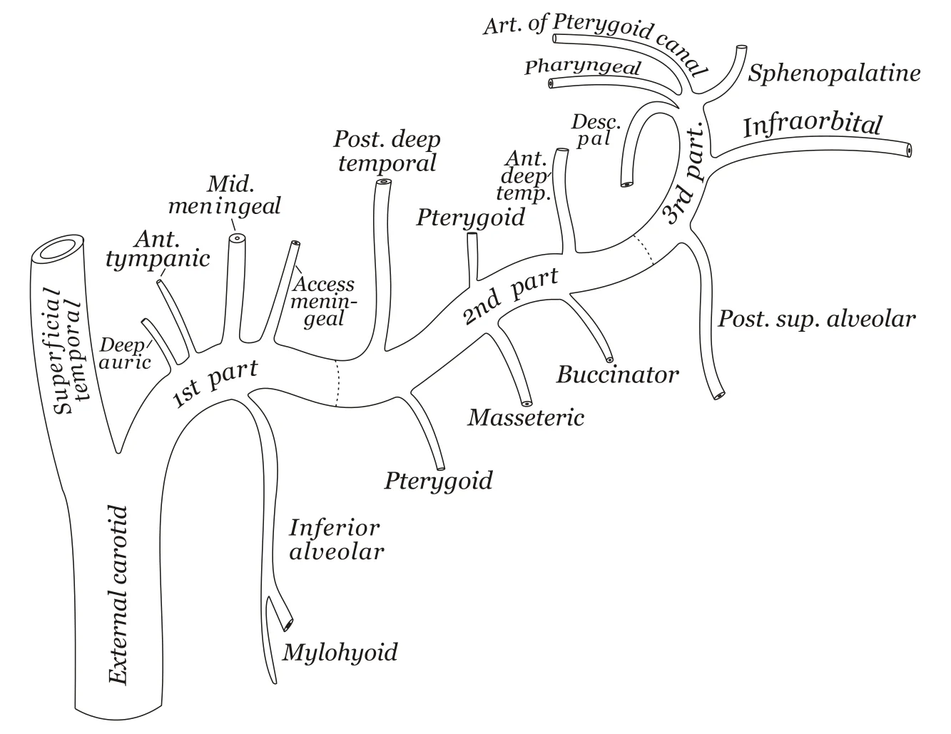
Long description
Plan of branches of internal maxillary artery.

Figure 7: Plan of branches of internal maxillary artery.
| Branch | Origin | Course | Distribution |
|---|---|---|---|
| Internal Carotid Artery | Bifurcation of common carotid | Ascends through carotid sheath, enters skull via carotid canal | Supplies brain, orbits, forehead |
| External Carotid Artery | Bifurcation of common carotid | Ascends anterior to internal carotid, divides into terminal branches | Supplies face, scalp, neck, dura mater |
The linguofacial trunk (LFT), where the lingual and facial arteries share a common origin, has a pooled prevalence of 16.41%. [2]
The lingual artery typically has four consistent branches: the suprahyoid, dorsal lingual, sublingual, and deep lingual arteries. [2]
The extensive collateral circulation and dense network of anastomoses involving these arteries contribute to the rich blood supply of the tongue and its tendency to bleed profusely from injuries. [2]
The jugular veins, including the internal and external jugular veins, drain blood from the head and neck. The internal jugular vein forms the terminal part of the intracranial venous system and receives blood from the dural venous sinuses, including the cavernous sinus. The cavernous sinus is a dural venous sinus located on either side of the sella turcica, receiving blood from the ophthalmic veins and draining into the internal jugular vein via the superior and inferior petrosal sinuses.
Figure 8Gray557
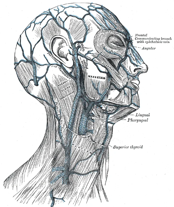
Long description
Veins of the head and neck. Labelled illustration from Wikimedia Commons, used under its open licence.

Figure 8: Veins of the head and neck.
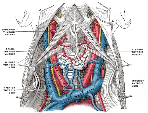
Figure 9Gray562
Long description
The fascia and middle thyroid veins. The veins here designated the inferior thyroid are called by Kocher the thyroidea ima. (Poirier and Charpy.)

Figure 9: The fascia and middle thyroid veins.
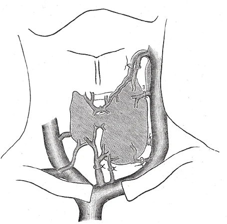
Figure 10Gray561
Long description
Diagram showing common arrangement of thyroid veins. (Kocher.)

Figure 10: Diagram showing common arrangement of thyroid veins (Kocher.).
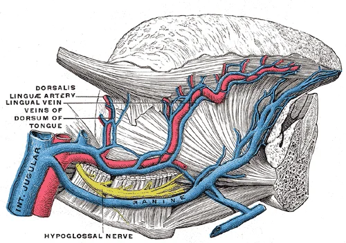
Figure 11Gray559
Long description
Veins of the tongue. The hypoglossal nerve has been displaced downward in this preparation. (Testut after Hirschfeld.)

Figure 11: Veins of the tongue.
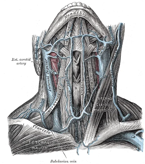
Figure 12Gray558
Long description
The veins of the neck, viewed from in front. (Spalteholz.)

Figure 12: The veins of the neck, viewed from in front (Spalteholz.).
The blood supply to the head and neck is characterised by extensive anastomoses, ensuring robust collateral circulation. This redundancy is critical because the ventropharyngeal region’s midline location ensures a robust, bilateral blood supply that supports collateral circulation. In cases of proximal lingual artery (LA) obstruction, blood flow is maintained through sublingual routes. The submental artery (STA) may also provide additional collateral support in the linguofacial area when both the LAs and facial arteries (FA) are compromised. Furthermore, collateral flow can be supplemented by reversed flow in vessels such as the superior masseteric and proximal occipital arteries. [2]
The lingual artery arises from the external carotid and supplies the tongue and floor of the mouth. The sublingual artery, a branch of the lingual artery, not uncommonly arises from the facial artery. [2]
Clinical pearlCollateral Circulation
The midline location of the ventropharyngeal region ensures a robust, bilateral blood supply that supports collateral circulation.
The sublingual artery usually originates at the anterior border of the hyoglossus muscle and courses between the mylohyoid and genioglossus muscles to the sublingual glands. [2]
In the context of the shoulder, the humeral head is supplied via an anastomosing intraosseous network including a major contribution from the arcuate artery (from the ACHA).
| Vessel | Branches/Connections | Anastomoses With |
|---|---|---|
| Sublingual Artery | Mandibular and Submental branches | Dental and deep LAs; Submental artery |
| Lingual Artery | Sublingual artery (usually) | Facial artery (sometimes) |
| Facial Artery | Submental artery | Sublingual artery (sometimes) |
DefinitionAnastomosis
A connection between two blood vessels, allowing collateral circulation if one vessel is blocked.
Similarly, in the head and neck, the bilateral supply to the ventropharyngeal region ensures that unilateral obstruction does not lead to tissue death. Understanding these territories and their interconnections is essential for surgical planning and managing vascular emergencies. [1]
The vascular architecture of the head and neck relies on extensive anastomotic networks to ensure perfusion redundancy. These connections allow blood to bypass occluded segments, a principle critical for surgical planning and understanding ischaemic complications. The primary arterial supply derives from the external and internal carotid arteries, with significant cross-talk occurring at the level of the face, scalp, and intracranial compartments.
The external carotid artery gives rise to multiple branches that anastomose with branches of the internal carotid and vertebral systems. The facial artery, for instance, courses over the mandible and anastomoses with the angular artery, a terminal branch of the ophthalmic artery. This connection provides a collateral pathway between the external and internal carotid circulations. Similarly, the superficial temporal artery anastomoses with the occipital artery and the posterior auricular artery, creating a robust network across the scalp.
Figure 13Gray515
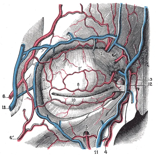
Long description
Bloodvessels of the eyelids, front view. 1, supraorbital artery and vein; 2, nasal artery; 3, angular artery, the terminal branch of 4, the facial artery; 5, suborbital artery; 6, anterior branch of the superficial temporal artery; 6’, malar branch of the transverse artery of the face; 7, lacrimal a

Figure 13: Bloodvessels of the eyelids, front view.
Figure 14Gray510
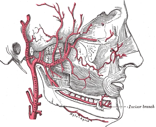
Long description
Plan of branches of internal maxillary artery.

Figure 14: Plan of branches of internal maxillary artery.
Within the orbit, the ophthalmic artery (a branch of the internal carotid) forms critical anastomoses with the external carotid system. The dorsal nasal branch of the ophthalmic artery anastomoses with the angular artery, while the lacrimal artery may communicate with the superficial temporal artery. These connections are clinically significant in cases of carotid occlusion, where retrograde flow can potentially maintain ocular perfusion, although this is variable and often insufficient.
Venous drainage mirrors the arterial supply but includes unique dural sinus connections. The facial vein lacks valves, allowing for potential retrograde flow into the cavernous sinus. This anatomical feature explains the risk of cavernous sinus thrombosis following infections in the 'danger triangle' of the face. The superior ophthalmic vein drains into the cavernous sinus, connecting the facial venous system with the intracranial dural sinuses.
Understanding these anastomoses is vital for procedures involving ligation or embolisation. For example, ligation of the external carotid artery may not result in immediate tissue necrosis due to collateral flow from the internal carotid and vertebral arteries. Conversely, inadvertent injury to these collateral pathways during neck dissection can lead to unexpected ischaemic deficits. The variability in these connections, as noted in anatomical studies, underscores the need for careful preoperative imaging and intraoperative vigilance.
Clinical pearlCollateral Circulation
The head and neck possess rich anastomotic networks, allowing for significant collateral flow between the external and internal carotid systems, which is crucial for maintaining perfusion during vascular compromise.
The vascular anatomy of the head and neck is characterised by significant interindividual variation. This variability is not merely academic; it has direct clinical consequences for surgical planning and interventional procedures. Understanding these deviations from the standard model is essential to avoid iatrogenic injury, particularly in complex neurovascular and orthopaedic contexts.
The lingual artery (LA) typically arises from the external carotid artery (ECA). However, a common anatomical variation involves the formation of a shared trunk with other branches of the ECA.
Figure 15Gray514
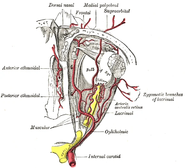
Long description
The ophthalmic artery and its branches. Labelled illustration from Wikimedia Commons, used under its open licence.

Figure 15: The ophthalmic artery and its branches.
DefinitionLinguofacial Trunk
A variant where the lingual and facial arteries share a common origin from the external carotid artery.
A systematic review by Triantafyllou et al. reported that the pooled prevalence of LFTs is 16.41% across studied cases[1]. These trunks are more often unilateral than bilateral. The morphologic characteristics of LFTs, including the side of origin relative to the ECA, height of origin, course, shape, and length, can vary between cases and are challenging to predict[1]. [2]
| Feature | Description |
|---|---|
| Prevalence | 16.41% (pooled) |
| Laterality | More often unilateral |
| Clinical Risk | Injury at the trunk affects both LA and FA |
In carotid endarterectomies, recognising the superficial temporal artery (STA) as a landmark is standard. However, variations such as a proximally originating LA or a common trunk with the STA are essential for accurate navigation[1]. In endovascular interventions for malignant tongue tumours, patients with an LFT variant require precise catheter tip positioning within the common trunk to deliver anticancer agents adequately to the LA or FA[1]. A common LFT implies that an in [2]
The LA generally courses medially to the hyoglossus muscle. However, variations can include lateral or superficial pathways, sometimes even penetrating the mylohyoid[1]. The artery branches into suprahyoid, dorsal lingual, sublingual, and deep LAs. It maintains rich collateral circulation with contributions from the superior thyroid, occipid, and facial arteries[1]. [2]
Appreciating LA anatomy is essential for optimal surgical planning, minimizing complications, and refining the interpretation of head-and-neck angiography[1]. Given this anatomical complexity, familiarity with the LA’s anatomy and variation is crucial, particularly in neurovascular procedures where accurate vascular mapping can impact surgical planning and outcomes[1]. [2]
The major concern raised by vascular variation is the risk of postoperative complications.
Exam trapOsteonecrosis Risk
Reduced intraosseous supply to high-risk areas is linked to osteonecrosis, which almost always develops initially in this specific region. [1]
The blood supply of the head and neck is clinically significant due to the high vascularity of the region and the potential for life-threatening haemorrhage. Understanding arterial anatomy is essential for managing trauma, performing surgical ligation, and interpreting imaging.
The external carotid artery gives off the superior thyroid, lingual, facial, and occipital arteries. The internal carotid artery ascends without branches in the neck.
Clinical pearlPulse Points
Palpate the facial artery as it crosses the inferior border of the mandible and the superficial temporal artery anterior to the tragus for rapid assessment of perfusion.
Exam trapLigation Risk
Ligation of the external carotid artery proximal to its branches may not stop bleeding if collateral circulation from the contralateral side or vertebral system is robust.
This course is considered the normal variant, representing 93% of cases in large cadaveric studies. [2]
A less common variation involves the artery running laterally. For the third course variation, the LA initially runs laterally and superficially along the posterior aspect of the hyoglossus, then turns medially into the muscle and reemerges at its anterior border (representing 1.8% of cases). This superficially coursing variant in relation to the hyoglossus may facilitate extraoral ligation of the LA in appropriate circumstances. In this variant, the dorsal lingual branch arises from the LA but can also arise from the FA proximal to the origin of the submental artery. [2]
| Variant | Course | Frequency | Clinical Relevance |
|---|---|---|---|
| Normal | Medial to hyoglossus | 93% | Standard surgical approach |
| Lateral/Superficial | Lateral to hyoglossus | 1.8% | Facilitates extraoral ligation |
Aneurysms may occur at the carotid bifurcation due to turbulent flow. Access points for intervention include the common femoral artery for endovascular procedures. Pulse points include the facial artery at the mandible and the superficial temporal artery. Ligation of the external carotid artery is a last-resort measure for uncontrollable haemorrhage. It emerges from the LA as the LA descends from its loop and travels along the upper margin of the hyoid bone to nourish the muscles surrounding the hyoid bone. It forms anastomoses with the ipsilateral STA and contralateral suprahyoid artery. [2]
Course variants can also have significant implications in surgical intervention. A LA that runs lateral to the hyoglossus or through the sublingual region is more exposed and, consequently, may be more susceptible to injury during transoral surgeries or floor-of-mouth procedures due to its unexpected lateral position. The occurrence (and recognition thereof) of such a lateral course may, conversely, facilitate extra oral ligation of the LA. [2]
The anterior cerebral artery (ACA) supplies the medial aspects of the frontal and parietal lobes. A critical anatomical landmark is the anterior communicating artery, which connects the left and right ACAs. Variations in this circle are common, but the constant feature is the entry of the anterolateral branch into the proximal humerus just inferior to the transverse humeral ligament. This branch supplies most of the epiphysis as the arcuate artery, the largest intraosseous branch. The posterior circumflex humeral artery (PCHA) most commonly originates as a single branch from the axillary artery. It travels through the quadrangular space with the axillary nerve. Less common origins include a common trunk with the anterior circumflex humeral artery or collectively from the subscapular artery. [1]
In multislice cardiac CT, 34% of patients had the vein of Marshall. [3]
The term anterolateral branch is used by the majority of authors. Duparc et al. describe a lateral ascending branch following the lateral edge of the intertubercular groove. In coronary computed tomographic angiographies, an obtuse angle (133 ± 14º) was present in 79% of cases from a lateral view. The mitro-aortic annular vein (MAAV) emerges from the GCV before the GCV-AIV junction at 2 o’clock on the mitral ring. In approximately three quarters of angiograms, one of these veins was identified. [1][3]
Exam trapCommon Trap
Do not confuse the anterolateral branch of the humerus with branches of the lingual artery. The anterolateral branch enters the proximal humerus inferior to the transverse humeral ligament.
Clinical pearlKey Fact
The valve of Vieussens is present in about 60% of cases. It can cause difficulties when advancing the catheter and positioning the LV lead in CRT procedures. This would include women over 60 years of age with dominant hand involvement and surgical interventions requiring additional procedures performed at the shoulder during typical RC repair (e.g., biceps tenotomy/tenodesis, acromioplasty and debridement). [1][3]
| Artery | Common Origin | Course |
|---|---|---|
| PCHA | Axillary artery | Quadrangular space with axillary nerve |
| ACHA | Axillary artery | Anterolateral branch enters proximal humerus |
| GCV | Oblique vein of Marshall | Left atrioventricular groove |
DefinitionAnatomical Constant
The one constant concerning the anterolateral branch is that it enters the proximal humerus just inferior to the transverse humeral ligament. [1]
Students frequently confuse the arterial supply of the head and neck with the venous drainage of the heart, particularly when studying coronary anatomy. A primary error is assuming the anterior interventricular vein (AIV) is the sole drainage route for the anterior septum. The AIV originates at the apex and runs into the anterior interventricular sulcus, accompanied by the left anterior descending coronary artery. However, it specifically receives septal tributaries draining the anterior portion of the interventricular septum [3]
A significant anatomical misconception concerns the circumflex humeral artery (ACHA). Many assume it remains superficial, but it continues within the intertubercular groove by passing deep to the long head of biceps tendon. Within this groove, it can give off up to four branches. One such branch is an anterolateral branch that courses superiorly along the lateral edge of the intertubercular groove. This branch continues as the arcuate artery when it enters the bone. Furthermore, before entering [1]
In venous anatomy, students often misinterpret the dimensions and clinical relevance of the coronary sinus (CS). The diameter of the CS ostium varies between 5–15 mm. This diameter is influenced by gender and underlying heart disease. The CS ostium tends to be larger in male patients. It is also larger in the presence of significant mitral regurgitation. Additionally, it is larger in patients with coronary artery disease. The size is correlated with the left ventricular (LV) size and has an inverse correlation with the LV ejection fraction. [3]
Another common error relates to the length of the CS. The length ranged between 15–70 mm in Ludinghausen's morphological study. In an angiographic study by Simoon et al., the average length was 71.70 ± 9.71 mm when formed from the great cardiac vein (GCV) and left marginal vein. It was 70.18 ± 14.98 mm when formed from the GCV and posterolateral vein. Students also struggle with tricuspid valve (TV) anatomy. The transverse diameter of the CS was 12.2 ± 3.5 mm. The TV was present in 82.1% of the patients. [3]
Finally, the vein of Marshall is often misunderstood as a simple remnant. It is an embryonic remnant of the left superior vena cava enclosed within the ligament of Marshall. It is the main vein of the left atrium, draining the middle and lateral part of the posterior wall. Its presence varies, reaching 99% in necrotic studies. It has a mean length of 2–3 cm and an average diameter of 1 mm. Venous angiographies showed presence in 73% to 92.7% of cases. [3]
This anatomical knowledge is critical at the bedside and in the operating theatre for managing cardiac arrhythmias and vascular access. The AIV is currently used as an alternative pacing site in patients with tricuspid valve valvopathies or prosthetic tricuspid valve. It is also used for mapping and ablation of idiopathic ventricular arrhythmias originating from the left ventricular summit (LVS). The MCV flows along the posterior interventricular groove and terminates near the CS. Side branches [3]
In the context of atrial fibrillation, the CV tree is more than just a passive conduit. Myocardial sleeves of the CS wall have similarities to that of Purkinje fibers. The significant presence of complex fractionated atrial electrograms (CFAEs) in the CS indicates that the CS may play a part in the recurrence of atrial fibrillation after conventional ablation. Understanding the VOM is vital for ablation techniques. Discrepancies in VOM presence between necrotic studies and imaging studies might [3]
On imaging, recognizing the ACHA's path is essential for shoulder surgery. The ACHA passes deep to the long head of biceps tendon. Its anterolateral branch courses superiorly along the lateral edge of the intertubercular groove. This branch continues as the arcuate artery when it enters the bone. A small branch runs superiorly to the rotator cuff before entering the humerus. For head and neck procedures, knowing the deep LA courses ventrally near the frenulum is key. It reaches the tip of the tongue, supplying the body via a submucosal arterial network. [1][2]
In examinations, students must recall specific measurements. The CS ostium diameter varies between 5–15 mm. The CS length averages 71.70 ± 9.71 mm in specific formations. Obstructive TV covering 100% of the ostium occurs in 2.6% of cases. CS cannulation failure rates range between 2.87% to 3.7%. The VOM has a mean length of 2–3 cm and an average diameter of 1 mm. [3]
CS Cannulation Anatomy
- Assess CS OstiumDiameter 5–15 mm
- Question: TV present?
Yes
- Check Coverage100% coverage = Obstructive (2.6%)
- Question: Obstructive?
Yes
- High Failure Risk2.87–3.7% failure rate
No
- Standard CannulationProceed with caution
No
- Standard CannulationProceed normally
The ACHA passes deep to the long head of biceps tendon within the intertubercular groove. The anterolateral branch of the ACHA becomes the arcuate artery upon entering the bone. The deep LA courses ventrally near the frenulum to the tongue tip, forming a submucosal network. The CS ostium diameter varies between 5–15 mm, influenced by gender and heart disease. The CS length averages 71.70 ± 9.71 mm when formed from the GCV and left marginal vein. Only TV covering 100% of the CS ostium is considered obstructive, occurring in 2.6% of cases. [3] The AIV is used for pacing in tricuspid valvopathies and ablation of LVS arrhythmias. The VOM drains the middle and lateral posterior left atrium, with presence varying by imaging method.
Answer each one in your head first, then open the answer.
What structure does the ACHA pass deep to within the intertubercular groove?
Show answerHide answer
The long head of biceps tendonWhat is the typical diameter range of the CS ostium?
Show answerHide answer
5–15 mmWhich artery supplies the tongue’s body via a submucosal arterial network?
Show answerHide answer
The deep lingual arteryWhat percentage of cases have a TV covering 100% of the CS ostium?
Show answerHide answer
2.6%What is the average length of the CS when formed from the GCV and left marginal vein?
Show answerHide answer
71.70 ± 9.71 mm
- Keough N, Lorke DE. The humeral head: A review of the blood supply and possible link to osteonecrosis following rotator cuff repair. Journal of anatomy 2021;239(5):973-982. doi:10.1111/joa.13496. Accessed .
- Na J, Kakazu A, Muthana A, et al. Lingual artery: Angiographic anatomy and variations review for neurosurgeons. Surgical neurology international 2025;16:156. doi:10.25259/sni_282_2025. Accessed .
- Zehan IG, Eötvös CA, Moldovan MP, et al. Angiographic Anatomy of the Left Coronary Veins: Beyond Conventional Cardiac Resynchronization Therapy. Current cardiology reports 2025;27(1):58. doi:10.1007/s11886-025-02204-z. Accessed .
Figure credits
- Figure 1: Henry Vandyke Carter, via Wikimedia Commons, Public domain, Public domain.
- Figure 2: Henry Vandyke Carter, via Wikimedia Commons, Public domain, Public domain.
- Figure 3: Henry Vandyke Carter, via Wikimedia Commons, Public domain, Public domain.
- Figure 4: Henry Vandyke Carter, via Wikimedia Commons, Public domain, Public domain.
- Figure 5: Henry Vandyke Carter, via Wikimedia Commons, Public domain, Public domain.
- Figure 6: Henry Vandyke Carter, via Wikimedia Commons, Public domain, Public domain.
- Figure 7: Henry Vandyke Carter, via Wikimedia Commons, Public domain, Public domain.
- Figure 8: Henry Vandyke Carter, via Wikimedia Commons, Public domain, Public domain.
- Figure 9: Henry Vandyke Carter, via Wikimedia Commons, Public domain, Public domain.
- Figure 10: Henry Vandyke Carter, via Wikimedia Commons, Public domain, Public domain.
- Figure 11: Henry Vandyke Carter, via Wikimedia Commons, Public domain, Public domain.
- Figure 12: Henry Vandyke Carter, via Wikimedia Commons, Public domain, Public domain.
- Figure 13: Henry Vandyke Carter, via Wikimedia Commons, Public domain, Public domain.
- Figure 14: Henry Vandyke Carter, via Wikimedia Commons, Public domain, Public domain.
- Figure 15: Henry Vandyke Carter, via Wikimedia Commons, Public domain, Public domain.
Educational content for medical students and doctors. It is not a substitute for clinical judgement or local guidelines.