Lymphatic drainage of the head and neck
Lymphatic drainage of head and neck via superficial and deep nodes to jugular trunks.
The lymphatic system in the head and neck consists of lymphatic vessels and lymph nodes that drain interstitial fluid back into the circulation. Lymphatic vessels begin as blind-ending capillaries that merge into larger vessels, eventually emptying into the bloodstream via ducts. Unlike the cardiovascular system, lymph is not actively pumped by the heart but moves through body movements, skeletal muscle contractions, and breathing, with one-way valves ensuring flow toward the heart. [1]
The lymphatic drainage of the head and neck is organized into superficial and deep cervical lymph node groups. Superficial lymphatics generally follow venous routes, while deep lymphatics typically accompany arteries. The scalp drains primarily to the occipital, parotid, and submental nodes. The face drains to the submandibular and submental nodes. The mouth drains to submandibular and submental nodes, while the tongue drains to submandibular and deep cervical nodes. The pharynx drains to the deep cervical nodes.
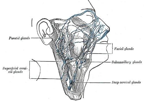
Figure 1Gray604
Long description
The lymphatics of the face. (After Küttner.)

Figure 1: The lymphatics of the face (After Küttner.).
Cancer spread in the head and neck follows predictable lymphatic pathways. For example, oral cavity cancers typically spread to submandibular nodes before progressing to deep cervical nodes. Lymph node examination is crucial in head and neck cancer staging, with palpation of the cervical chain being a standard part of the physical examination.
The lymphatic trunks of the head and neck eventually merge to form the jugular trunks, which drain into the subclavian veins. On the right side, lymph from the head and neck drains into the right lymphatic duct, while on the left side, it enters the thoracic duct. This asymmetrical drainage pattern is important to understand when evaluating lymphatic spread of infections or malignancies. [1]
Lymph nodes function as filters of the lymph, removing debris and pathogens. They are surrounded by a connective tissue capsule and contain lymphoid follicles with germinal centers where B cells proliferate. Understanding these lymphatic pathways is essential for diagnosing infections, planning surgical procedures, and staging head and neck cancers. [1]
The lymphatic system of the head and neck originates as lymphatic capillaries, also called terminal lymphatics, which are vessels where interstitial fluid enters the lymphatic system to become lymph fluid[1]. These capillaries are formed by a one cell-thick layer of endothelial cells and represent the open end of the system, allowing interstitial fluid to flow into them via overlapping cells[1]. When interstitial pressure is low, the endothelial flaps close to prevent "backflow," but as interstitial pressure increases, the spaces between the cells open up, allowing fluid to enter[1]. Entry of fluid into lymphatic capillaries is also enabled by collagen filaments that anchor the capillaries to surrounding structures[1].
In the head and neck region, lymphatic capillaries are interlaced among the arterioles and venules of the circulatory system in the soft connective tissues[1]. These capillaries drain into larger lymphatic vessels, which are similar to veins in terms of their three-tunic structure and the presence of valves[1]. The one-way valves in these vessels are located fairly close to one another, causing bulges that give the vessels a beaded appearance[1].
The lymphatic vessels of the head and neck follow specific pathways that generally accompany the blood vessels but drain toward the lymph nodes. The lymphatic drainage can be divided into regions based on the location of the structures being drained. The superficial lymphatic vessels of the scalp and face drain primarily to the superficial cervical lymph nodes, while deeper structures drain to the deep cervical lymph nodes.
Figure 2Gray606
Long description
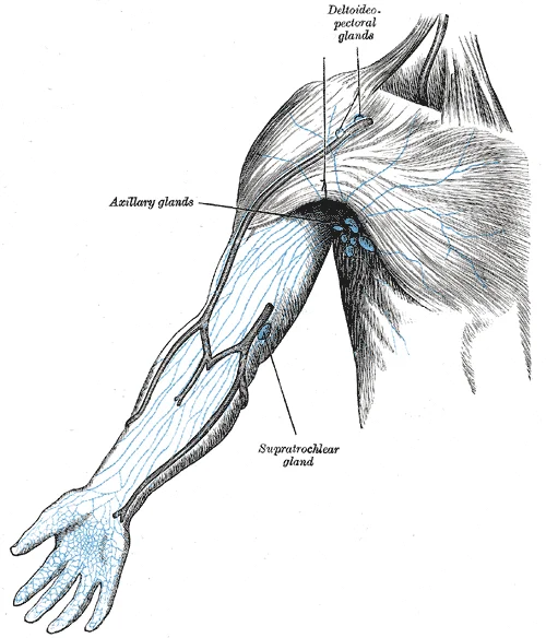
The superficial lymph glands and lymphatic vessels of the upper extremity.

Figure 2: The superficial lymph glands and lymphatic vessels of the upper extremity.
The lymphatic vessels eventually empty into larger lymphatic trunks, which then converge to form the thoracic duct or the right lymphatic duct[1]. The thoracic duct, which is the larger of the two ducts, typically originates from the cisterna chyli in the abdomen and ascends through the thoracic cavity to drain into the venous system at the junction of the left subclavian and left internal jugular veins. The right lymphatic duct, which is smaller, drains lymph from the right side of the head and neck, the right upper limb, and the right thorax into the venous system at the junction of the right subclavian and right internal jugular veins.
The lymph then travels through larger trunks to either the thoracic duct or the right lymphatic duct, depending on the side of origin, before emptying into the venous system at the jugular-subclavian junctions.
Figure 3Cervical lymph nodes and levels
Long description
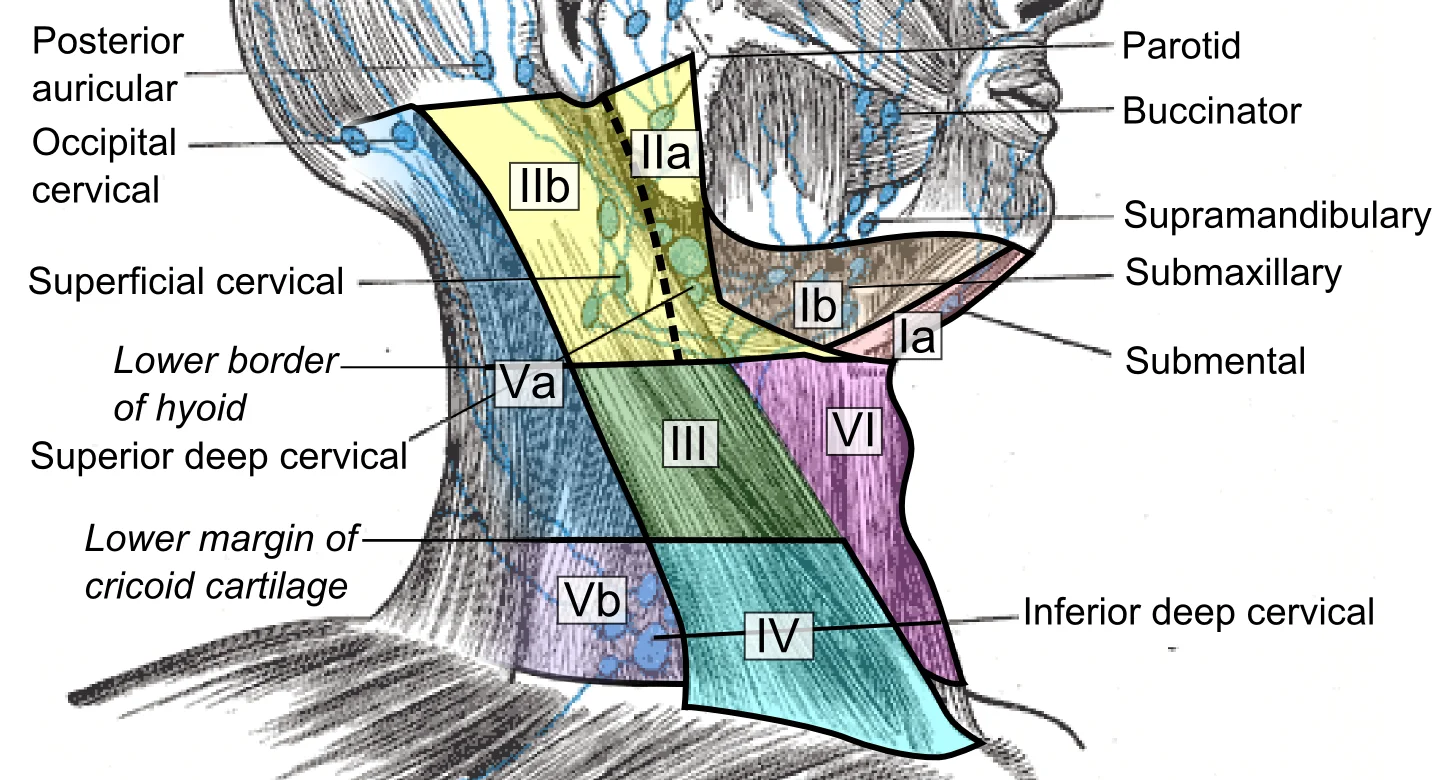
Superficial cervical lymph nodes and lymph node levels, initially described by the Memorial Sloan-Kettering Cancer Center (New York, NY) and further specified by the Committee for Head and Neck Surgery and Oncology of th

Figure 3: Superficial cervical lymph nodes and lymph node levels, initially described by the Memorial Sloan-Kettering Cancer Center (New York, NY) and further specified by the Committee for Head and Neck Surgery and Oncology of th.
The lymphatic system serves as a critical conduit for immune cell trafficking and the transport of dietary lipids and fat-soluble vitamins absorbed from the gut. When this system is compromised, for instance by obstruction from cancer cells or destruction via injury, protein-rich interstitial fluid accumulates in tissue spaces, a condition known as lymphedema. Understanding the specific branches of lymphatic drainage in the head and neck is essential for mapping this transport network. [1]
| Structure | Primary Drainage Territory | Key Relations |
|---|---|---|
| Submental Nodes | Tip of tongue, floor of mouth, central lower lip | Located in the submental triangle, anterior to the mylohyoid muscle. |
| Submandibular Nodes | Lateral lower lip, cheek, nose, upper lip, lateral tongue | Situated along the inferior border of the mandible, deep to the platysma. |
| Superficial Cervical Nodes | Auricle, external auditory meatus, scalp above the ear | Located along the external jugular vein, superficial to the sternocleidomastoid muscle. |
| Deep Cervical Nodes | Pharynx, larynx, thyroid, tongue, nasal cavity | Arranged along the internal jugular vein, extending from the skull base to the thorax. |
The submental nodes are situated in the midline, just anterior to the hyoid bone. They receive lymph from the tip of the tongue and the central part of the lower lip. These vessels typically drain into the submandibular nodes. The submandibular nodes lie along the lower border of the mandible. They collect lymph from the lateral aspects of the lower lip, the cheek, the nose, and the upper lip. The lateral tongue also drains into these nodes. This group is clinically significant because it is often the first site of metastasis for oral cancers.
The superficial cervical nodes are located along the external jugular vein. They drain the auricle, the external auditory meatus, and the scalp above the ear. These nodes are superficial to the sternocleidomastoid muscle. They ultimately drain into the deep cervical nodes. The deep cervical nodes form a chain along the internal jugular vein. They receive lymph from a wide variety of structures, including the pharynx, larynx, thyroid gland, tongue, and nasal cavity. The deep cervical nodes are the final common pathway for most lymphatic drainage from the head and neck. They converge to form the jugular lymphatic trunk.
The jugular lymphatic trunk drains into the thoracic duct on the left side or the right lymphatic duct on the right side. The thoracic duct empties into the junction of the left internal jugular and left subclavian veins. The right lymphatic duct empties into the junction of the right internal jugular and right subclavian veins. This arrangement ensures that lymph from the head and neck returns to the venous circulation.
DefinitionLymphedema
Inappropriate accumulation of protein-rich interstitial fluid in tissue spaces, often resulting from damage or obstruction of the lymphatic system.
NoteClinical Relevance
The deep cervical nodes are the final common pathway for most lymphatic drainage from the head and neck.
Figure 4Gray602
Long description
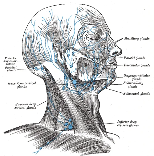
Superficial lymph glands and lymphatic vessels of head and neck.

Figure 4: Superficial lymph glands and lymphatic vessels of head and neck.
The lymphatic drainage of the head and neck is organized into distinct regional groups that follow a predictable pattern from superficial to deep, and from lateral to medial. Understanding these territories is essential for predicting the spread of pathology, particularly malignancies. The drainage generally flows towards the deep cervical lymph nodes, which serve as the final common pathway before lymph enters the venous system via the thoracic duct on the left and the right lymphatic duct on the right. [1]
The submental nodes drain the central lower lip, the floor of the mouth, and the tip of the tongue. These nodes are small and numerous, lying between the anterior bellies of the digastric muscles. They drain into the submandibular nodes or directly into the deep cervical nodes.
The submandibular nodes are larger and more clinically significant. They receive lymph from the lateral lower lip, the cheek, the nose, the upper lip, the lateral aspect of the tongue, and the gums. These nodes are located in the submandibular triangle, deep to the platysma and superficial to the mylohyoid muscle. They drain into the upper deep cervical nodes. The superficial cervical nodes lie along the external jugular vein and drain the posterior scalp and the posterior aspect of the auricle. These nodes are often overlooked but are important in the drainage of skin cancers of the posterior head.
The deep cervical nodes form a chain along the internal jugular vein, extending from the skull base to the root of the neck. This group is divided into upper and lower segments. The upper deep cervical nodes, also known as the jugulodigastric nodes, are located just below the angle of the mandible. They receive drainage from the tonsils, the posterior third of the tongue, and the nasal cavity. These nodes are frequently enlarged in tonsillitis and are a key landmark in clinical examination. The lower deep cervical nodes, including the supraclavicular nodes, drain the thorax and abdomen via the thoracic duct (on the left) or the right lymphatic duct (on the right). The left supraclavicular node, known as Virchow’s node, is particularly notable as it may receive lymph from abdominal malignancies, such as gastric cancer, via the thoracic duct.
DefinitionDeep Cervical Chain
A vertical chain of lymph nodes along the internal jugular vein, serving as the primary drainage pathway for most of the head and neck.
The convergence of these various territories into the deep cervical chain ensures that lymph from almost the entire head and neck region passes through these nodes before entering the venous circulation. This anatomical arrangement has significant clinical implications for the staging of head and neck cancers and the planning of surgical interventions.
| Region | Primary Nodal Group | Secondary Drainage |
|---|---|---|
| Central Lower Lip | Submental Nodes | Submandibular / Deep Cervical |
| Lateral Lower Lip | Submandibular Nodes | Deep Cervical Nodes |
| Tonsils | Jugulodigastric Nodes | Deep Cervical Nodes |
| Posterior Scalp | Superficial Cervical Nodes | Deep Cervical Nodes |
| Abdomen (via Thoracic Duct) | Left Supraclavicular Node | Venous System |
Figure 5Gray603
Long description
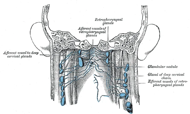
Lymphatics of pharynx. (Poirier and Charpy.

Figure 5: Lymphatics of pharynx (Poirier and Charpy.
The lymphatic system of the head and neck relies on a dense network of interconnecting channels to ensure drainage even if primary pathways are obstructed. These connections, known as anastomoses, allow lymph to bypass blocked nodes or vessels, maintaining fluid homeostasis. Understanding these collaterals is vital for predicting the spread of malignancy and managing infections.
The submandibular nodes sit along the inferior border of the mandible and receive drainage from the face, nose, and lips. Lateral to these, the parotid nodes are situated within or deep to the parotid gland, draining the scalp and external ear. The deep cervical nodes form a vertical chain along the internal jugular vein, receiving input from the submandibular, parotid, and occipital nodes. The occipital nodes, located at the base of the skull, drain the posterior scalp and anastomose with the posterior auricular nodes. These posterior auricular nodes lie just behind the ear and connect with the mastoid nodes. The flowchart of drainage demonstrates that while most lymph eventually converges into the jugular trunks, the initial pathways are highly redundant.
DefinitionLymphatic Anastomosis
A direct connection between two lymphatic vessels or channels, allowing collateral flow when primary routes are compromised.
The superficial lymphatic vessels of the head and neck are numerous and form a subcutaneous plexus. These vessels follow the major veins and arteries, ensuring that drainage is closely associated with vascular structures. The anastomoses between the facial, temporal, and occipital regions create a robust network that can compensate for local obstruction. For instance, if the submandibular nodes are removed or blocked, lymph from the lower lip and chin may reroute through the submental nodes and then directly into the deep cervical chain. [1]
| Node Group | Location | Primary Drainage Area |
|---|---|---|
| Submental | Beneath the chin | Tip of tongue, lower lip, chin |
| Submandibular | Along mandible | Upper lip, cheek, nose, lateral lower lip |
| Parotid | Within parotid gland | Lateral eyelid, external ear, scalp |
| Occipital | Base of skull | Posterior scalp |
| Posterior Auricular | Behind ear | External ear, scalp behind ear |
Deep lymphatic vessels accompany the major arteries and veins. The deep cervical nodes, arranged along the internal jugular vein, receive lymph from the tongue, larynx, thyroid, and pharynx. These nodes are further divided into anterior, lateral, and posterior groups. The anterior group drains the thyroid and larynx, while the lateral group drains the deeper structures of the neck and head. The posterior group receives drainage from the occipital and posterior auricular nodes. The convergence of these deep channels forms the jugular lymphatic trunk, which typically empties into the thoracic duct on the left and the right lymphatic duct on the right. However, variations exist, and anastomoses between the left and right jugular trunks can occur, allowing for cross-drainage.
Figure 6Gray605
Long description
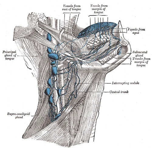
Lymphatics of the tongue. (Poirier and Charpy.)

Figure 6: Lymphatics of the tongue (Poirier and Charpy.).
Clinical pearlClinical Relevance
Enlargement of the jugulodigastric node often indicates infection in the tonsils or tongue, making it a key clinical landmark.
The redundancy of these pathways means that unilateral disease can sometimes present with bilateral nodal involvement. This is particularly relevant in cancers of the midline structures, such as the tongue or larynx, where lymphatic channels cross the midline. Surgeons must therefore consider the potential for bilateral spread when planning neck dissections. The anastomotic networks ensure that lymphatic drainage is not a simple linear process but a complex, interconnected system designed to maintain efficiency under varying physiological conditions.
The lymphatic drainage of the head and neck is not a rigid, fixed map but a dynamic network subject to significant anatomical variation. Understanding these variations is critical for predicting the spread of malignancy and planning surgical interventions. While standard textbooks describe a predictable pattern, individual differences in vessel size, confluence, and collateral pathways are common and clinically relevant.
The cervical lymph nodes are broadly divided into superficial and deep groups, with variations in their precise boundaries and connectivity. The superficial cervical nodes lie along the external jugular vein and often drain into the deep cervical chain. However, some individuals possess prominent pre-auricular nodes that may drain directly into the upper deep cervical nodes, bypassing intermediate stations. The deep cervical nodes, particularly the jugulodigastric node, are frequently enlarged in tonsillitis, but their exact position relative to the internal jugular vein can vary. Some students may find that the chain extends further inferiorly than depicted in standard diagrams, reaching the thoracic inlet.
The occipital nodes, located at the apex of the sternocleidomastoid muscle, typically drain the posterior scalp. Variations include the presence of accessory nodes that may receive drainage from the mastoid region. The post-auricular nodes, situated behind the ear, usually drain the scalp and external ear. In some cases, these nodes may communicate directly with the deep cervical chain, providing an alternative route for metastatic spread. It is important to note that the size and number of these nodes can vary significantly between individuals, affecting palpation and imaging findings.
The parotid lymph nodes are embedded within the parotid gland and are divided into superficial and deep groups. Variations in the extent of the parotid gland itself can alter the drainage pattern. Some individuals have nodes located anterior to the ear that drain the lateral eyelid and cheek, which may not be immediately obvious on physical examination. The submandibular nodes, located within the submandibular triangle, drain the face, nose, and lips. Variations include the presence of intraglandular nodes that may receive drainage from the tongue. The relationship between the submandibular nodes and the facial vein can also vary, influencing the surgical approach to node dissection.
Recognising these variations is essential for accurate staging of head and neck cancers. For instance, a tumour of the scalp may metastasise to the occipital nodes, but if accessory pathways exist, it may also spread to the deep cervical chain. Similarly, oral cancers may bypass the submandibular nodes and drain directly to the jugulodigastric node. Surgeons must be aware of these potential routes to ensure complete lymph node clearance. Imaging studies, such as CT or MRI, should be interpreted with caution, as standard atlases may not reflect individual anatomical nuances. Palpation techniques must also be adapted to account for variations in node size and position.
Clinical pearlClinical Pearl
Always consider collateral lymphatic pathways when a primary tumour drains to an unexpected nodal group.
| Structure | Typical Drainage | Common Variation |
|---|---|---|
| Occipital Nodes | Posterior scalp | Direct drainage to deep cervical chain |
| Parotid Nodes | Lateral eyelid, cheek | Accessory nodes anterior to ear |
| Submandibular Nodes | Face, nose, lips | Intraglandular nodes draining tongue |
| Cervical Chain | Deep neck structures | Extension to thoracic inlet |
The clinical significance of lymphatic drainage in the head and neck lies in its role as a pathway for metastatic disease and infection. Understanding the specific drainage territories allows clinicians to predict the spread of pathology from primary sites. The image presents a lateral view of the head and neck, oriented with the anterior aspect to the left. Key labelled structures include the submandibular nodes, located beneath the mandible; the jugulodigastric node, situated at the angle of the jaw where the digastric muscle crosses the internal jugular vein; and the supraclavicular nodes, found in the supraclavicular fossa above the clavicle. The submandibular nodes receive drainage from the face, nose, and anterior tongue. The jugulodigastric node, often enlarged in tonsillitis, lies deep to the sternocleidomastoid muscle. The supraclavicular nodes drain the thorax and abdomen, with left-sided enlargement (Virchow’s node) suggesting abdominal malignancy. These structures are arranged in a hierarchical chain, with superficial nodes draining into deep cervical nodes, which then empty into the jugular trunk. The jugular trunk typically drains into the right lymphatic duct on the right side and the thoracic duct on the left side. Recognising these relationships is crucial for physical examination and imaging interpretation.
Clinical pearlClinical Pearl
Enlargement of the jugulodigastric node is a classic sign of acute tonsillitis, reflecting its drainage of the palatine tonsils.
Aneurysm sites in the head and neck are rare but critical to identify. The carotid artery is a common site for aneurysm formation, particularly at the bifurcation. Access points for surgical intervention include the anterior triangle of the neck, bounded by the sternocleidomastoid, mandible, and midline. Pulse points are palpable at the carotid artery in the neck and the superficial temporal artery anterior to the ear. Ligation of the external carotid artery may be performed in cases of severe haemorrhage, though it carries risks of collateral circulation failure.
| Node Group | Location | Primary Drainage |
|---|---|---|
| Submandibular | Beneath mandible | Face, nose, anterior tongue |
| Jugulodigastric | Angle of jaw | Palatine tonsils |
| Supraclavicular | Supraclavicular fossa | Thorax, abdomen |
| Occipital | Back of head | Scalp |
Exam trapCommon Trap
Do not confuse the jugulodigastric node with the submental node; the latter is located below the chin and drains the tip of the tongue.
The deep cervical nodes form a vertical chain along the internal jugular vein. They receive lymph from the head and neck via the jugular trunk. The thoracic duct, which drains the majority of the body, terminates near the junction of the left internal jugular and subclavian veins. On the right side, the right lymphatic duct drains the right upper quadrant of the body. Knowledge of these pathways is essential for staging cancers and planning radiotherapy fields. For instance, squamous cell carcinoma of the oral cavity often metastasises to the submandibular and upper deep cervical nodes. Early detection of nodal enlargement can significantly impact prognosis and treatment strategy.
DefinitionVirchow’s Node
A left supraclavicular lymph node enlarged due to metastasis from abdominal malignancies, particularly gastric cancer.
Assessment of Neck Mass
- Identify Mass LocationDetermine if superficial or deep
- Question: Is it in the anterior triangle?
Yes
- Consider submandibular or jugulodigastric nodes
No
- Consider posterior triangle or supraclavicular nodes
- Assess ConsistencyHard suggests malignancy, soft suggests infection
- Order ImagingUltrasound or CT to confirm drainage pathway
Clinical pearlMaturation sites
While B cells mature in red bone marrow and T cells mature in the thymus, they both initially develop from bone marrow. [1]
Clinical pearlThymic cortex
The cortex is densely packed so it stains more intensely than the rest of the thymus. [1]
Clinical pearlSpleen dimensions
It is about 12 cm (5 in) long and is attached to the lateral border of the stomach via the gastrosplenic ligament.
Clinical pearlTonsil immunity
This seems to be the major function of tonsils—to help children’s bodies recognize, destroy, and develop immunity to common environmental pathogens so that they will be protected in their later lives.
Clinical pearlTonsillectomy criteria
Tonsils are often removed in those children who have recurring throat infections, especially those involving the palatine tonsils on either side of the throat, whose swelling may interfere with their breathing and/or swallowing.
Clinical pearlSuprahyoid action
The suprahyoid muscles raise the hyoid bone, the floor of the mouth, and the larynx during deglutition.
Clinical pearlDigastric mechanics
These include the digastric muscle, which has anterior and posterior bellies that work to elevate the hyoid bone and larynx when one swallows; it also depresses the mandible. [2]
Clinical pearlNK defense
NK cells are among the body’s first lines of defense against viruses and certain types of cancer. [1]
Clinical pearlThymic medulla
The medulla, where thymocytes migrate before leaving the thymus, contains a less dense collection of thymocytes, epithelial cells, and dendritic cells. [1]
Clinical pearlLymphocyte circulation
B cells and T cells are found in many parts of the body, circulating in the bloodstream and lymph, and residing in secondary lymphoid organs, including the spleen and lymph nodes, which will be described later in this section.
Clinical pearlT cell migration
T cells migrate from bone marrow to the thymus gland where they further mature. [1]
Clinical pearlNK mechanism
It shares this mechanism with the cytotoxic T cells of the adaptive immune response. [1]
Clinical pearlThymic trabeculae
The connective tissue capsule further divides the thymus into lobules via extensions called trabeculae. [1]
Clinical pearlThymic cortex cells
The outer region of the organ is known as the cortex and contains large numbers of thymocytes with some epithelial cells, macrophages, and dendritic cells (two types of phagocytic cells that are derived from monocytes). [1]
Clinical pearlSpleen fragility
The spleen is a fragile organ without a strong capsule, and is dark red due to its extensive vascularization. [1]
Clinical pearlLymphocyte location
They are distinguished from each other by their surface protein markers as well as by the molecules they secrete. [1]
Clinical pearlNaïve functionality
Naïve lymphocytes are fully functional immunologically, but have yet to encounter an antigen to respond to. [1]
Clinical pearlSecondary organs
In addition to circulating in the blood and lymph, lymphocytes concentrate in secondary lymphoid organs, which include the lymph nodes, spleen, and lymphoid nodules. [1]
Clinical pearlPalatine tonsils
Tonsils are often removed in those children who have recurring throat infections, especially those involving the palatine tonsils on either side of the throat, whose swelling may interfere with their breathing and/or swallowing.
Clinical pearlFacial expression
Additional muscles of facial expression are presented in. [2]
The lymphatic drainage of the head and neck is a critical anatomical pathway for immune surveillance and a potential route for metastatic spread. Understanding these pathways requires distinguishing between primary and secondary lymphoid organs. Lymphocytes develop and mature in the primary lymphoid organs, but they mount immune responses from the secondary lymphoid organs. Naïve lymphocytes are fully functional immunologically, but have yet to encounter an antigen to respond to. In addition to circulating in the blood and lymph, lymphocytes concentrate in secondary lymphoid organs, which include the lymph nodes, spleen, and lymphoid nodules. All of these tissues have many features in common, including the following: [1]
The scalp exhibits distinct drainage patterns based on region. The anterior scalp drains primarily to the preauricular and parotid lymph nodes, while the posterior scalp drains to the occipital lymph nodes. The face has a more complex drainage pattern, with the central face, including the nose and upper lip, draining to the submandibular lymph nodes. The lateral aspects of the face drain to the preauricular nodes. This central facial drainage is clinically significant because the area around the nose and upper lip, often termed the "danger area," can drain via ophthalmic veins towards the cavernous sinus, creating a potential pathway for intracranial infection.
The oral cavity drains to both submandibular and submental lymph nodes. The tongue demonstrates a dual drainage pattern: the anterior two-thirds drain to submandibular nodes, while the posterior third drains to deep cervical nodes. The pharynx drains to the retropharyngeal and deep cervical lymph nodes. These nodes form part of Waldeyer's ring, which includes the palatine, lingual, and pharyngeal tonsils. The tonsils serve a major function in helping children’s bodies recognize, destroy, and develop immunity to common environmental pathogens so that they will be protected in their later lives. Tonsils are often removed in those children who have recurring throat infections, especially those involving the palatine tonsils on either side of the throat, whose swelling may interfere with their breathing and/or swallowing.
The deep cervical lymph nodes form a chain along the internal jugular vein and are divided into upper, middle, and lower groups. These nodes receive drainage from all the superficial regions of the head and neck and ultimately drain into the jugular lymph trunk, which empties into the venous system at the junction of the internal jugular and subclavian veins.
These muscles raise the hyoid bone, the floor of the mouth, and the larynx during deglutition. [2]
Facial muscles also contribute to the structural integrity and function of the head. Place your finger on your eyebrows at the point of the bridge of the nose. Raise your eyebrows as if you were surprised and lower your eyebrows as if you were frowning. With these movements, you can feel the action of the corrugator supercilii. Additional muscles of facial expression are presented in. [2]
These cells are found in many parts of the body, circulating in the bloodstream and lymph, and residing in secondary lymphoid organs, including the spleen and lymph nodes, which will be described later in this section. While B cells mature in red bone marrow and T cells mature in the thymus, they both initially develop from bone marrow. T cells migrate from bone marrow to the thymus gland where they further mature. They are distinguished from each other by their surface protein markers as well as by the molecules they secrete. [1]
A fourth important lymphocyte is the natural killer cell, a participant in the innate immune response. It shares this mechanism with the cytotoxic T cells of the adaptive immune response. NK cells are among the body’s first lines of defense against viruses and certain types of cancer. [1]
The connective tissue capsule further divides the thymus into lobules via extensions called trabeculae. The outer region of the organ is known as the cortex and contains large numbers of thymocytes with some epithelial cells, macrophages, and dendritic cells (two types of phagocytic cells that are derived from monocytes). The cortex is densely packed so it stains more intensely than the rest of the thymus. The medulla, where thymocytes migrate before leaving the thymus, contains a less dense collection of thymocytes, epithelial cells, and dendritic cells.
Students frequently confuse the functional anatomy of the head and neck with the systemic immunology of lymphoid organs. A primary error is assuming that all lymphoid tissues possess a complete fibrous capsule. Tonsils are lymphoid nodules located along the inner surface of the pharynx and are important in developing immunity to oral pathogens. Histologically, tonsils do not contain a complete capsule, and the epithelial layer invaginates deeply into the interior of the tonsil to form tonsillar crypts. This lack of a complete capsule allows pathogens to penetrate deep into the tonsillar tissues where they are acted upon by numerous lymphoid follicles and eliminated. Students must remember that the absence of a capsule is a defining histological feature, not a pathological anomaly. [1]
Another frequent mistake involves the origin and action of the extrinsic muscles of the tongue, which are often conflated with intrinsic movements. The extrinsic muscles all include the word root glossus, and the muscle names are derived from where the muscle originates. For instance, the genioglossus originates on the mandible and allows the tongue to move downward and forward. In contrast, the styloglossus originates on the styloid bone, and allows upward and backward motion. Confusing the styloid origin with the hyoid origin leads to incorrect predictions of tongue movement; the hyoglossus originates on the hyoid bone to move the tongue downward and flatten it. [2]
Students also often misattribute the site of lymphocyte maturation. It is critical to distinguish between the primary lymphoid organs and secondary sites. The primary lymphoid organs are the bone marrow and thymus gland. The B cell undergoes nearly all of its development in the red bone marrow, whereas the immature T cell, called a thymocyte, leaves the bone marrow and matures largely in the thymus gland. A common error is to suggest that B cells mature in the thymus or that T cells complete their development in the bone marrow. The structural support of the lymph node is provided by a series of reticular fibers laid down by fibroblasts, but this is distinct from the hematopoietic function of the bone marrow. [1]
Exam trapCapsule Confusion
Tonsils lack a complete capsule, unlike lymph nodes which have a fibrous capsule and trabeculae. Do not assume all lymphoid tissues are encapsulated.
DefinitionExtrinsic Tongue Muscles
These muscles originate outside the tongue (mandible, styloid, hyoid, soft palate) and insert into the tongue, controlling its gross movement.
Knowledge of lymphatic drainage and lymphoid anatomy is essential for interpreting imaging and managing infections. In the operating theatre, understanding the relationship between the tonsils and the pharynx is vital. The tonsil located at the back of the throat, the pharyngeal tonsil, is sometimes referred to as the adenoid when swollen. Such swelling is an indication of an active immune response to infection. Surgeons must recognize that the deep invagination of the epithelial layer forms tonsillar crypts, which accumulate materials taken into the body through eating and breathing. This anatomical feature explains why tonsillitis can be recurrent and why complete removal requires careful dissection of the crypts. [1]
On imaging, distinguishing between the functional zones of the spleen helps in assessing trauma or systemic disease. The red pulp primarily functions as a filtration system of the blood, using cells of the relatively nonspecific immune response, and white pulp is where adaptive T and B cell responses are mounted. Radiologists look for enlargement of the white pulp in viral infections or changes in the red pulp in haemolytic conditions. Similarly, in the gastrointestinal tract, Peyer’s patches, a type of MALT in the small intestine, are especially important for immune responses against ingested substances. These structures contain specialized endothelial cells called M cells that sample material from the intestinal lumen and transport it to nearby follicles so that adaptive immune responses to potential pathogens can be mounted. [1]
At the bedside, the examination of the tongue relies on understanding its muscular anatomy. The tongue is obviously important for tasting food, it is also necessary for mastication, deglutition, and speech. Because it is so moveable, the tongue facilitates complex speech patterns and sounds. A lesion of the hypoglossal nerve will cause deviation of the tongue towards the side of the lesion when protruded, as the genioglossus muscle (originating on the mandible) will be weak on that side. This clinical sign directly tests the function of the extrinsic muscles derived from the mandible, styloid, hyoid, and soft palate.
| Muscle | Origin | Action |
|---|---|---|
| Genioglossus | Mandible | Moves tongue downward and forward |
| Styloglossus | Styloid bone | Moves tongue upward and backward |
| Hyoglossus | Hyoid bone | Moves tongue downward and flattens it |
| Palatoglossus | Soft palate | Elevates the back of the tongue |
Tongue Movement Assessment
- Patient protrudes tongueObserve direction of deviation
- Question: Does tongue deviate to the left?
Yes
- Left genioglossus weakLeft CN XII lesion
No
- Check right sideIf deviates right, right CN XII lesion
Clinical pearlSpleen Zones
Red pulp filters blood nonspecifically; white pulp mounts adaptive T and B cell responses.
- Tonsils lack a complete capsule and contain deep crypts for pathogen exposure.
- B cells mature in red bone marrow; T cells mature in the thymus.
- The genioglossus originates on the mandible and protrudes the tongue.
- The styloglossus originates on the styloid bone and retracts the tongue.
- The hyoglossus originates on the hyoid bone and depresses the tongue.
- Red pulp filters blood; white pulp handles adaptive immunity.
- M cells in Peyer's patches sample intestinal luminal content.
- Lymph nodes are supported by reticular fibers from fibroblasts.
Answer each one in your head first, then open the answer.
Which lymphoid tissue lacks a complete capsule?
Show answerHide answer
TonsilsWhere do B cells undergo nearly all of their development?
Show answerHide answer
Red bone marrowWhat is the origin of the genioglossus muscle?
Show answerHide answer
MandibleWhich spleen zone mounts adaptive T and B cell responses?
Show answerHide answer
White pulpWhat cells in Peyer's patches sample material from the intestinal lumen?
Show answerHide answer
M cells
- Betts JG, Young KA, Wise JA, et al. Anatomy of the Lymphatic and Immune Systems. OpenStax, Anatomy and Physiology 2e (CC BY-NC-SA 4.0). Accessed .
- Betts JG, Young KA, Wise JA, et al. Axial Muscles of the Head, Neck, and Back. OpenStax, Anatomy and Physiology 2e (CC BY-NC-SA 4.0). Accessed .
Figure credits
- Figure 1: Henry Vandyke Carter, via Wikimedia Commons, Public domain, Public domain.
- Figure 2: Henry Vandyke Carter, via Wikimedia Commons, Public domain, Public domain.
- Figure 3: Mikael Häggström, M.D., via Wikimedia Commons, CC0, CC0.
- Figure 4: Henry Vandyke Carter, via Wikimedia Commons, Public domain, Public domain.
- Figure 5: Henry Vandyke Carter, via Wikimedia Commons, Public domain, Public domain.
- Figure 6: Henry Vandyke Carter, via Wikimedia Commons, Public domain, Public domain.
Educational content for medical students and doctors. It is not a substitute for clinical judgement or local guidelines.