Applied head and neck anatomy
A concise guide to head and neck anatomy, imaging, and normal variants for medical students.
The head and neck region is a complex anatomical zone where the skull attaches to the vertebral column, allowing for balance, movement, and rotation. This area is primarily mobilised by the neck muscles, which act to flex, extend, and rotate the head depending on whether they contract unilaterally or bilaterally. The sternocleidomastoid muscle is the major muscle responsible for lateral flexion and rotation of the head, while bilateral contraction of these muscles serves as the primary flexors. When viewed from the side, this muscle divides the neck into anterior and posterior triangles, providing key landmarks for clinical examination. [1]
DefinitionSternocleidomastoid
The major muscle that laterally flexes and rotates the head, dividing the neck into anterior and posterior triangles.
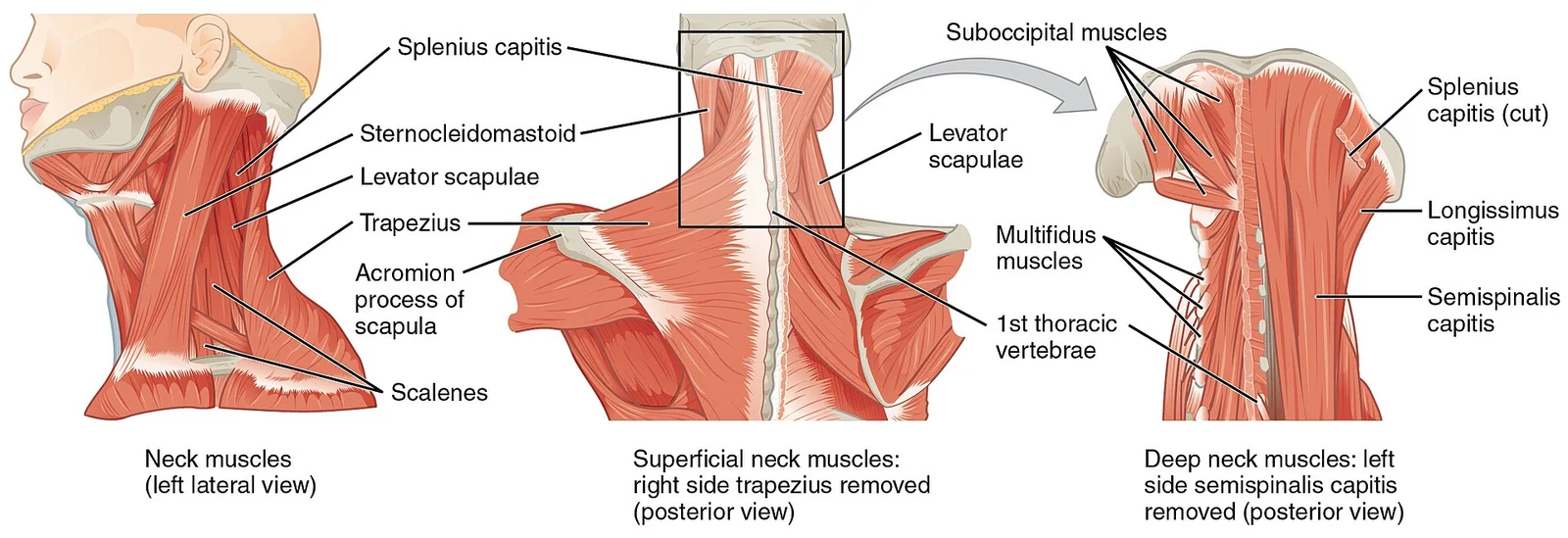
The splenius capitis inserts onto the head region, extending the head, laterally flexing it, and rotating it. Similarly, the longissimus capitis and spinalis capitis are associated with the head region, contributing to extension and stability. These muscles work in concert to maintain posture and facilitate the wide range of motion required for visual and auditory orientation.
| Group | Head Region Component | Action |
|---|---|---|
| Splenius | Splenius capitis | Extend, laterally flex, rotate |
| Longissimus | Longissimus capitis | Extend, rotate |
| Spinalis | Spinalis capitis | Extend |
Imaging plays a crucial role in evaluating this region. Computed tomography (CT) and magnetic resonance imaging (MRI) are standard modalities for assessing soft tissue and bony structures. Panoramic dental films are frequently used to evaluate the dentition and jaw bones, providing a broad view of the maxilla and mandible. Normal variants in this area include the presence of the epicranial aponeurosis, or galea aponeurosis, which connects the frontal and occipital bellies of the occipitofrontalis muscle. This broad tendon, resembling a helmet, allows for the movement of the scalp and eyebrows without a muscle crossing the top of the head.
NoteNormal Variant
The galea aponeurosis connects the frontalis and occipitalis muscles, with no muscle across the top of the head.
Understanding these boundaries and structures is essential for interpreting imaging studies and performing physical examinations. The integration of muscular anatomy with radiological findings allows for accurate diagnosis of pathologies ranging from trauma to malignancy. The clear delineation of triangles and muscle groups provides a framework for locating nerves, vessels, and lymph nodes during clinical assessment.
The sternocleidomastoid muscle is the most prominent feature, dividing the neck into anterior and posterior triangles. Its sternal head arises from the manubrium and its clavicular head from the medial third of the clavicle, converging to insert on the mastoid process and superior nuchal line. Palpate this muscle by asking the patient to rotate their head against resistance; it becomes taut and easily identifiable.
DefinitionSternocleidomastoid
The sternocleidomastoid divides the neck into anterior and posterior triangles, serving as the primary landmark for regional anatomy.
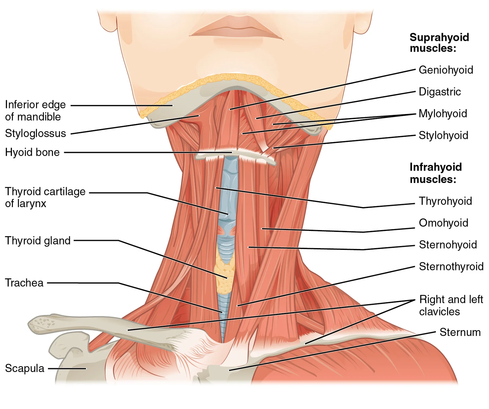
Superior to the sternocleidomastoid, the hyoid bone is a horseshoe-shaped bone that functions as a solid foundation on which the tongue can move. It is palpable at the level of the third cervical vertebra. The suprahyoid muscles are superior to it, and the infrahyoid muscles are located inferiorly. The infrahyoid muscles, also known as the strap muscles, include the sternohyoid, omohyoid, sternothyroid, and thyrohyoid. These muscles of the anterior neck assist in deglutition and speech by controlling the positions of the larynx and the hyoid bone. [1]
| Muscle | Origin | Insertion | Action | Nerve Supply |
|---|---|---|---|---|
| Sternohyoid | Manubrium | Hyoid bone | Depresses hyoid | Ansa cervicalis |
| Omohyoid | Scapula | Hyoid bone | Depresses hyoid | Ansa cervicalis |
| Sternothyroid | Manubrium | Thyroid cartilage | Depresses larynx | Ansa cervicalis |
| Thyrohyoid | Thyroid cartilage | Hyoid bone | Depresses hyoid | C1 via hypoglossal |
The carotid pulse is palpable anterior to the sternocleidomastoid muscle, at the level of the upper border of the thyroid cartilage. This is the surface marking for the common carotid artery bifurcation. The vagus nerve lies within the carotid sheath, posterior to the artery. The masseter muscle is palpable when the patient clenches their teeth, indicating the action of mastication.
Exam trapHyoid Bone
The hyoid bone is unique as it does not articulate with any other bone, relying on muscle attachments for stability.
The trachea is midline and palpable below the cricoid cartilage. The thyroid gland lies anterior to the trachea, with its lobes on either side of the tracheal rings. The recurrent laryngeal nerve runs in the tracheoesophageal groove, a critical relation during thyroid surgery. The spinal accessory nerve crosses the posterior triangle, supplying the sternocleidomastoid and trapezius muscles. Injury to this nerve results in weakness of shoulder elevation.
Clinical pearlNeck Triangles
The sternocleidomastoid divides the neck into anterior and posterior triangles, which are further subdivided by the omohyoid and occipital arteries.
The submandibular gland lies in the submandibular triangle, bounded by the mandible and the anterior and posterior bellies of the digastric muscle. It is palpable beneath the mandible. The sublingual gland lies superior to the mylohyoid muscle, in the floor of the mouth. The parotid gland lies anterior to the ear, extending down to the angle of the mandible. Its duct crosses the masseter to enter the oral cavity opposite the second upper molar.
Figure 1Gray1201
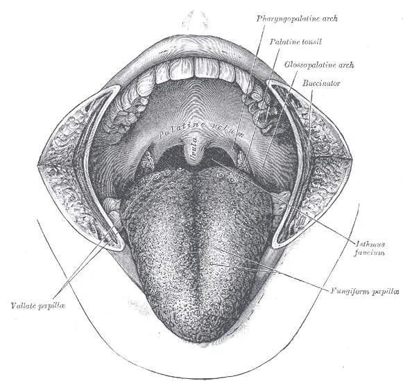
Long description
The mouth cavity. The cheeks have been slit transversely and the tongue pulled forward.

Figure 1: The mouth cavity.
NoteGlandular Landmarks
Palpate the submandibular gland by placing fingers beneath the mandible and the parotid gland anterior to the earlobe.
The cavernous sinus is a paired dural venous sinus located on either side of the sella turcica. [1]
DefinitionCutaneous Innervation
The cervical plexus provides cutaneous innervation to the skin of the neck via the lesser occipital, great auricular, transverse cervical, and supraclavicular nerves. [1]
The jugular vein is palpable lateral to the carotid artery, within the carotid sheath. It is often used for central venous access. The internal jugular vein drains blood from the brain, face, and neck. It joins the subclavian vein to form the brachiocephalic vein. The external jugular vein runs superficially, crossing the sternocleidomastoid muscle. It drains into the subclavian vein. [1]
Exam trapJugular Veins
The internal jugular vein is deep within the carotid sheath, while the external jugular vein is superficial and crosses the sternocleidomastoid.
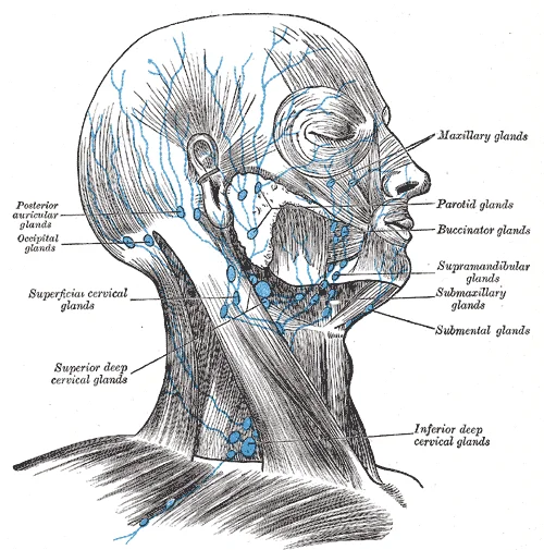
The lymph nodes of the neck are grouped into cervical chains. The anterior cervical nodes lie along the anterior jugular vein. The lateral cervical nodes lie along the internal jugular vein. The submandibular nodes lie beneath the mandible. The submental nodes lie beneath the chin. These nodes drain specific regions of the head and neck.
Clinical pearlLymphatic Drainage
Lymph from the head and neck drains into the deep cervical lymph nodes, which ultimately empty into the jugular trunk.
The surface anatomy of the head includes the temporalis muscle, which is palpable on the side of the head. It inserts on the coronoid process of the mandible. The masseter muscle is palpable on the angle of the mandible. The buccinator muscle lies deep to the cheek, forming the cheek wall. The orbicularis oculi surrounds the eye, closing the eyelids. The orbicularis oris surrounds the mouth, closing the lips. [1]
DefinitionFacial Muscles
Facial muscles insert into the skin rather than onto bone, allowing for facial expression. [1]
The scalp is vascular and innervated by the supraorbital, supratrochlear, zygomaticotemporal, auriculotemporal, greater auricular, and lesser occipital nerves. The galea aponeurotica connects the frontalis and occipitalis muscles. The epicranial aponeurosis provides a strong layer for surgical incisions. The temporal fascia covers the temporalis muscle. The parotid fascia covers the parotid gland.
Figure 2Gray1207
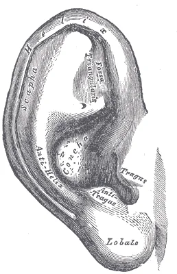
Long description
The auricula or pinna. Lateral surface. Labelled illustration from Wikimedia Commons, used under its open licence.

Figure 2: The auricula or pinna (lateral surface).
NoteScalp Layers
The scalp consists of five layers: Skin, Connective tissue, Aponeurosis, Loose areolar tissue, and Pericranium. [1]
The surface anatomy of the neck is essential for clinical examination and surgical procedures. The sternocleidomastoid muscle is the key landmark. The hyoid bone is the key bony landmark. The carotid pulse is the key vascular landmark. The lymph nodes are the key pathological landmarks. Understanding these landmarks allows for accurate diagnosis and treatment.
Exam trapClinical Relevance
Surface anatomy is critical for identifying pathological changes and planning surgical approaches in the head and neck region.
The trachea is midline and palpable below the cricoid cartilage. The thyroid gland lies anterior to the trachea. The recurrent laryngeal nerve runs in the tracheoesophageal groove. The spinal accessory nerve crosses the posterior triangle. The jugular vein is lateral to the carotid artery. The lymph nodes are grouped into cervical chains. The scalp is vascular and innervated by multiple nerves. The facial muscles insert into the skin. The muscles of the anterior neck assist in deglutition. The p [1]
DefinitionMuscle Function
The muscles of the head and neck are all axial, grouped based on location, function, or both. [1]
The sternocleidomastoid divides the neck into anterior and posterior triangles. The hyoid bone is a horseshoe-shaped bone. The infrahyoid muscles are located inferiorly. The carotid pulse is palpable anterior to the sternocleidomastoid. The vagus nerve lies within the carotid sheath. The facial nerve branches emerge from the parotid gland. The masseter muscle is palpable when the patient clenches their teeth. The trachea is midline and palpable below the cricoid cartilage. The thyroid gland lies anterior to the trachea. The recurrent laryngeal nerve runs in the tracheoesophageal groove. The spinal accessory nerve crosses the posterior triangle. The jugular vein is lateral to the carotid artery. The lymph nodes are grouped into cervical chains. The scalp is vascular and innervated by multiple nerves. The facial muscles insert into the skin. The muscles of the anterior neck assist in deglutition. The posterior muscles of the neck are primarily concerned with head movements. [1]
Figure 3Gray1204
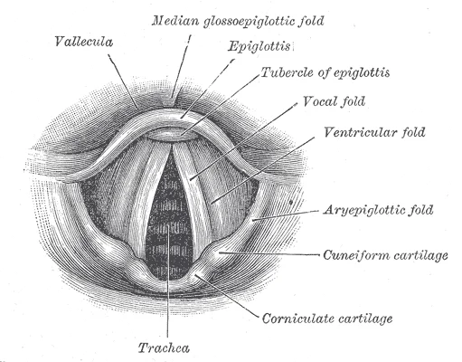
Long description
Laryngoscopic view of interior of larynx

Figure 3: Laryngoscopic view of interior of larynx.
Clinical pearlAnatomical Relations
Understanding the relations between structures is essential for safe surgical intervention and accurate clinical examination.
The surface anatomy of the head and neck is complex but systematic. The sternocleidomastoid muscle is the primary landmark. The hyoid bone is the secondary landmark. The carotid pulse is the tertiary landmark. The lymph nodes are the quaternary landmark. The scalp is the quinary landmark. The facial muscles are the senary landmark. The muscles of the anterior neck are the septenary landmark. The posterior muscles of the neck are the octonary landmark. The trachea is the nonary landmark. The thyroid gland is the denary landmark. The recurrent laryngeal nerve is the undecary landmark. The spinal accessory nerve is the duodecary landmark. The jugular vein is the tredecary landmark. The lymph nodes are the quattuordecary landmark. The scalp is the quindecary landmark. The facial muscles are the sexdecary landmark. The muscles of the anterior neck are the septendecary landmark. The posterior muscles of the neck are the octodecary landmark.
Figure 4Gray1210
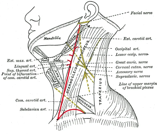
Long description
Side of neck, showing chief surface markings.

Figure 4: Side of neck, showing chief surface markings.
NoteSystematic Approach
A systematic approach to surface anatomy ensures that no structure is overlooked during clinical examination.
The surface anatomy of the head and neck is essential for medical students. It provides the foundation for clinical practice. The sternocleidomastoid muscle is the key landmark. The hyoid bone is the key bony landmark. The carotid pulse is the key vascular landmark. The lymph nodes are the key pathological landmark. The scalp is the key integumentary landmark. The facial muscles are the key muscular landmark. The muscles of the anterior neck are the key functional landmark. The posterior muscles of the neck are the key stabilising landmark. The trachea is the key airway landmark. The thyroid gland is the key endocrine landmark. The recurrent laryngeal nerve is the key neural landmark. The spinal accessory nerve is the key motor landmark. The jugular vein is the key venous landmark. The lymph nodes are the key immune landmark. The scalp is the key protective landmark. The facial muscles are the key expressive landmark. The muscles of the anterior neck are the key deglutition landmark. The posterior muscles of the neck are the key movement landmark.
DefinitionSummary
Surface anatomy provides the visual and tactile guide for understanding the deeper structures of the head and neck.
The frontal bone forms the forehead and the superior part of the orbits. It articulates with the nasal bones anteriorly and the zygomatic bones laterally. This landmark is crucial for local anaesthesia of the forehead.
The zygomatic bone forms the prominence of the cheek and the lateral wall of the orbit. It articulates with the frontal, sphenoid, maxilla, and temporal bones. The zygomaticofacial foramen, located on the lateral surface, transmits the zygomaticofacial nerve. The zygomatic arch, formed by the zygomatic process of the temporal bone and the temporal process of the zygomatic bone, serves as an attachment for the masseter muscle.
The maxilla is the central bone of the face, forming the upper jaw, the floor of the orbit, and the lateral nasal cavity. It contains the maxillary sinus. The infraorbital foramen, located below the orbit, transmits the infraorbital nerve and vessels. This is a key landmark for anaesthesia of the mid-face. The alveolar process of the maxilla holds the upper teeth.
Figure 5Gray1203
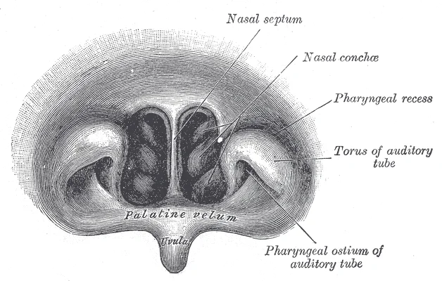
Long description
Front of nasal part of pharynx, as seen with the laryngoscope.

Figure 5: Front of nasal part of pharynx, as seen with the laryngoscope.
Figure 6Gray1199
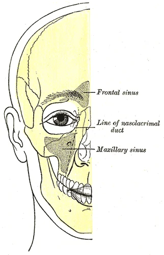
Long description
Outline of bones of face, showing position of air sinuses.

Figure 6: Outline of bones of face, showing position of air sinuses.
The mandible is the largest and strongest bone of the face. It consists of the body and the ramus. The ramus terminates superiorly in the coronoid process and the condylar process. The condylar process articulates with the temporal bone to form the temporomandibular joint. The mandibular foramen, located on the medial surface of the ramus, transmits the inferior alveolar nerve and vessels. This is the site for inferior alveolar nerve block anaesthesia. The mental foramen, located on the lateral surface of the body, transmits the mental nerve.
The temporal bone houses the structures of the ear and forms part of the skull base. The styloid process, projecting inferiorly, serves as an attachment for several muscles and ligaments. The external acoustic meatus is located in the tympanic part of the temporal bone.
Figure 7Gray1209
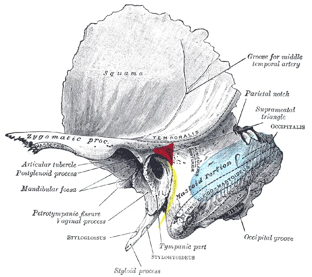
Long description
Left temporal bone showing surface markings for the tympanic antrum (red), transverse sinus (blue), and facial nerve (yellow).

Figure 7: Left temporal bone showing surface markings for the tympanic antrum (red), transverse sinus (blue), and facial nerve (yellow).
The sphenoid bone is a complex bone at the base of the skull. The greater wing of the sphenoid forms part of the middle cranial fossa and the lateral wall of the orbit. The foramen rotundum, foramen ovale, and foramen spinosum are located in the greater wing. The foramen ovale transmits the mandibular nerve (V3). The foramen spinosum transmits the middle meningeal artery.
Figure 8Gray1197
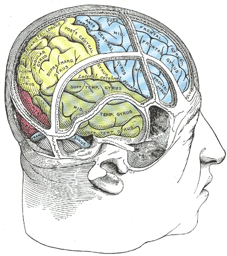
Long description
Drawing of a cast by Cunningham to illustrate the relations of the brain to the skull.

Figure 8: Drawing of a cast by Cunningham to illustrate the relations of the brain to the skull.
The ethmoid bone is located between the orbits. The cribriform plate forms the roof of the nasal cavity and transmits the olfactory nerves. The perpendicular plate forms the superior part of the nasal septum.
The vomer forms the inferior part of the nasal septum. The palatine bones form the posterior part of the hard palate and the lateral wall of the nasal cavity. The inferior nasal conchae are separate bones attached to the lateral nasal wall.
The hyoid bone is a U-shaped bone in the neck. It does not articulate with any other bone. It serves as an attachment for the muscles of the tongue and the floor of the mouth. The greater horns of the hyoid bone are palpable landmarks in the neck.
DefinitionKey Landmark
The mandibular foramen is the entry point for the inferior alveolar nerve into the mandibular canal, essential for dental anaesthesia.
| Foramen | Nerve Transmitted | Clinical Significance |
|---|---|---|
| Mental | Mental nerve | Anaesthesia of lower lip and chin |
| Mandibular | Inferior alveolar nerve | Anaesthesia of lower teeth |
| Infraorbital | Infraorbital nerve | Anaesthesia of mid-face |
| Supraorbital | Supraorbital nerve | Anaesthesia of forehead |
Lateral to the spinous processes lie the transverse processes, connected by the intertransversarii muscles. The scalene muscles are prominent on the lateral aspect. The anterior scalene lies anterior to the middle scalene. The posterior scalene is the smallest, located posterior to the middle scalene. These muscles work together to flex, laterally flex, and rotate the head. They also contribute to deep inhalation. The fascia encloses these muscles, creating distinct compartments that guide the spread of infection or tumour. Understanding these boundaries is crucial for surgical access and diagnostic imaging interpretation. [1]
DefinitionSegmental Muscles
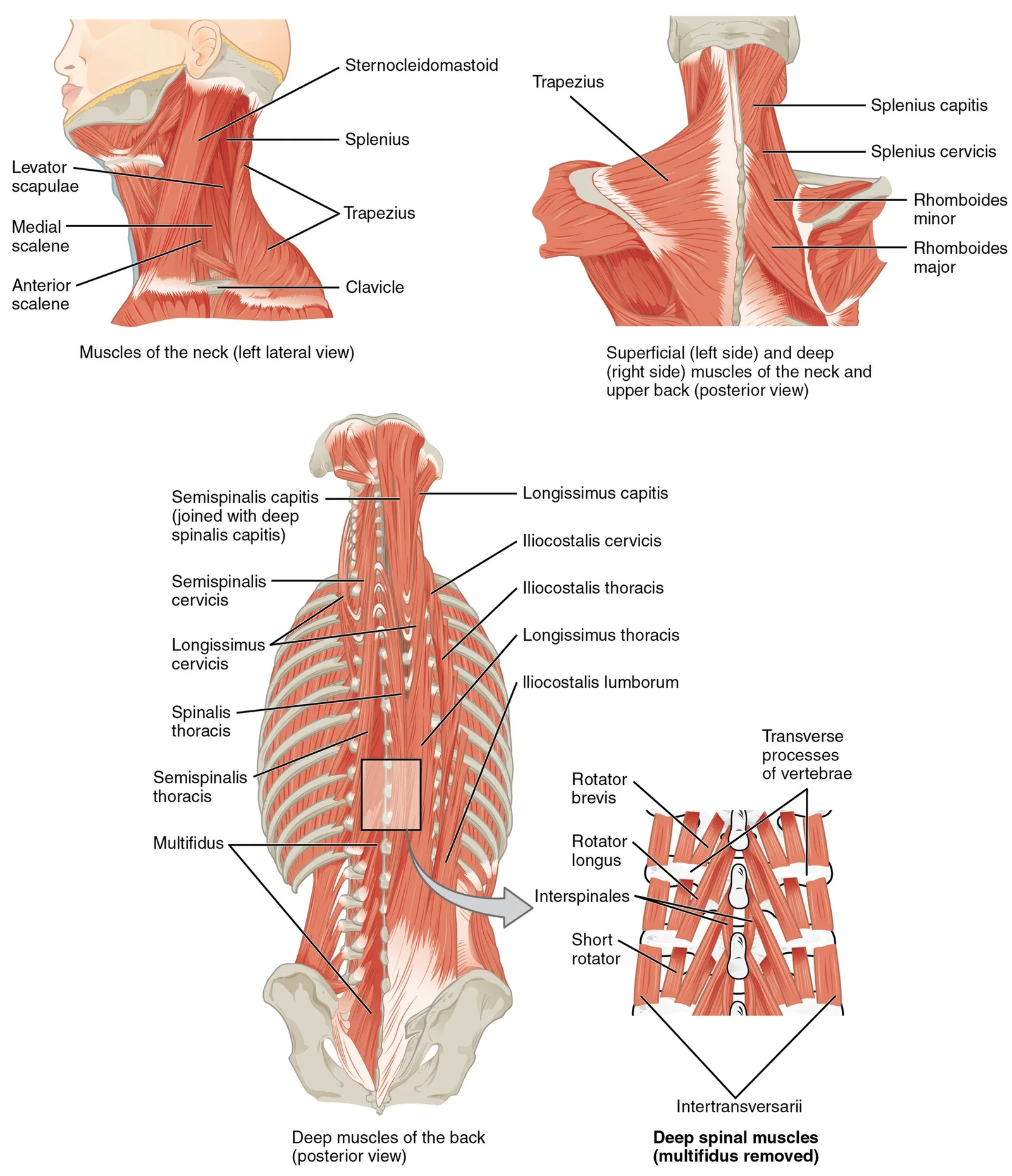
The interspinales and intertransversarii muscles stabilize the vertebral column by connecting consecutive spinous and transverse processes. [1]
The scalene muscles are key landmarks in the neck. Their anterior, middle, and posterior positions define the posterior triangle boundaries. The middle scalene forms the floor of the posterior triangle. This anatomical arrangement is vital for vascular access and understanding neurovascular compression syndromes. The fascia surrounding these muscles provides a pathway for the spread of pathology. Infections can track along these fascial planes, leading to deep neck space infections. Knowledge of these compartments allows clinicians to predict the spread of disease and plan appropriate interventions. The stability provided by the segmental muscles is essential for maintaining posture and protecting the spinal cord. The scalenes' role in respiration highlights the integration of musculoskeletal and respiratory systems.
| Muscle | Position | Function |
|---|---|---|
| Anterior | Anterior to middle | Flex, laterally flex, rotate head; deep inhalation |
| Middle | Intermediate, longest | Flex, laterally flex, rotate head; deep inhalation |
| Posterior | Posterior to middle | Flex, laterally flex, rotate head; deep inhalation |
The relationships between these structures are precise. The anterior scalene is anterior to the middle scalene. The middle scalene is intermediate between the anterior and posterior scalenes. The posterior scalene is posterior to the middle scalene. This order is consistent and reliable for anatomical identification. The fascia provides a sheath for these muscles, allowing smooth movement during head rotation and flexion. Compartment syndrome in the neck is rare but serious, requiring immediate decompression. The stability of the vertebral column depends on the integrity of these segmental muscles and their fascial coverings. Any disruption can lead to instability and neurological compromise.
Figure 91111 Posterior and Side Views of the Neck
Long description
The superficial and deep muscles of the neck are responsible for moving the head, cervical vertebrae, and scapulas.

Figure 9: The superficial and deep muscles of the neck are responsible for moving the head, cervical vertebrae, and scapulas.
Figure 101117 Muscles of the Neck and Back
Long description
The large, complex muscles of the neck and back move the head, shoulders, and vertebral column.

Figure 10: The large, complex muscles of the neck and back move the head, shoulders, and vertebral column.
Figure 111110 Muscle of the Anterior Neck
Long description
The anterior muscles of the neck facilitate swallowing and speech. The suprahyoid muscles originate from above the hyoid bone in the chin region. The infrahyoid muscles originate below the hyoid bone in the lower neck.

Figure 11: The anterior muscles of the neck facilitate swallowing and speech.
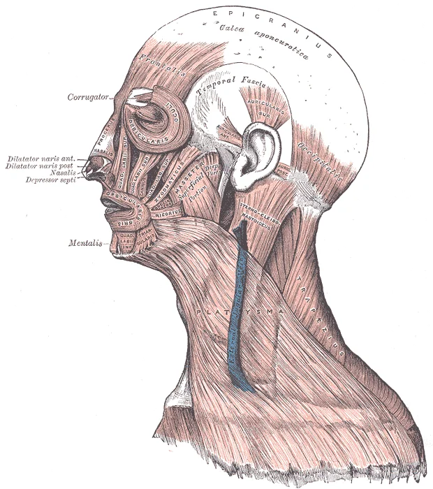
Figure 12Gray378
Long description
Muscles of the head, face, and neck. Labelled illustration from Wikimedia Commons, used under its open licence.

Figure 12: Muscles of the head, face, and neck.
The muscles of the head and neck are grouped by function and compartment. The muscles of facial expression originate on the surface of the skull and insert into the dermis, allowing skin movement for expression[1]. The buccinator compresses the cheek, aiding whistling and chewing[1]. The corrugator supercilii is the prime mover of the eyebrows[1].
Mastication involves the masseter, which elevates the mandible, and the temporalis, which retracts it[1]. You can feel the temporalis move at the temple during chewing[1].
Tongue muscles are extrinsic or intrinsic[1]. Extrinsic muscles move the whole tongue, while intrinsic muscles change its shape[1]. The genioglossus originates on the mandible, moving the tongue downward and forward[1]. The styloglossus originates on the styloid bone, allowing upward and backward motion[1]. The palatoglossus originates on the soft palate, elevating the back of the tongue[1]. The hyoglossus originates on the hyoid bone, moving the tongue downward and flattening it[1].
The suprahyoid muscles raise the hyoid bone, floor of the mouth, and larynx during deglutition[1]. The digastric has anterior and posterior bellies, elevating the hyoid and larynx, and depressing the mandible[1]. The stylohyoid moves the hyoid posteriorly, elevating the larynx[1]. The mylohyoid lifts the hyoid and presses the tongue to the mouth[1]. The geniohyoid depresses the mandible and raises the hyoid anteriorly[1].
The infrahyoid muscles generally depress the hyoid bone and control larynx position[1]. The omohyoid has superior and inferior bellies, depressing the hyoid with the sternohyoid and thyrohyoid[1].
Extrinsic eye muscles originate outside the eye and insert onto the sclera, controlling coordinated eyeball movement[1].
The back muscles move the vertebral column. The erector spinae is the primary extensor, comprising iliocostalis, longissimus, and spinalis groups[1]. The transversospinales run from transverse to spinous processes, including semispinalis capitis, cervicis, and thoracis, plus multifidus and rotatores[1]. The splenius group includes capitis and cervicis[1]. The scalenes include anterior, middle, and posterior muscles[1].
| Muscle | Origin | Insertion | Action | Nerve |
|---|---|---|---|---|
| Buccinator | Maxilla/mandible | Orbicularis oris | Compresses cheek | Facial |
| Masseter | Zygomatic arch | Mandible | Elevates mandible | Trigeminal |
| Genioglossus | Mandible | Tongue | Protrudes tongue | Hypoglossal |
| Sternohyoid | Sternum | Hyoid | Depresses hyoid | Ansa cervicalis |
| Iliocostalis | Ilium/Ribs | Ribs/Cervical vertebrae | Extends spine | Dorsal rami |
The arterial supply of the head and neck is derived primarily from the common carotid arteries, which bifurcate into the internal and external carotid arteries. The internal carotid artery supplies the brain and orbit, while the external carotid artery supplies the face, neck, and scalp. This diagram shows the bifurcation of the common carotid artery at the level of the upper border of the thyroid cartilage. These branches include the superior thyroid, lingual, facial, occipital, posterior auricular, maxillary, and superficial temporal arteries. Each branch supplies specific regions of the head and neck. The superficial temporal artery, the other terminal branch, supplies the scalp and the parotid gland. Understanding the course and branches of these arteries is essential for surgical procedures and diagnosing vascular pathologies.
Figure 13Gray1200
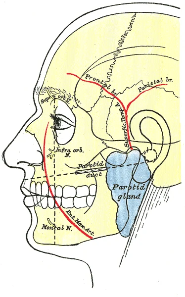
Long description
Outline of side of face, showing chief surface markings.

Figure 13: Outline of side of face, showing chief surface markings.
Figure 14Gray1198
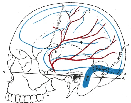
Long description
Relations of the brain and middle meningeal artery to the surface of the skull. 1. Nasion. 2. Inion. 3. Lambda. 4. Lateral cerebral fissure. 5. Central sulcus. AA. Reid’s base line. B. Point for trephining the anterior branch of the middle meningeal artery. C. Suprameatal triangle. D. Sigmoid bend o

Figure 14: Relations of the brain and middle meningeal artery to the surface of the skull (1).
DefinitionCarotid Bifurcation
The point where the common carotid artery divides into the internal and external carotid arteries, typically at the level of the upper border of the thyroid cartilage.
The superior thyroid artery supplies the thyroid gland and larynx. The lingual artery supplies the tongue and floor of the mouth. The facial artery supplies the face and palatine tonsil. The ascending pharyngeal artery supplies the pharynx and meninges. The occipital artery supplies the scalp and sternocleidomastoid muscle. The posterior auricular artery supplies the scalp behind the ear. The two terminal branches are the maxillary and superficial temporal arteries. The superficial temporal artery supplies the scalp and parotid gland.
Figure 15Gray1202
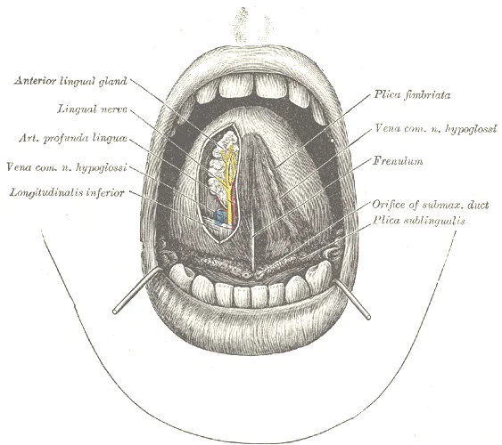
Long description
The mouth cavity. The apex of the tongue is turned upward, and on the right side a superficial dissection of its under surface has been made.

Figure 15: The mouth cavity.
| Branch | Primary Supply |
|---|---|
| Superior Thyroid | Thyroid gland, larynx |
| Lingual | Tongue, floor of mouth |
| Facial | Face, palatine tonsil |
| Occipital | Scalp, sternocleidomastoid |
| Maxillary | Deep face, teeth, brain |
| Superficial Temporal | Scalp, parotid gland |
Clinical relevance of this anatomy is significant. Injury to the external carotid artery or its branches can lead to significant bleeding. Knowledge of the course of these arteries is crucial for surgical approaches to the head and neck. For example, during a thyroidectomy, the superior thyroid artery must be ligated to control bleeding. Similarly, during a facelift, the facial artery must be identified and preserved to maintain blood supply to the face. The internal carotid artery is also clinically important. It is susceptible to atherosclerosis, which can lead to stroke. Understanding the anatomy of the carotid bifurcation is essential for carotid endarterectomy, a procedure to remove plaque from the carotid artery.
Figure 16Gray AnatomyOfHumanBody1918-P644 Figure557
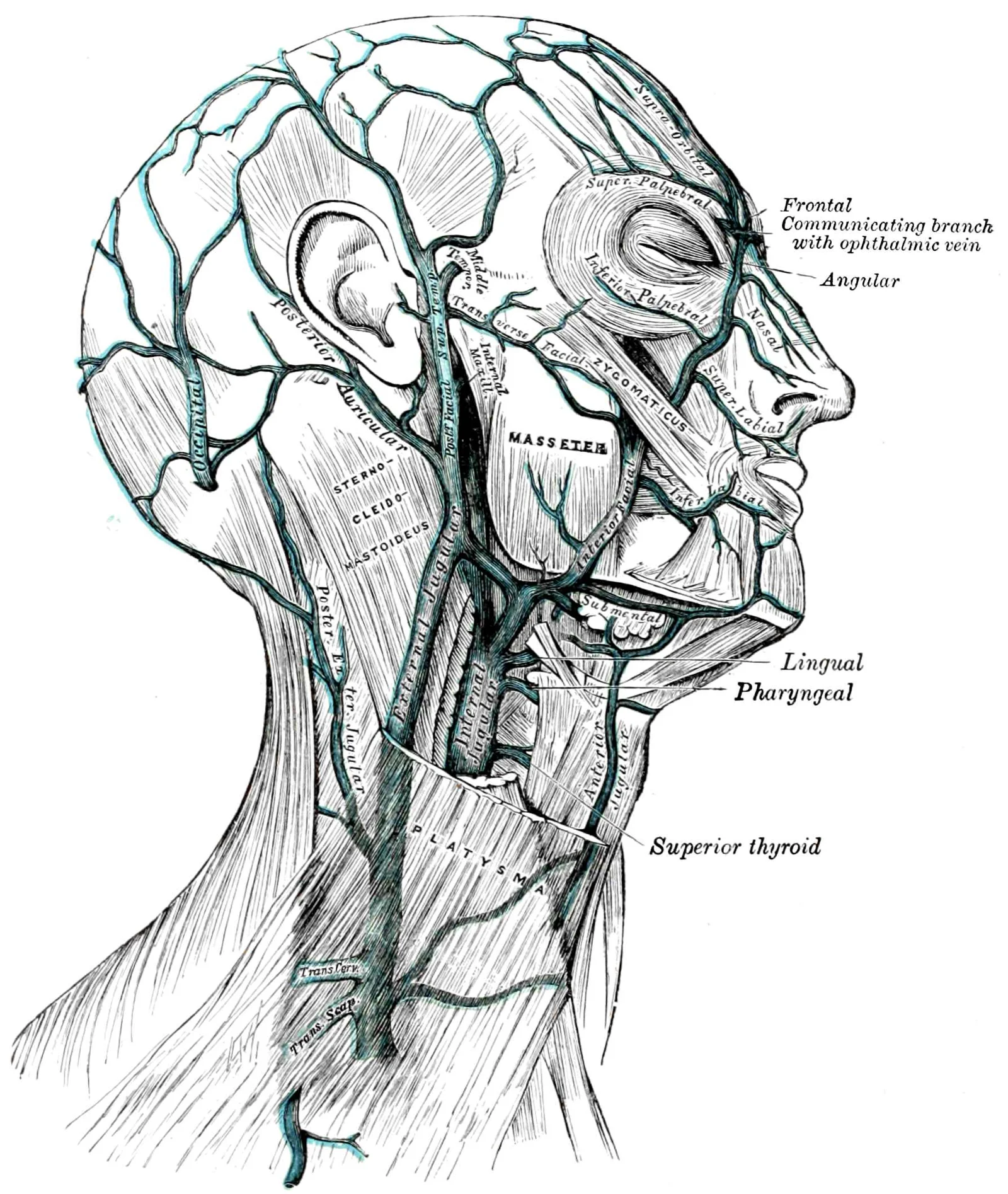
Long description
Lateral view of the side of the head and neck, showing outermost / external veins as well as some of the musculature

Figure 16: Lateral view of the side of the head and neck, showing outermost / external veins as well as some of the musculature.
The venous drainage of the head and neck is primarily through the internal jugular vein, which receives blood from multiple tributaries. The internal jugular vein originates at the jugular foramen as a continuation of the sigmoid sinus. It descends vertically through the neck within the carotid sheath, lying lateral to the common carotid artery and internal carotid artery. At the root of the neck, it joins the subclavian vein to form the brachiocephalic vein.
The facial vein is a major tributary that drains the face. It begins at the medial angle of the eye as the angular vein, then descends posterior to the facial artery. It communicates with the pterygoid plexus through the deep facial vein and ultimately drains into the internal jugular vein. The anterior division joins the facial vein to form the common facial vein, which empties into the internal jugular vein. [1]
It crosses the sternocleidomastoid muscle obliquely and pierces the deep fascia to drain into the subclavian vein.
The external jugular vein crosses the sternocleidomastoid muscle superficially.
The dural venous sinuses are specialized venous channels located within the dura mater.
The dural venous sinuses are specialized venous channels located within the dura mater.
The emissary veins connect the extracranial veins with the dural venous sinuses. [1]
It is divided into three major branches: the ophthalmic (V1), maxillary (V2), and mandibular (V3) nerves. V1 and V2 are purely sensory, supplying the skin of the face, the mucosa of the nasal and oral cavities, and the teeth. V3 is the only branch that carries motor fibres, in addition to sensory ones.
Immediately after exiting, it gives off the meningeal branch and the medial pterygoid nerve. The main trunk then divides into anterior and posterior divisions. The anterior division gives rise to the buccal nerve and the motor branches to the muscles of mastication. The posterior division yields the auriculotemporal, lingual, and inferior alveolar nerves.
| Muscle | Origin | Insertion | Action | Nerve Supply |
|---|---|---|---|---|
| Masseter | Zygomatic arch | Lateral surface of ramus and angle of mandible | Elevates and closes jaw | Mandibular nerve (V3) |
| Temporalis | Temporal fossa | Coronoid process of mandible | Elevates and retracts jaw | Mandibular nerve (V3) |
| Medial Pterygoid | Medial surface of lateral pterygoid plate | Medial surface of ramus and angle of mandible | Elevates jaw, assists in side-to-side movement | Mandibular nerve (V3) |
| Lateral Pterygoid | Lateral surface of lateral pterygoid plate | Neck of mandible and TMJ disc | Depresses jaw, protrudes mandible | Mandibular nerve (V3) |
Although the masseter and temporalis are responsible for elevating and closing the jaw to break food into digestible pieces, the medial pterygoid and lateral pterygoid muscles provide assistance in chewing and moving food within the mouth. These muscles are all supplied by the motor branch of the mandibular nerve. Damage to this nerve results in paralysis of these muscles, leading to difficulty in mastication. [1]
Clinical pearlClinical Pearl
Trismus, temporomandibular disease, and fibrosis limiting function of the lips and tongue may develop as late RT side effects. [3]
RT to the muscles of mastication (masseter, temporalis, and the medial and lateral pterygoids) and region of the temporomandibular joint causes inflammatory changes which can lead to muscle fibrosis. Trismus can lead to difficulty in eating, swallowing, speech, general oral hygiene and access for use of dental prosthesis and delivery of dental care. While international evidence‐based guidelines for patients with HNC experiencing RTIT are lacking, clinical regional guidelines have been developed, suggesting that RTIT may be prevented by active, passive and supportive stretching of the muscles of mastication during RT. [3]
Figure 17Gray602
Long description
Superficial lymph glands and lymphatic vessels of head and neck.

Figure 17: Superficial lymph glands and lymphatic vessels of head and neck.
Lymph nodes lie along its lateral aspect. The sternocleidomastoid muscle overlies these structures anteriorly. The spinal accessory nerve crosses the posterior triangle. It runs superficial to the levator scapulae. The transverse cervical artery provides blood supply nearby. Understanding this spatial arrangement is vital for surgical access. The nodes drain the scalp, face, and oral cavity. Efferent vessels pass to the jugular trunk. This trunk may join the subclavian trunk. Together they form the bronchomediastinal trunk. On the right, this drains into the right lymphatic duct. On the left, it enters the thoracic duct. The thoracic duct ascends through the superior mediastinum. It arches over the subclavian artery. It empties into the junction of the left subclavian and internal jugular veins. Knowledge of this pathway aids in staging head and neck cancers. Metastasis often follows these predictable routes. Palpation of the supraclavicular nodes is a key clinical skill. Enlargement here suggests pathology in the thorax or abdomen.
DefinitionCisterna Chyli
The cisterna chyli receives the right and left lumbar lymphatic trunks. The cisterna chyli lies in front of the first and second lumbar vertebrae behind the crus of the diaphragm. It is a single sac, usually 1–2 cm wide and up to 5 cm long. Despite this classic description, the cisterna chyli has a highly variable appearance. This wide variation of the cisterna chyli has led some authors to prefer the descriptive term “abdominal confluence of the lymphatic trunks” . [4]
The crura of the diaphragm protect it posteriorly. Its dimensions are typically one to two centimetres wide. It can extend up to five centimetres in length. However, anatomical variation is common. Some texts refer to it as the abdominal confluence of the lymphatic trunks. This reflects its inconsistent morphology. It collects lymph from the lower limbs and pelvic organs. The lumbar trunks empty directly into this sac. From here, lymph ascends through the aortic hiatus. It enters the posterior mediastinum. This crossing is a critical landmark for surgeons. Injury here causes chylothorax. Chyle leakage can lead to significant metabolic disturbance.
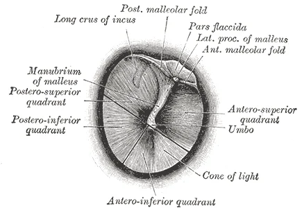
Figure 18Gray1208
Long description
The right tympanic membrane as seen through a speculum.

Figure 18: The right tympanic membrane as seen through a speculum.
| Structure | Location | Relation |
|---|---|---|
| Cisterna Chyli | Anterior to L1-L2 | Behind diaphragmatic crus |
| Thoracic Duct | Posterior Mediastinum | Ascends to left of aorta |
| Right Lymphatic Duct | Right Neck | Drains right upper quadrant |
Lymphatic anomalies in the head and neck region present distinct challenges. It usually appears as a cystic multiseptated mass with thin walls. These lesions are benign but can cause mass effect. They may compress adjacent airways or vessels. Imaging reveals multiloculated cystic structures. The content of cystic dilatation is usually homogeneous, but a fluid-fluid level may be observed related to sedimentation caused by haemorrhagic debris or by fatty content of the formation (Fig. ). Therefore, a signal intensity of lymphatic aneurysmal dilatation may be variable in the same patient at different MR examinations (Fig. ). These dilatations are unilateral in most cases, but bilateral location may also be observed (Fig. ). Lymphangiomas are classified as capillary, cavernous or cystic depending on the size of the lymphatic spaces. This classification guides management. Cystic lesions often require sclerotherapy or surgical excision. Capillary forms are more diffuse and harder to treat. Early recognition prevents complications such as infection or bleeding. [4]
It contains three major structures: the common carotid artery, the internal jugular vein, and the vagus nerve. These relations are crucial for surgical access and understanding potential complications during procedures such as central line insertion or neck dissection.
Lateral to the carotid sheath lies the sternocleidomastoid muscle. This large muscle originates from the manubrium of the sternum and the medial third of the clavicle. It inserts onto the mastoid process of the temporal bone and the lateral half of the superior nuchal line. Its primary actions include ipsilateral lateral flexion and contralateral rotation of the head. The muscle is supplied by the spinal accessory nerve (cranial nerve XI). Understanding its course is essential for identifying the cervical plexus branches that emerge from its posterior border.
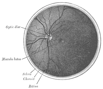
Figure 19Gray1206
Long description
The interior of the posterior half of the left eyeball.

Figure 19: The interior of the posterior half of the left eyeball.
Deep to the sternocleidomastoid and medial to the carotid sheath are the prevertebral muscles and the cervical spine. The longus colli and longus capitis muscles lie directly anterior to the vertebral bodies. These muscles are responsible for flexion of the neck. They are innervated by the anterior rami of the cervical spinal nerves. The prevertebral fascia encloses these muscles, forming a barrier that separates the visceral compartment from the vertebral column.
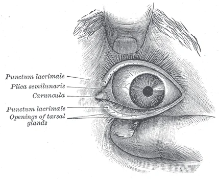
Figure 20Gray1205
Long description
Front of left eye with eyelids separated to show medial canthus.

Figure 20: Front of left eye with eyelids separated to show medial canthus.
The visceral compartment contains the trachea, oesophagus, and thyroid gland. The trachea lies anterior to the oesophagus and is midline. The thyroid gland straddles the trachea, with its lobes connected by the isthmus. The recurrent laryngeal nerves run in the tracheoesophageal groove, making them vulnerable during thyroid surgery.
| Muscle | Origin | Insertion | Action | Nerve Supply |
|---|---|---|---|---|
| Sternocleidomastoid | Manubrium, Clavicle | Mastoid process | Head rotation, flexion | Spinal Accessory (XI) |
| Longus Colli | Cervical Vertebrae | Cervical Vertebrae | Neck flexion | Anterior Rami C3-C8 |
| Trapezius | Occipital Bone, Ligamentum Nuchae | Clavicle, Scapula | Shoulder elevation | Spinal Accessory (XI) |
It inserts into the inferior border of the mandible and the skin of the lower face. Its action is to tense the skin of the neck and depress the mandible. It is innervated by the cervical branch of the facial nerve. This muscle is often used in reconstructive surgery due to its reliable blood supply and pliability.
Clinical pearlSurgical Landmark
The posterior border of the sternocleidomastoid is a key landmark for identifying the cervical plexus cutaneous branches, which emerge at the midpoint of this border.
DefinitionCarotid Sheath
A vertical fascial compartment in the neck containing the common carotid artery, internal jugular vein, and vagus nerve.
The central structure is the airway, appearing as a dark, air-filled lumen. Surrounding this are the soft tissue densities of the tongue base and pharyngeal walls. The parotid glands lie anterior and lateral to the mandibular rami. The sternocleidomastoid muscles form the lateral boundaries of the neck. Understanding these relationships is crucial for interpreting pathology.
DefinitionRadiological Anatomy
The study of anatomical structures as visualised by imaging modalities such as X-ray, CT, MRI and ultrasound. It relies on recognising normal appearances to identify deviations.
Skull radiographs provide a two-dimensional projection of the complex three-dimensional skull. They are useful for initial assessment of fractures but lack the detail of cross-sectional imaging. Computed tomography (CT) offers superior bone detail and is the modality of choice for trauma. Magnetic resonance imaging (MRI) provides excellent soft tissue contrast, making it ideal for evaluating the brain, cranial nerves, and soft tissue masses. Ultrasound is particularly valuable for superficial structures such as the thyroid and lymph nodes, allowing for dynamic assessment and guided biopsy.
| Modality | Primary Strength | Key Application |
|---|---|---|
| X-ray | Bone density | Initial fracture screening |
| CT | Bone detail | Trauma, sinus disease |
| MRI | Soft tissue contrast | Tumours, nerve pathology |
| Ultrasound | Superficial structures | Thyroid, lymph nodes |
Asymmetry may indicate pathology such as a mass or swelling. The airway should be patent and midline. The bony structures should be intact without discontinuity. The soft tissues should show uniform density without focal lesions. Familiarity with these normal variants and landmarks allows for accurate diagnosis and management of head and neck conditions. Always correlate imaging findings with clinical presentation for a comprehensive assessment.
Figure 21Illu01 head neck
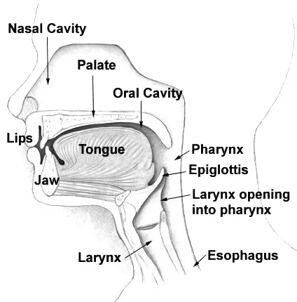
Long description
A diagram of a midsagittal section of a human head and neck.

Figure 21: A diagram of a midsagittal section of a human head and neck.
The clinical management of head and neck pathology relies on precise anatomical knowledge to guide imaging, nerve blocks, and surgical intervention. Surface landmarks are critical for locating deep structures without invasive exploration.
Imaging of neck masses must consider these anatomical compartments to determine origin and spread.
DefinitionClinical Landmarks
Surface landmarks provide a map for locating deep neurovascular structures, guiding procedures like nerve blocks and surgical incisions.
The facial nerve divides the gland into superficial and deep lobes. Knowledge of these relations is essential for managing parotid tumours or trauma. Obstruction here leads to sialolithiasis and gland swelling.
| Structure | Landmark | Clinical Relevance |
|---|---|---|
| Common Carotid Artery | Anterior border of sternocleidomastoid, C4 level | Palpation for pulse; site for block |
| Facial Nerve | Stylomastoid foramen to parotid gland | Risk in parotid surgery; Bell’s palsy |
| Hypoglossal Nerve | Deep to mylohyoid, C1-C2 | Injury causes tongue deviation |
| Submandibular Duct | Sublingual caruncle | Sialolithiasis site |
Exam trapRadiation Caries
Radiation caries are not a distinct pathological entity but rather rapid-onset caries due to hyposalivation, affecting atypical tooth surfaces.
NoteNeuropathic Pain
Neuropathic pain in the head and neck can be delayed and persistent, complicating long-term survivorship care.
NoteORN Classification
Osteoradionecrosis classification considers HBO response, clinical behavior, and anatomical extent to guide management.
- The central sulcus is located by identifying a point 1.25 cm posterior to the midpoint of the nasion-inion line and drawing a line that forms a 70-degree angle opening anteriorly with the midsagittal plane.
- The suprameatal triangle serves as the surface landmark for locating the tympanic antrum, bounded by the zygomatic arch root, the posterior border of the external acoustic meatus, and the meatus's upper margin.
- The hyoid bone projects at the level of the third or fourth cervical vertebra, serving as a primary landmark for evaluating the anterior neck and suprahyoid and infrahyoid regions.
- The choanæ align horizontally with the atlas, while the palatine velum lies opposite the body of the axis.
- Palpation immediately anterior to the sternocleidomastoid muscle at the superior border of the thyroid cartilage locates the common carotid artery bifurcation and its corresponding pulse.
- The mobile portion of the nasal septum is formed by the skin and the medial crura of the greater alar cartilages, readily distinguishable between the nares during physical inspection.
A common mistake is underestimating the tongue's multifunctional role beyond taste. The tongue is essential for mastication (chewing), deglutition (swallowing), and speech. Students often focus solely on gustatory functions while neglecting these critical physiological roles. [1]
Another pitfall involves misunderstanding the relationship between radiation therapy and head and neck cancers. While prior radiation therapy is associated with an increased incidence of thyroid cancer, salivary gland tumors, squamous cell carcinomas, and sarcomas, there is a considerable time delay before these adverse effects manifest, and the overall risk remains relatively low. Students may incorrectly perceive this risk as immediate or more common than it actually is. [2]
Misinterpreting oral lesions is another frequent error. Early malignant lesions can mimic periodontitis, and expert evaluation by an oral health professional may be necessary for proper diagnosis. Students should be cautious when assessing oral lesions and not dismiss suspicious areas as simply periodontal disease. [3]
Confusion about topical antifungal medications is also common. While topical clotrimazole is available in a lozenge and cream, the assumption that its use as a topical agent completely avoids concerns of systemic exposure and drug-drug interactions is not supported by reports of such complications. Similarly, students may not be aware that topical miconazole is available in cream form and as a muco-adhesive tablet (Loramyc® EU; Oravig® USA) that does not contain sugar sweetening and is used once daily. [3]
Exam trapRadiation timing misconception
The effects of radiation therapy are cumulative with an initially slow onset, and radiation-induced thyroid dysfunction may begin soon after the end of treatment or at any time up to 2 years posttreatment. [3]
Head and neck anatomy knowledge is crucial at the bedside for examining patients and identifying abnormalities. When assessing a patient with dysphagia, understanding the anatomical structures involved in swallowing is essential. Similarly, in patients with speech difficulties, knowledge of tongue musculature and innervation helps localize potential neurological deficits.
In the operating theatre, precise anatomical knowledge is vital for safe surgical procedures. Surgeons must be familiar with the origin, course, relations, branches, and termination of structures like the facial nerve to prevent iatrogenic injury. For head and neck cancer patients, integrated pretreatment oral and dental evaluation is the best approach to achieving optimal clinical outcomes. This includes identifying teeth with periodontal attachment loss that may need extraction before radiation therapy, particularly in regions planned for high-dose radiation. [3]
Head and neck cancer pretreatment dental evaluation
- Initial dental examinationComprehensive assessment of oral health
- Question: Severe periodontal disease or high-risk teeth?
Yes
- Extract teeth with compromised periodontiumEspecially in planned high-dose RT areas
- Continue with cancer treatment
No
- Provide preventive dental careBefore, during, and after cancer therapy
- Continue with cancer treatment
On imaging, anatomical knowledge allows for accurate interpretation of scans. For example, when using 68Ga DOTATATE for imaging neuroendocrine tumors, it's important to recognize normal physiological uptake patterns in organs like the pancreas and spleen to avoid false positives. The pancreas may demonstrate heterogeneous activity related to islet cell hypertrophy, which is usually low grade and should not be mistaken for disease. [5]
In examinations, students must demonstrate knowledge of head and neck anatomy through various assessment methods. This includes identifying structures on diagrams, describing their relationships, and understanding clinical correlations. For instance, students should be able to explain how radiation therapy affects periodontal health in a dose-dependent manner and how pre-existing periodontitis may worsen with cancer treatment.
| Muscle | Origin | Insertion | Action | Nerve supply |
|---|---|---|---|---|
| Masseter | Zygomatic arch | Mandibular ramus and body | Elevates mandible | Trigeminal nerve (V3) |
| Temporalis | Temporal fossa | Coronoid process of mandible | Elevates and retracts mandible | Trigeminal nerve (V3) |
| Medial pterygoid | Medial surface of lateral pterygoid plate | Medial surface of ramus of mandible | Elevates and protrudes mandible | Trigeminal nerve (V3) |
| Lateral pterygoid | Greater wing of sphenoid and lateral pterygoid plate | Neck of condyle and disc | Protrudes and depresses mandible | Trigeminal nerve (V3) |
The tongue is essential for mastication, deglutition, and speech, not just taste. Prior radiation therapy increases risk of certain head and neck cancers, but with considerable delay and relatively low overall risk. Early malignant lesions can mimic periodontitis, requiring expert evaluation. Integrated pretreatment dental evaluation is crucial for head and neck cancer patients. Topical antifungals can have systemic effects and drug interactions. Radiation-induced thyroid dysfunction may occur up to 2 years posttreatment. Knowledge of head and neck anatomy is vital for bedside examination, surgery, imaging interpretation, and examinations.
Answer each one in your head first, then open the answer.
What are the four primary functions of the tongue?
Show answerHide answer
Mastication, deglutition, speech, and tasteWhat is the typical time delay before radiation therapy may lead to thyroid dysfunction?
Show answerHide answer
Up to 2 years posttreatmentWhich oral condition can mimic early malignant lesions?
Show answerHide answer
PeriodontitisWhat is the recommended approach for dental evaluation in head and neck cancer patients?
Show answerHide answer
Integrated pretreatment oral and dental evaluationWhat is a common false positive finding on 68Ga DOTATATE imaging?
Show answerHide answer
Heterogeneous activity in the pancreas related to islet cell hypertrophy
- Betts JG, Young KA, Wise JA, et al. Axial Muscles of the Head, Neck, and Back. OpenStax, Anatomy and Physiology 2e (CC BY-NC-SA 4.0). Accessed .
- Barsouk A, Aluru JS, Rawla P, et al. Epidemiology, Risk Factors, and Prevention of Head and Neck Squamous Cell Carcinoma. Medical sciences (Basel, Switzerland) 2023;11(2):42. doi:10.3390/medsci11020042. Accessed .
- Sroussi HY, Epstein JB, Bensadoun RJ, et al. Common oral complications of head and neck cancer radiation therapy: mucositis, infections, saliva change, fibrosis, sensory dysfunctions, dental caries, periodontal disease, and osteoradionecrosis. Cancer medicine 2017;6(12):2918-2931. doi:10.1002/cam4.1221. Accessed .
- Derhy S, El Mouhadi S, Ruiz A, et al. Non-contrast 3D MR lymphography of retroperitoneal lymphatic aneurysmal dilatation: a continuous spectrum of change from normal variants to cystic lymphangioma. Insights into imaging 2013;4(6):753-758. doi:10.1007/s13244-013-0290-4. Accessed .
- Malan N, Vangu MD. Normal Variants, Pitfalls and Artifacts in Ga-68 DOTATATE PET/CT Imaging. Frontiers in nuclear medicine 2022;2:825486. doi:10.3389/fnume.2022.825486. Accessed .
Figure credits
- Figure 1: Henry Vandyke Carter, via Wikimedia Commons, Public domain, Public domain.
- Figure 2: Henry Vandyke Carter, via Wikimedia Commons, Public domain, Public domain.
- Figure 3: Henry Vandyke Carter, via Wikimedia Commons, Public domain, Public domain.
- Figure 4: Henry Vandyke Carter, via Wikimedia Commons, Public domain, Public domain.
- Figure 5: Henry Vandyke Carter, via Wikimedia Commons, Public domain, Public domain.
- Figure 6: Henry Vandyke Carter, via Wikimedia Commons, Public domain, Public domain.
- Figure 7: Henry Vandyke Carter, via Wikimedia Commons, Public domain, Public domain.
- Figure 8: Henry Vandyke Carter, via Wikimedia Commons, Public domain, Public domain.
- Figure 9: OpenStax, via Wikimedia Commons, CC BY 4.0, CC BY 4.0. resized.
- Figure 10: OpenStax, via Wikimedia Commons, CC BY 4.0, CC BY 4.0. resized.
- Figure 11: OpenStax, via Wikimedia Commons, CC BY 4.0, CC BY 4.0. resized.
- Figure 12: Henry Vandyke Carter, via Wikimedia Commons, Public domain, Public domain.
- Figure 13: Henry Vandyke Carter, via Wikimedia Commons, Public domain, Public domain.
- Figure 14: Henry Vandyke Carter, via Wikimedia Commons, Public domain, Public domain.
- Figure 15: Henry Vandyke Carter, via Wikimedia Commons, Public domain, Public domain.
- Figure 16: Henry Vandyke Carter; Henry Gray, via Wikimedia Commons, Public domain, Public domain. resized.
- Figure 17: Henry Vandyke Carter, via Wikimedia Commons, Public domain, Public domain.
- Figure 18: Henry Vandyke Carter, via Wikimedia Commons, Public domain, Public domain.
- Figure 19: Henry Vandyke Carter, via Wikimedia Commons, Public domain, Public domain.
- Figure 20: Henry Vandyke Carter, via Wikimedia Commons, Public domain, Public domain.
- Figure 21: Arcadian, via Wikimedia Commons, Public domain, Public domain.
Educational content for medical students and doctors. It is not a substitute for clinical judgement or local guidelines.