Ear anatomy
Anatomical structures of the ear involved in sound transduction and transmission.
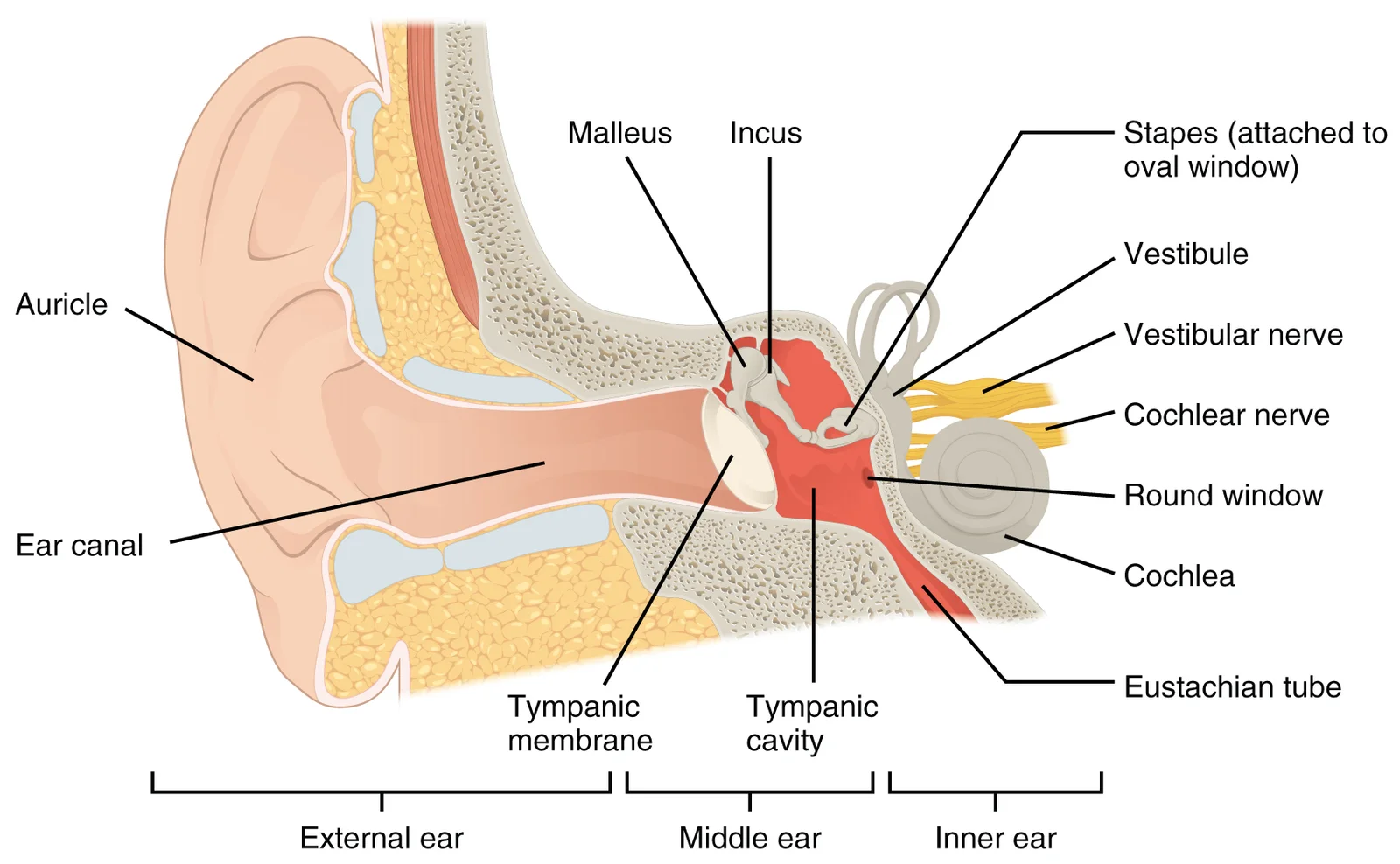
The external ear comprises the auricle, also known as the pinna, and the external auditory canal. The auricle is a large, fleshy structure on the lateral aspect of the head, featuring C-shaped curves that direct sound waves toward the auditory canal. The canal enters the skull through the external auditory meatus of the temporal bone. At the distal end lies the tympanic membrane, or ear drum, which vibrates in response to sound waves. These components constitute the external ear. [1]
The middle ear is a space spanned by three ossicles: the malleus, incus, and stapes. The malleus attaches to the tympanic membrane and articulates with the incus. The incus articulates with the stapes, which attaches to the inner ear. This cavity connects to the pharynx via the Eustachian tube, which equilibrates air pressure across the tympanic membrane. The tube remains normally closed but opens during swallowing or yawning. [1]
Figure 1Gray911
Long description
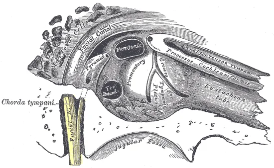
View of the inner wall of the tympanum (enlarged.)

Figure 1: View of the inner wall of the tympanum (enlarged.).
DefinitionOssicles
The malleus, incus, and stapes are Latin names translating to hammer, anvil, and stirrup respectively.
Frequencies in the higher ranges of 20 KHz are encoded by hair cells at the base of the cochlea, close to the round and oval windows. [1]
Otitis media involves inflammation of the middle ear, often due to Eustachian tube dysfunction. Cholesteatoma is a destructive growth of keratinizing squamous epithelium in the middle ear. The mastoid air cells are pneumatized spaces within the mastoid process of the temporal bone, communicating with the middle ear cavity. Infection can spread from the middle ear to the mastoid air cells, causing mastoiditis.
| Muscle | Origin | Insertion | Action | Nerve Supply |
|---|---|---|---|---|
| Tensor Tympani | Cartilaginous part of auditory tube | Neck of malleus | Tenses tympanic membrane | Mandibular nerve |
| Stapedius | Posterior wall of tympanic cavity | Neck of stapes | Dampens stapes vibration | Facial nerve |
Figure 21404 The Structures of the Ear
Long description
The external ear contains the auricle, ear canal, and tympanic membrane. The middle ear contains the ossicles and is connected to the pharynx by the Eustachian tube. The inner ear contains the cochlea and vestibule, which are responsible for audition and equilibrium, respectively.

Figure 2: The external ear contains the auricle, ear canal, and tympanic membrane.
The tympanic membrane, or eardrum, separates the external ear from the middle ear cavity. It is a thin, semi-transparent membrane that vibrates in response to sound pressure changes. [2]
DefinitionExternal Acoustic Meatus
The canal leading from the auricle to the tympanic membrane, conducting sound waves to the middle ear.
The middle ear contains the ossicles: the malleus, incus, and stapes. These bones transmit vibrations from the tympanic membrane to the oval window of the inner ear. The inner ear consists of the bony labyrinth, embedded within the petrous part of the temporal bone. This labyrinth contains the cochlea for hearing and the vestibular apparatus for balance. The cochlea is a spiral-shaped structure responsible for transducing sound waves into neural signals. The vestibular apparatus includes the utr [2]
Figure 31405 Sound Waves and the Ear
Long description
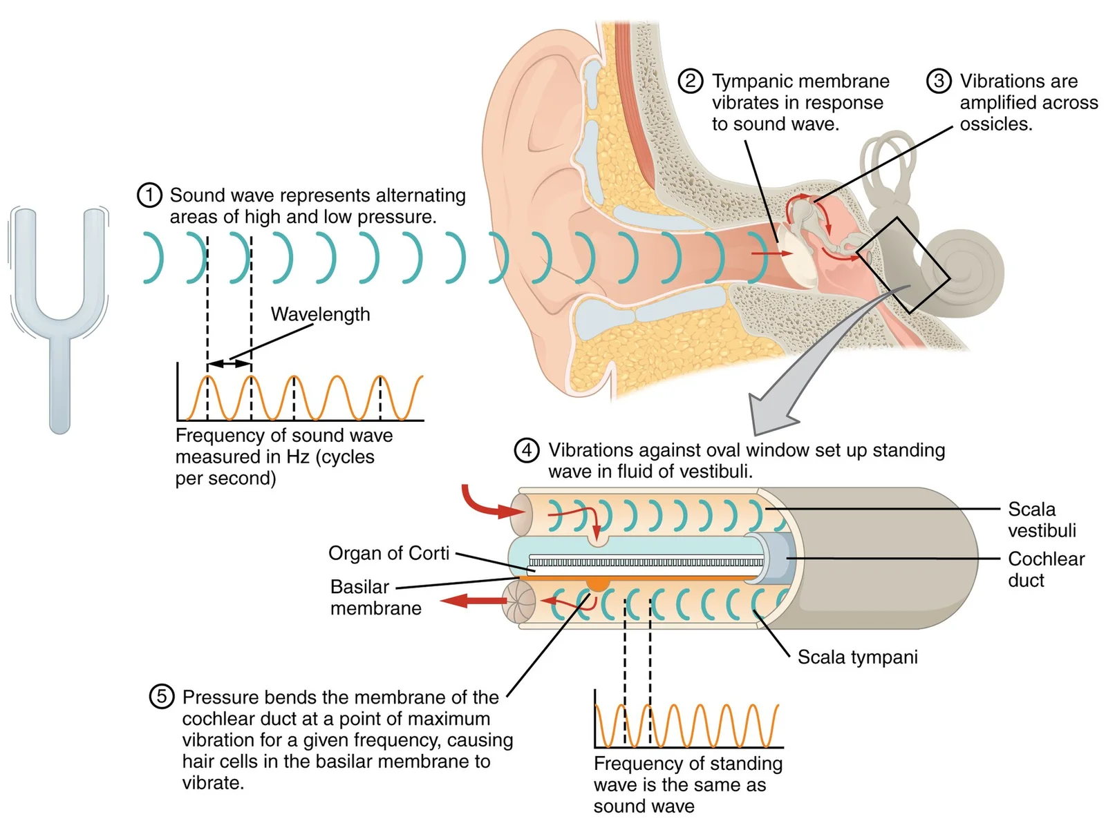
A sound wave causes the tympanic membrane to vibrate. This vibration is amplified as it moves across the malleus, incus, and stapes. The amplified vibration is picked up by the oval window causing pressure waves in the fluid of the scala vestibuli and scala tympani. The complexity of the pressure wa

Figure 3: A sound wave causes the tympanic membrane to vibrate.
Figure 4Gray922
Long description
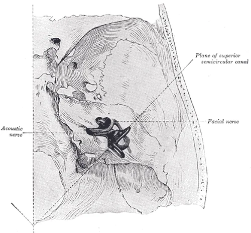
Position of the right bony labyrinth of the ear in the skull, viewed from above. The temporal bone is considered transparent and the labyrinth drawn in from a corrosion preparation. (Spalteholz.)

Figure 4: Position of the right bony labyrinth of the ear in the skull, viewed from above.
Figure 5Gray918
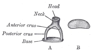
Long description
A. Left stapes. B. Base of stapes, medial surface.

Figure 5: A (left stapes).
Figure 6Gray917
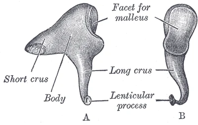
Long description
Left incus. A. From within. B. From the front.

Figure 6: Left incus (a).
Figure 7Gray916
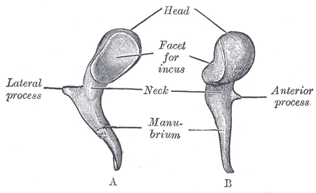
Long description
Left malleus. A. From behind. B. From within.

Figure 7: Left malleus (a).
| Structure | Function |
|---|---|
| Cochlea | Transduces sound waves into neural signals |
| Utricle and Saccule | Sense head position |
| Semicircular Canals | Sense head movement |
| Vestibulocochlear Nerve | Transmits equilibrium and auditory sensations to the medulla |
The vestibulocochlear nerve (CN VIII) carries both equilibrium and auditory sensations from the inner ear to the medulla. The sensory nerves from the cochlea and vestibule travel side-by-side as this nerve, emerging from the inner ear and passing through the internal auditory meatus. The facial nerve also passes through the temporal bone, running close to the middle and inner ear structures. Inflammation or infection, such as otitis media, can affect these structures, potentially leading to hearing deficits or balance issues. Ménière's disease, for example, results from fluid accumulation in the inner ear, causing vertigo and hearing loss. Understanding the anatomical relationships between these structures is crucial for diagnosing and treating ear pathologies. [2] [1]
The ear is a complex sensory organ situated within the temporal bone, serving as the primary apparatus for audition and equilibrium. Its anatomical position is defined by its intimate relationships with surrounding structures, which are critical for both function and surgical access. The ear is broadly divided into the external, middle, and internal components, each with distinct relations to adjacent tissues.
The external ear, comprising the auricle and the external acoustic meatus, is often referred to as the external ear.
It is medial to the middle ear and is separated from it by the medial wall of the tympanic cavity. The internal ear consists of the bony labyrinth, which contains the membranous labyrinth. The bony labyrinth is filled with perilymph, while the membranous labyrinth contains endolymph. The vestibulocochlear nerve is responsible for transmitting auditory and equilibrium signals.
Figure 8Gray900
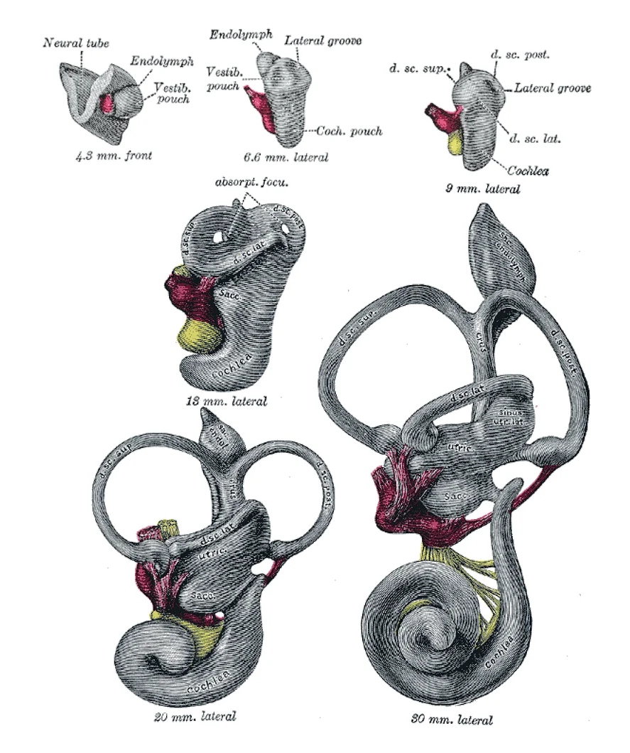
Long description
Lateral views of membranous labyrinth and acoustic complex. X 25 dia. (Streeter.) absorpt. focu, area of wall where absorption is complete; amp., ampulla membranacea; crus, crus commune; d. sc. lat., ductus semicircularis lateralis; d. sc. post., ductus semicircularis posterior; d. sc. sup., ductus

Figure 8: Lateral views of membranous labyrinth and acoustic complex (x 25 dia).
Figure 9Gray925
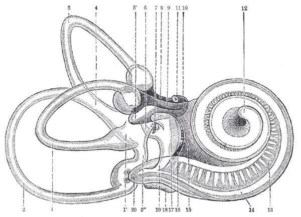
Long description
Right human membranous labyrinth, removed from its bony enclosure and viewed from the antero-lateral aspect. (G. Retzius.)

Figure 9: Right human membranous labyrinth, removed from its bony enclosure and viewed from the antero-lateral aspect (G.
Figure 10Gray914
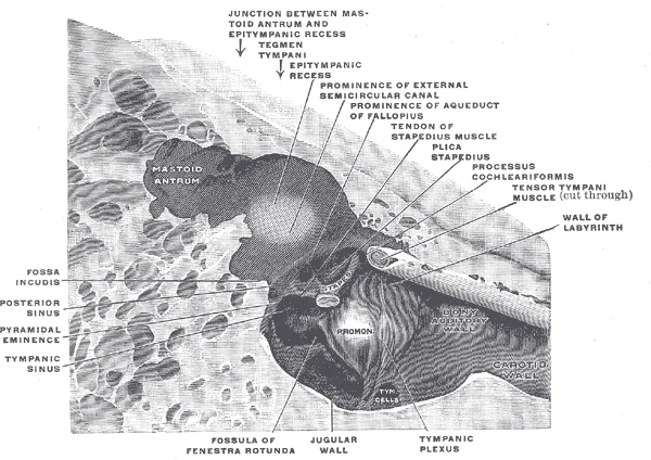
Long description
The medial wall and part of the posterior and anterior walls of the right tympanic cavity, lateral view. (Spalteholz.)

Figure 10: The medial wall and part of the posterior and anterior walls of the right tympanic cavity, lateral view (Spalteholz.).
Figure 11Gray912
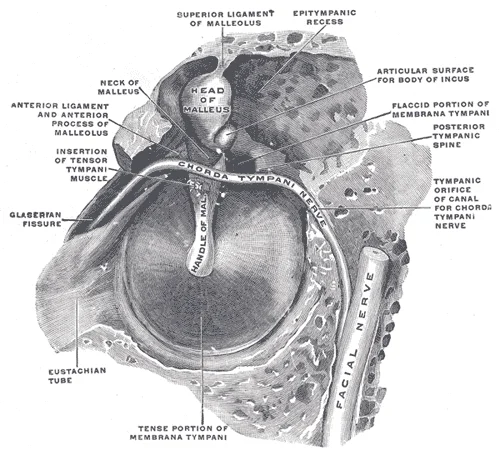
Long description
The right membrana tympani with the hammer and the chorda tympani, viewed from within, from behind, and from above. (Spalteholz.)

Figure 11: The right membrana tympani with the hammer and the chorda tympani, viewed from within, from behind, and from above (Spalteholz.).
Figure 12Gray909
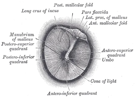
Long description
Right tympanic membrane as seen through a speculum.

Figure 12: Right tympanic membrane as seen through a speculum.
The relations of the ear are also significant for clinical examination. Understanding these relations is essential for localizing lesions and understanding the spread of infection or disease. For instance, infection of the middle ear can spread to the mastoid air cells, leading to mastoiditis. Infection can also spread to the meninges via the tegmen tympani, causing meningitis. Similarly, disease of the internal ear can affect the vestibulocochlear nerve, leading to hearing loss or vertigo. [1]
DefinitionAnatomical Planes
The ear is viewed in coronal, axial, and sagittal planes to understand its three-dimensional relationships with the skull base and brain.
The external ear is related to the parotid gland anteriorly and the sternocleidomastoid muscle posteriorly. These relationships are crucial for surgical approaches to the ear, such as mastoidectomy or cochlear implantation. Surgeons must be aware of the proximity of these vital structures to avoid iatrogenic injury.
Figure 13Gray906
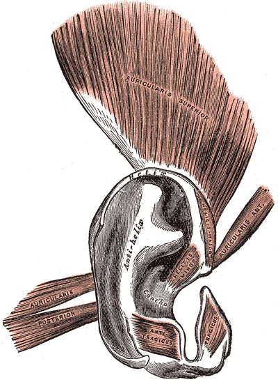
Long description
The muscles of the auricula. Labelled illustration from Wikimedia Commons, used under its open licence.

Figure 13: The muscles of the auricula.
| Component | Lateral Relation | Medial Relation | Superior Relation | Inferior Relation |
|---|---|---|---|---|
| Auricle | Skin | Temporalis fascia | Temporal bone | Parotid gland |
| External Acoustic Meatus | Auricle | Tympanic membrane | Temporal bone | Parotid gland |
| Middle Ear | Tympanic membrane | Lateral wall of internal ear | Tegmen tympani | Internal jugular vein |
| Internal Ear | Lateral wall of middle ear | Brainstem | Temporal bone | Internal jugular vein |
The vestibulo-ocular reflex involves the cranial nerves for gaze control, linking the internal ear to the brainstem. The cochlea, part of the internal ear, is responsible for hearing. It converts sound waves into neural signals that are transmitted to the brain via the cochlear nerve. The anatomical integrity of these structures is essential for normal hearing and balance.
NoteClinical Relevance
Damage to the structures in the internal ear can lead to sensorineural hearing loss or vestibular dysfunction, affecting both hearing and balance.
The ear's relations also extend to the lymphatic system. Lymph from the auricle drains into the preauricular and superficial parotid nodes. Lymph from the external acoustic meatus drains into the deep cervical nodes. Understanding these lymphatic relations is important for diagnosing and managing infections and malignancies of the ear. [1]
In summary, the ear's anatomy is defined by its complex relationships with surrounding structures. These relationships are critical for understanding the function of the ear and for diagnosing and treating ear diseases. The external, middle, and internal ears each have distinct relations that must be considered in clinical practice.
The ear is a complex sensory organ responsible for hearing and balance, composed of three distinct regions: the external, middle, and internal ear. In this view, the external ear is shown laterally, leading into the auditory canal which terminates at the tympanic membrane. This membrane separates the external environment from the air-filled middle ear cavity. [1]
DefinitionAuditory Canal
A tube that conducts sound waves from the pinna to the tympanic membrane.
The middle ear contains the ossicular chain, a series of three small bones that transmit vibrations from the tympanic membrane to the inner ear. These bones are the malleus, incus, and stapes. This arrangement acts as an impedance matcher, amplifying sound pressure to overcome the resistance of the fluid in the inner ear.
| Bone | Attachment Point | Action |
|---|---|---|
| Malleus | Tympanic membrane | Transmits vibration |
| Incus | Malleus and Stapes | Transfers vibration |
| Stapes | Oval window | Pushes into inner ear |
The internal ear, or labyrinth, is embedded within the temporal bone and consists of bony and membranous structures. The bony labyrinth contains perilymph, while the membranous labyrinth contains endolymph. The cochlea is the spiral-shaped portion responsible for hearing. It is divided into three fluid-filled chambers: the scala vestibuli, scala tympani, and scala media. These signals are then relayed via the vestibulocochlear nerve to the central nervous system.
Figure 14Gray929
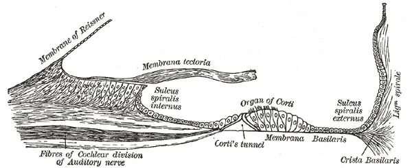
Long description
Floor of ductus cochlearis. Labelled illustration from Wikimedia Commons, used under its open licence.

Figure 14: Floor of ductus cochlearis.
Figure 15Gray928
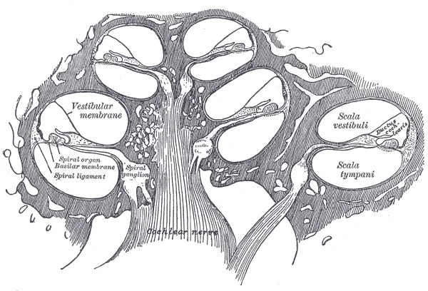
Long description
Diagrammatic longitudinal section of the cochlea.

Figure 15: Diagrammatic longitudinal section of the cochlea.
Figure 16Gray926
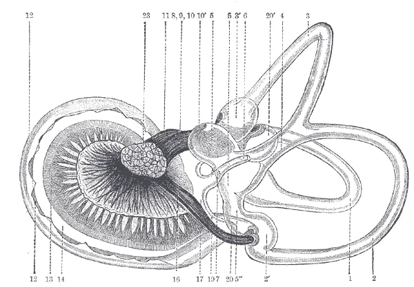
Long description
The same from the postero-medial aspect. 1. Lateral semicircular canal; 1’, its ampulla; 2. Posterior canal; 2’, its ampulla. 3. Superior canal; 3’, its ampulla. 4. Conjoined limb of superior and posterior canals ( sinus utriculi superior ). 5. Utricle. 5’. Recessus utriculi. 5”. Sinus utriculi post

Figure 16: The same from the postero-medial aspect (1).
Figure 17Gray924
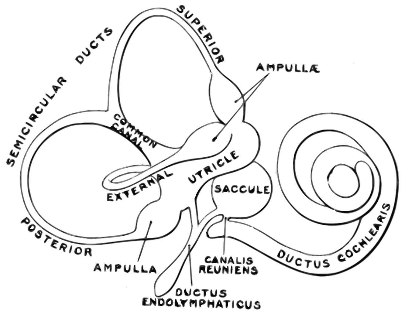
Long description
The membranous labyrinth. (Enlarged.). Labelled illustration from Wikimedia Commons, used under its open licence.

Figure 17: The membranous labyrinth (Enlarged.).
Figure 18Gray923
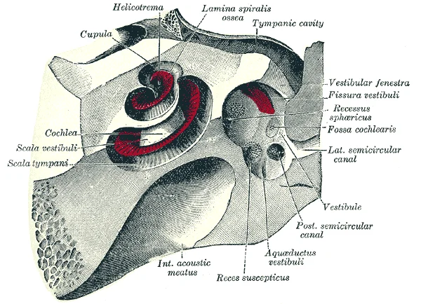
Long description
The cochlea and vestibule, viewed from above. All the hard parts which form the roof of the internal ear have been removed with the saw.

Figure 18: The cochlea and vestibule, viewed from above.
Figure 19Gray921
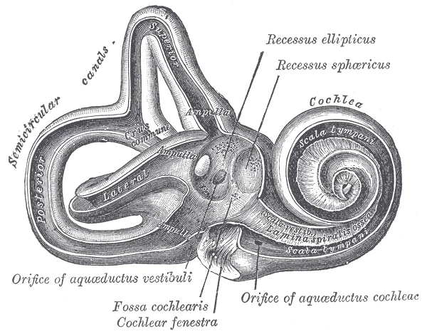
Long description
Interior of right osseous labyrinth. Labelled illustration from Wikimedia Commons, used under its open licence.

Figure 19: Interior of right osseous labyrinth.
Figure 20Gray920
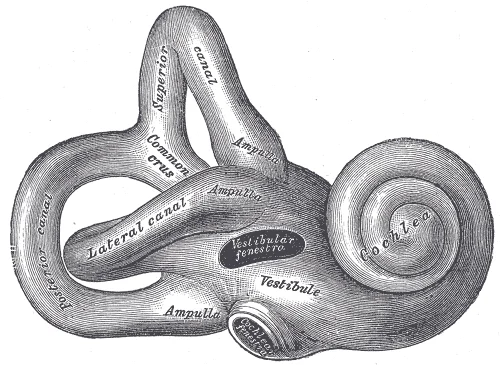
Long description
Right osseous labyrinth. Lateral view. Labelled illustration from Wikimedia Commons, used under its open licence.

Figure 20: Right osseous labyrinth (lateral view).
Figure 21Gray913
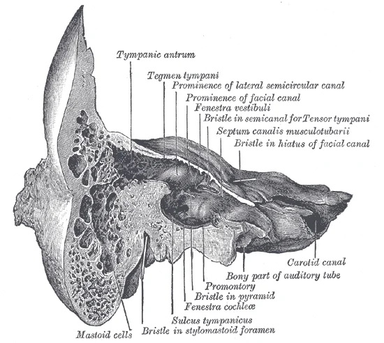
Long description
Coronal section of right temporal bone. Labelled illustration from Wikimedia Commons, used under its open licence.

Figure 21: Coronal section of right temporal bone.
Clinical pearlImpedance Matching
The ossicles amplify sound pressure to efficiently transfer energy from air to fluid.
Balance is mediated by the vestibular system, which includes the semicircular canals and the otolith organs. These sensory inputs are integrated in the brainstem to maintain posture and eye position. [1]
Figure 22Gray927
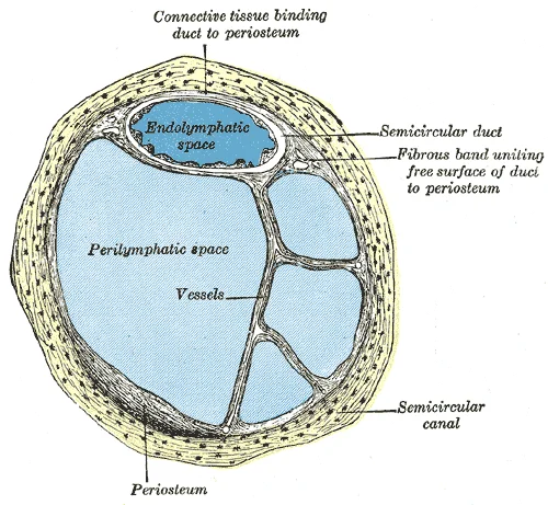
Long description
Transverse section of a human semicircular canal and duct (after Rüdinger).

Figure 22: Transverse section of a human semicircular canal and duct (after Rüdinger).
NoteFluid Compartments
Perilymph surrounds the membranous labyrinth; endolymph is inside it.
The vascular supply to the ear is critical for maintaining the electrochemical gradients necessary for sensory transduction. Any disruption to this blood flow can lead to sensorineural hearing loss or vestibular dysfunction. Understanding the precise anatomical layout is essential for diagnosing pathologies such as otitis media or labyrinthitis. [1]
The external ear receives its blood supply from a rich anastomotic network derived primarily from branches of the external carotid artery. This vascular arrangement ensures robust perfusion to the cartilaginous framework and overlying skin, which is critical given the ear's exposure to environmental stressors. The primary contributors are the posterior auricular artery and the superficial temporal artery, with additional support from the occipital artery. Understanding this supply is vital for surgical planning and understanding the spread of infection or malignancy. [1]
Figure 23Gray905
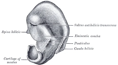
Long description
Cranial surface of cartilage of right auricula.

Figure 23: Cranial surface of cartilage of right auricula.
This contribution is often variable but can be significant in cases where other sources are diminished. [1]
| Artery | Origin | Supply Area |
|---|---|---|
| Posterior auricular | External carotid | Posterior auricle, scalp behind ear |
| Superficial temporal | External carotid | Anterior auricle, scalp anterior to ear |
| Occipital | External carotid | Posterior scalp, posterior auricle |
| Facial | External carotid | Minor contribution to anterior auricle |
The external ear, comprising the auricle and the external acoustic meatus, is often referred to as the external ear.
The venous drainage of the ear is a complex network that mirrors the arterial supply, ensuring efficient removal of deoxygenated blood from both external and internal structures. Understanding this system is crucial for appreciating how infections or thromboses may spread to intracranial compartments. The drainage pathways are divided into superficial and deep systems, which communicate extensively but have distinct terminations.
The external ear, comprising the auricle and the external acoustic meatus, is often referred to as the external ear.
The middle ear consists of a space spanned by three small bones called the ossicles. [1]
The external ear, comprising the auricle and the external acoustic meatus, is often referred to as the external ear.
Clinical pearlClinical Pearl
Infections in the middle ear can spread to the cavernous sinus via the emissary veins, leading to cavernous sinus thrombosis.
| Structure | Primary Drainage | Termination |
|---|---|---|
| Auricle | Superficial temporal vein | External jugular vein |
| External acoustic meatus | Posterior auricular vein | External jugular vein |
| Middle ear | Pterygoid plexus | Internal jugular vein |
| Figure 5 (F-B1-2406-5) in 'Venous drainage': venous drainage of ear anatomy. This figure illustrates the venous drainage of the ear, showing the superficial temporal vein, posterior auricular vein, and their connections to the external jugular vein. It also depicts the pterygoid plexus and its communication with the emissary veins and the dural venous sinuses. The view is a lateral dissection of the head and neck, with the orientation showing the right side of the patient. The labelled structure [1] | ||
| Inner ear | Labyrinthine vein | Inferior petrosal sinus |
The lymphatic drainage of the ear is divided into distinct territories based on anatomical boundaries, primarily the external auditory meatus and the tympanic membrane. Understanding these pathways is critical for predicting the spread of infection or malignancy.
The pinna and the external auditory canal drain anteriorly and inferiorly. Lymph from the upper and anterior parts of the pinna flows into the preauricular nodes, also known as the parotid nodes. These nodes are situated anterior to the ear, superficial to the parotid gland. From here, efferent vessels pass to the deep cervical lymph nodes.
Lymph from the lower and posterior parts of the pinna drains into the postauricular nodes, located behind the ear on the mastoid process. These nodes also receive drainage from the scalp posterior to the pinna. Efferents from the postauricular nodes join those from the preauricular nodes to drain into the upper deep cervical nodes.
The external auditory meatus has a dual drainage pattern. The lateral one-third, which is cartilaginous, drains into the preauricular and parotid nodes. The medial two-thirds, which is bony, drains posteriorly into the postauricular nodes. This distinction is vital when assessing swelling or infection in the ear canal.
The middle ear cavity drains primarily into the retropharyngeal nodes. These nodes are located in the space between the pharynx and the vertebral column. Lymph from the middle ear passes through the jugular foramen or via small channels to reach these deep cervical structures. Consequently, pathology in the middle ear, such as otitis media, may present with deep neck swelling or pain, although this is less common than local symptoms. [1]
When examining a patient with ear pathology, palpation of the preauricular and postauricular regions is essential. Enlargement of the preauricular nodes suggests pathology in the anterior pinna or lateral ear canal. Enlargement of the postauricular nodes indicates involvement of the posterior pinna or mastoid air cells.
Clinical pearlKey Drainage Pathway
Remember: anterior pinna drains to preauricular nodes; posterior pinna drains to postauricular nodes; middle ear drains to retropharyngeal nodes.
| Node Group | Location | Drainage Area |
|---|---|---|
| Preauricular | Anterior to ear, superficial to parotid | Upper/anterior pinna, lateral ear canal |
| Postauricular | Posterior to ear, on mastoid | Lower/posterior pinna, scalp posterior to ear |
| Retropharyngeal | Between pharynx and vertebrae | Middle ear cavity |
The ear receives its sensory innervation from a complex network of somatic and autonomic fibres. Somatic sensation is primarily conveyed by the trigeminal nerve (CN V) via its auriculotemporal branch for the anterior external auditory meatus and the facial nerve (CN VII) for the posterior aspect. The glossopharyngeal nerve (CN IX) supplies the middle ear mucosa via the tympanic branch. Autonomic parasympathetic fibres, originating from the facial and glossopharyngeal nerves, regulate glandular secretion and vascular tone. Referred pain is common; pathology in the middle ear may present as pain in the distribution of the trigeminal or glossopharyngeal nerves due to shared central pathways.
Figure 24Gray907
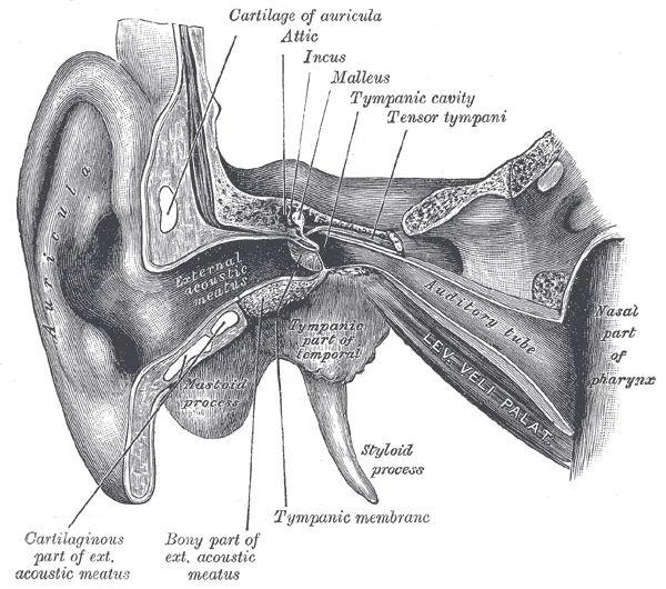
Long description
External and middle ear, opened from the front. Right side.

Figure 24: External and middle ear, opened from the front (right side).
Figure 25Gray904
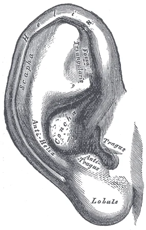
Long description
The auricula. Lateral surface. Labelled illustration from Wikimedia Commons, used under its open licence.

Figure 25: The auricula (lateral surface).
DefinitionReferred Pain
Pain perceived in a location other than the site of the painful stimulus, often due to convergence of sensory pathways in the spinal cord or brainstem.
The somatic innervation of the external ear is critical for localising pathology. The trigeminal nerve provides general sensation to the anterior part of the pinna and the external auditory canal. The vagus nerve (CN X) also contributes via its auricular branch, supplying a small area of the external auditory meatus. This overlapping innervation means that lesions may present with atypical pain patterns.
Figure 26Gray933
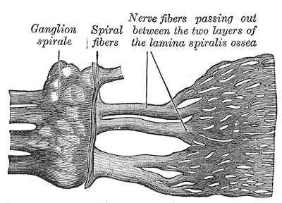
Long description
Part of the cochlear division of the acoustic nerve, highly magnified. (Henle.)

Figure 26: Part of the cochlear division of the acoustic nerve, highly magnified (Henle.).
| Structure | Nerve Supply | Source |
|---|---|---|
| Anterior Pinna | Auriculotemporal Nerve | Trigeminal (CN V3) |
| Posterior Pinna | Auricular Branch | Facial (CN VII) |
| External Auditory Meatus | Auriculotemporal & Vagus | Trigeminal (CN V3) & Vagus (CN X) |
| Tympanic Membrane | Tympanic Branch | Glossopharyngeal (CN IX) |
| Middle Ear Mucosa | Tympanic Branch | Glossopharyngeal (CN IX) |
Autonomic innervation is provided by the parasympathetic fibres of the facial and glossopharyngeal nerves. The facial nerve’s greater petrosal nerve contributes to lacrimation and salivation, while the glossopharyngeal nerve’s tympanic branch forms the lesser petrosal nerve, which synapses in the otic ganglion to supply the parotid gland. These fibres regulate blood flow and glandular activity in the ear region. Understanding this supply is essential for interpreting clinical signs such as Ramsay Hunt syndrome, where herpes zoster affects the geniculate ganglion of the facial nerve, causing pain, vesicles, and facial paralysis.
The inner ear contains a complex system of fluid-filled chambers responsible for transducing mechanical vibrations into neural signals. Central to this process is the cochlea, a spiral-shaped structure housing the sensory organs for hearing. The internal architecture of the cochlea is divided into three distinct compartments, each playing a specific role in sound transmission.
The scala vestibuli is the uppermost fluid-filled tube within the cochlea. It begins at the oval window, where vibrations from the stapes are transmitted into the inner ear. From this origin, the scala vestibuli travels above the cochlear duct. The cochlear duct itself is the central cavity of the cochlea and contains the sound-transducing neurons known as the organ of Corti. At the uppermost tip of the cochlea, the scala vestibuli curves over the top of the cochlear duct.
At this apex, the scala vestibuli transitions into the scala tympani. The scala tympani is the lower fluid-filled tube that returns to the base of the cochlea. It travels under the cochlear duct, running parallel to the scala vestibuli but separated by the cochlear duct and the basilar membrane. The scala tympani ends at the round window. This structure is covered by a membrane that contains the fluid within the scala, allowing for the displacement of fluid as pressure changes occur.
When vibrations of the ossicles travel through the oval window, they create pressure waves in the perilymph of the scala vestibuli. These waves travel through the fluid-filled tubes in a wave-like motion. The movement of the fluid causes the basilar membrane within the cochlear duct to vibrate. This vibration stimulates the hair cells within the organ of Corti, leading to the generation of action potentials in the auditory nerve. The frequency of the fluid waves matches the frequencies of the sound waves, allowing for pitch discrimination.
Figure 27Gray932
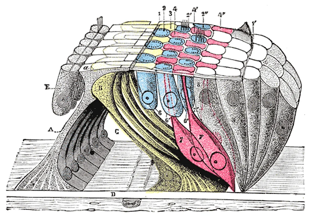
Long description
The lamina reticularis and subjacent structures. (Schematic.) A. Internal rod of Corti, with a, its plate. B. External rod (in yellow). C. Tunnel of Corti. D. Membrana basilaris. E. Inner hair cells. 1, 1’. Internal and external borders of the membrana reticularis. 2, 2’, 2”. The three rows of circu

Figure 27: The lamina reticularis and subjacent structures (Schematic.) A.
Figure 28Gray931
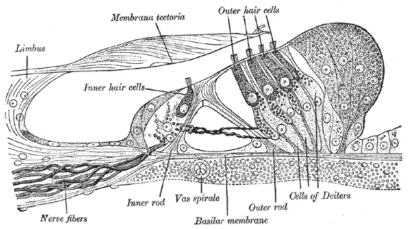
Long description
Section through the spiral organ of Corti. Magnified. (G. Retzius.)

Figure 28: Section through the spiral organ of Corti (magnified).
Figure 29Gray930
Long description
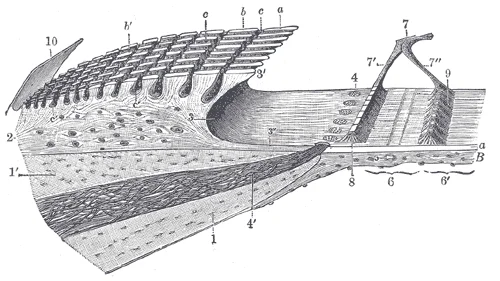
Limbus laminæ spiralis and membrana basilaris. (Schematic.) 1, 1’. Upper and lower lamellæ of the lamina spiralis ossea. 2. Limbus laminæ spiralis, with a, the teeth of the first row; b, b’, the auditory teeth of the other rows; c, c’, the interdental grooves and the cells which are lodged in them.

Figure 29: Limbus laminæ spiralis and membrana basilaris (Schematic.) 1, 1’.
DefinitionCochlear Duct
The central cavity of the cochlea that contains the sound-transducing neurons.
Clinical pearlFluid Dynamics
The frequency of the fluid waves matches the frequencies of the sound waves.
Link to the histology page.
The ear develops from multiple embryological sources, primarily the first and second pharyngeal arches and the surface ectoderm. Understanding this origin clarifies the adult anatomy. The external ear arises from six auricular hillocks, which are swellings of mesenchyme around the first pharyngeal cleft. These hillocks fuse to form the pinna. The external auditory meatus forms from the first pharyngeal cleft, which deepens to become the canal. The tympanic membrane separates the external meatus from the middle ear cavity.
Figure 30Gray899
Long description
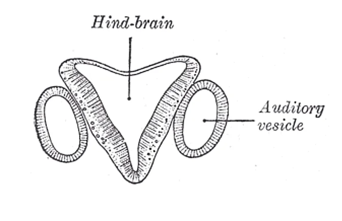
Section through hind-brain and auditory vesicles of an embryo more advanced than that of Fig. 898. (After His.)

Figure 30: Section through hind-brain and auditory vesicles of an embryo more advanced than that of Fig (898).
Figure 31Gray898
Long description
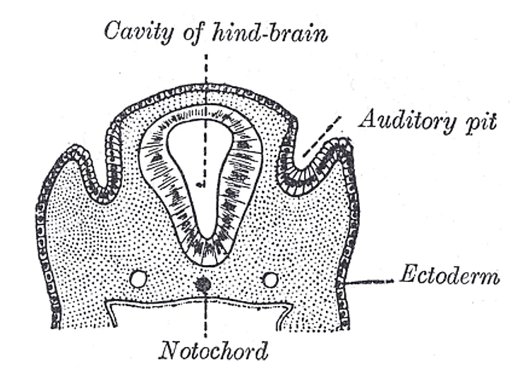
Section through the head of a human embryo, about twelve days old, in the region of the hind-brain. (Kollmann.)

Figure 31: Section through the head of a human embryo, about twelve days old, in the region of the hind-brain (Kollmann.).
DefinitionFirst Pharyngeal Cleft
The ectodermal groove that gives rise to the external auditory meatus and the lateral aspect of the tympanic membrane.
The middle ear cavity, or tympanic cavity, develops from the first pharyngeal pouch. This endodermal outpouching expands to form the tympanic cavity and the auditory tube. The ossicles within the middle ear have distinct origins. The malleus and incus derive from the first pharyngeal arch, specifically the cartilage of Meckel. The stapes originates from the second pharyngeal arch, associated with Reichert’s cartilage. This dual origin explains the different nerve supplies to the ossicular muscles.
Figure 32Gray919
Long description
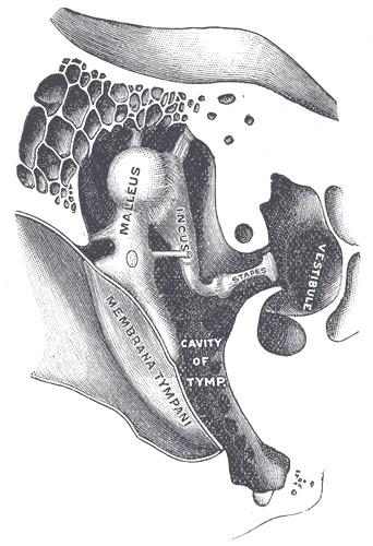
Chain of ossicles and their ligaments, seen from the front in a vertical, transverse section of the tympanum. (Testut.)

Figure 32: Chain of ossicles and their ligaments, seen from the front in a vertical, transverse section of the tympanum (Testut.).
Figure 33Gray915
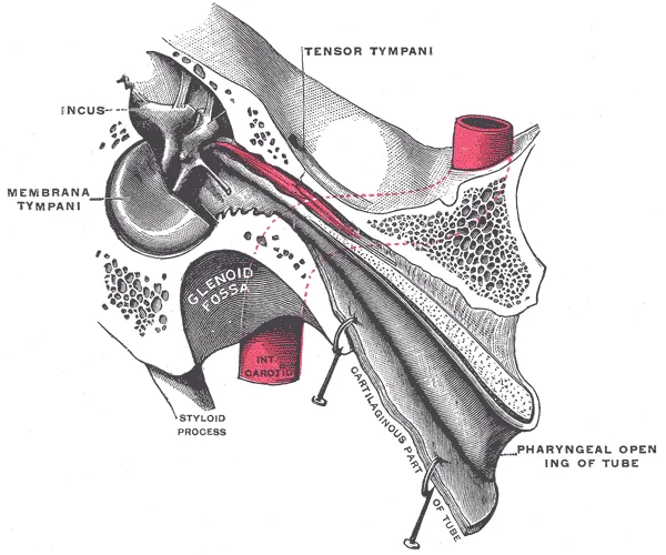
Long description
Auditory tube, laid open by a cut in its long axis. (Testut.)

Figure 33: Auditory tube, laid open by a cut in its long axis (Testut.).
Figure 34Gray910
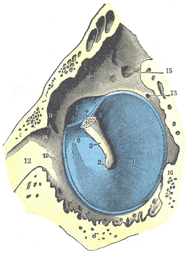
Long description
The tympanic membrane viewed from within. (Testut.) The malleus has been resected immediately beyond its lateral process, in order to show the tympanomalleolar folds and the membrana flaccida. 1. Tympanic membrane. 2. Umbo. 3. Handle of the malleus. 4. Lateral process. 5. Anterior tympanomalleolar f

Figure 34: The tympanic membrane viewed from within.
NoteArch Derivatives
Remember that the first arch forms the malleus and incus, while the second arch forms the stapes. This is a frequent examination point.
The inner ear, or labyrinth, develops from the otic placode, a thickening of the surface ectoderm. The placode invaginates to form the otic vesicle, or otocyst. This vesicle differentiates into the membranous labyrinth, which includes the cochlea, utricle, and saccule. The surrounding mesenchyme forms the bony labyrinth. The vestibulocochlear nerve (CN VIII) grows into the developing inner ear to innervate the sensory epithelia.
Figure 35Gray901
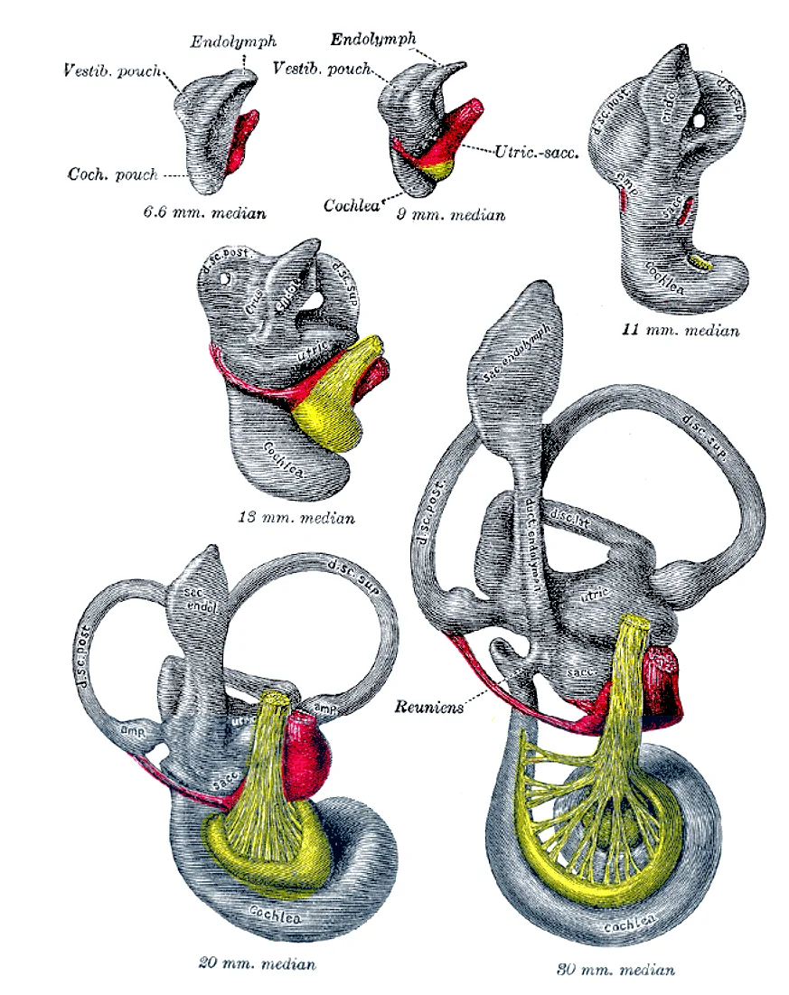
Long description
Median views of membranous labyrinth and acoustic complex in human embryos. X 25 dia. (Streeter.)

Figure 35: Median views of membranous labyrinth and acoustic complex in human embryos (x 25 dia).
Clinical pearlOtic Vesicle
The otocyst is the precursor to all sensory structures of the inner ear. It gives rise to both hearing and balance organs.
The development of the ear is closely linked to the development of the face and neck. The external ear moves from a low position on the neck to its final position on the side of the head. This migration occurs as the mandible grows. Congenital anomalies of the ear often reflect errors in this complex developmental process. For example, microtia results from incomplete fusion of the auricular hillocks. Atresia of the external auditory meatus may result from failure of the first pharyngeal cleft to canalise properly.
Exam trapCommon Misconception
Do not confuse the first pharyngeal cleft with the first pharyngeal pouch. The cleft forms the external ear canal, while the pouch forms the middle ear cavity.
The tympanic membrane develops from three layers. The outer layer is derived from the ectoderm of the first pharyngeal cleft. The inner layer is derived from the endoderm of the first pharyngeal pouch. The middle layer is fibrous, derived from mesoderm. This trilaminar structure is essential for its function as a sound conductor. The fibrous layer provides tensile strength, while the epithelial layers provide protection.
DefinitionTympanic Membrane Layers
The tympanic membrane consists of an outer ectodermal layer, a middle fibrous layer, and an inner endodermal layer.
Development continues postnatally. The external auditory meatus elongates and straightens. The middle ear cavity expands as the mastoid air cells develop. The mastoid process itself appears after birth as the sternocleidomastoid muscle attaches. This attachment requires the bony prominence for leverage. The inner ear reaches adult size relatively early in childhood. However, the neural connections continue to mature. This maturation is crucial for the development of hearing and balance skills.
NoteMastoid Development
The mastoid process is not present at birth. It develops in response to muscle attachment, highlighting the functional adaptation of bone.
Understanding these developmental origins helps in diagnosing congenital defects. It also explains the adult anatomical relationships. For instance, the proximity of the middle ear to the facial nerve is due to their shared arch origin. This anatomical relationship is clinically significant in otological surgery.
Radiological assessment of the ear relies primarily on otoscopic examination and computed tomography (CT). These modalities provide complementary views of the external auditory canal, tympanic membrane, and middle ear structures. Understanding the spatial relationships between these components is essential for accurate diagnosis of pathology.
In the otoscopic view, the external auditory canal is visible as a tubular structure leading to the tympanic membrane. The tympanic membrane appears as a pearly grey, semi-transparent membrane. It is tilted obliquely, with the handle of the malleus visible as a prominent landmark. The umbo, the tip of the malleus, is located at the centre of the membrane. Surrounding the tympanic membrane is the bony external auditory canal, which forms the lateral wall of the middle ear cavity.
The CT anatomy provides a cross-sectional view of the temporal bone. The temporal bone houses the structures of the inner and middle ear. The mastoid air cells are visible posterior to the middle ear cavity. These air cells are lined with mucosa and communicate with the middle ear via the aditus ad antrum. The aditus is a narrow passage that connects the epitympanum to the mastoid antrum.
The middle ear cavity is divided into three parts: the epitympanum, mesotympanum, and hypotympanum. The epitympanum is the superior part, containing the head of the malleus and the body of the incus. The hypotympanum is the inferior part, located below the level of the tympanic membrane.
The ossicles are the three small bones of the middle ear. They transmit sound vibrations from the tympanic membrane to the oval window of the inner ear. The malleus is attached to the tympanic membrane. The incus connects the malleus to the stapes. The stapes is the smallest bone in the body and fits into the oval window. [1]
DefinitionOssicular Chain
The malleus, incus, and stapes form the ossicular chain, which transmits sound vibrations from the tympanic membrane to the inner ear.
The facial nerve runs through the temporal bone, close to the middle ear structures. It is important to identify its course on CT scans to avoid injury during surgery. The nerve exits the skull via the stylomastoid foramen.
| Muscle | Origin | Insertion | Action | Nerve Supply |
|---|---|---|---|---|
| Tensor Tympani | Cartilaginous part of auditory tube | Handle of malleus | Tenses tympanic membrane | Mandibular nerve |
| Stapedius | Pyramid of posterior wall | Neck of stapes | Dampens stapes movement | Facial nerve |
The tensor tympani muscle originates from the cartilaginous part of the auditory tube and inserts onto the handle of the malleus. It is supplied by the mandibular nerve. The stapedius muscle originates from the pyramid of the posterior wall and inserts onto the neck of the stapes. It is supplied by the facial nerve.
Lesions of the middle ear can affect the ossicles or the surrounding structures. Cholesteatoma is a common pathology that can erode the ossicles and mastoid air cells. It is important to identify cholesteatoma on CT scans to plan appropriate treatment.
The ear is not merely a passive receiver of sound but a complex anatomical structure with distinct functional zones. Understanding its layout requires a clear distinction between the external, middle, and inner ear, each with specific clinical relevance. The external ear comprises the auricle and the external acoustic meatus. The auricle, or pinna, is composed of elastic cartilage covered by skin. It serves to collect sound waves and direct them into the ear canal. The external acoustic meatus i
The tympanic membrane, or eardrum, separates the external ear from the middle ear. It is an oval, semi-transparent membrane that vibrates in response to sound waves. The membrane is attached to the tympanic ring and the squamous part of the temporal bone. It has a concave shape, with the umbo pointing downwards and inwards. The handle of the malleus is attached to the inner surface of the membrane. The upper part of the membrane, the pars flaccida, is smaller and more horizontal than the larger, oblique pars tensa. Clinically, the position and colour of the tympanic membrane are key indicators of middle ear health. A bulging, red membrane suggests acute otitis media, while a retracted, dull membrane may indicate eustachian tube dysfunction.
The middle ear is an air-filled cavity within the temporal bone. It contains the auditory ossicles: the malleus, incus, and stapes. These bones transmit vibrations from the tympanic membrane to the inner ear. The malleus is attached to the tympanic membrane, the incus connects the malleus to the stapes, and the stapes footplate fits into the oval window of the cochlea. The middle ear is connected to the nasopharynx by the auditory tube, or eustachian tube. This tube helps equalise pressure on both sides of the tympanic membrane. Dysfunction of this tube can lead to negative pressure in the middle ear, causing fluid accumulation and hearing loss. [1]
The inner ear, or labyrinth, is embedded in the petrous part of the temporal bone. It consists of the bony labyrinth and the membranous labyrinth within it. The bony labyrinth contains perilymph, while the membranous labyrinth contains endolymph. The cochlea is responsible for hearing, converting sound vibrations into neural signals. The vestibular system, comprising the semicircular canals, utricle, and saccule, is responsible for balance. Lesions in the inner ear can cause vertigo, tinnitus, and sensorineural hearing loss. Understanding the precise anatomical relationships of these structures is crucial for diagnosing and managing ear disorders.
Medial to the tympanic membrane, the malleus is attached to its inner surface. The eustachian tube extends inferiorly and anteriorly towards the nasopharynx.
| Muscle | Action | Nerve Supply |
|---|---|---|
| Auricularis anterior | Draws auricle forward and upward | Facial nerve |
| Auricularis superior | Draws auricle upward | Facial nerve |
| Auricularis posterior | Draws auricle backward | Facial nerve |
Clinical pearlClinical Pearl
The external acoustic meatus is curved; examination requires pulling the auricle upwards and backwards in adults to straighten the canal.
This direct pathway explains why smell is a potent trigger for memories and emotion. These axons then project to the primary olfactory cortex in the inferior and medial temporal lobe, as well as to limbic structures. [1]
Students frequently confuse the actions of the extraocular muscles, particularly the obliques. A common error is assuming the superior oblique elevates the eye because of its name. The angle of the tendon through the trochlea means that contraction of the superior oblique rotates the eye laterally. This lateral rotation opposes the inferior oblique, which originates from the floor of the orbit and inserts into the inferolateral surface of the eye. When it contracts, it laterally rotates the eye, in opposition to the superior oblique. Understanding this opposition is necessary because the eye is not perfectly aligned on the sagittal plane. The superior rectus pulls at approximately a 20-degree angle, rather than straight up. Therefore, when the eye looks up, the inferior oblique compensates for this pull. Similarly, the inferior rectus is compensated by contraction of the inferior oblique during downward gaze. [1]
Another frequent mistake involves the anatomy of the retina and visual acuity. Students often assume the photoreceptors are the first layer light encounters. Note that the photoreceptors in the retina (rods and cones) are located behind the axons, RGCs, bipolar cells, and retinal blood vessels. A significant amount of light is absorbed by these structures before the light reaches the photoreceptor cells. This leads to confusion about the blind spot. Because these axons pass through the retina, there are no photoreceptors at the very back of the eye, where the optic nerve begins [1]
Students also misunderstand the fovea's structure. They may think the fovea has the most blood vessels for nourishment. However, at the fovea, the retina lacks the supporting cells and blood vessels, and only contains photoreceptors. Therefore, visual acuity, or the sharpness of vision, is greatest at the fovea. This is because the fovea is where the least amount of incoming light is absorbed by other retinal structures. In addition, each photoreceptor cell of the fovea is connected to a single RGC [1]
Exam trapOblique Action
The superior oblique does not elevate; it laterally rotates the eye due to the trochlear angle. The inferior oblique also laterally rotates, opposing the superior oblique.
DefinitionBlind Spot
The blind spot exists because retinal ganglion cell axons pass through the retina to form the optic nerve, leaving no photoreceptors at the optic disc. This creates a corresponding blind spot in our visual field. [1]
Knowledge of eye anatomy is critical at the bedside for testing visual fields and eye movements. Testing vision relies on the tests that are common in an optometry office. The Snellen chart demonstrates visual acuity by presenting standard Roman letters in a variety of sizes. The result of this test is a rough generalization of the acuity of a person based on the normal accepted acuity, such that a letter that subtends a visual angle of 5 minutes of an arc at 20 feet can be seen. To have 20/60 vision, for example, means that the smallest letters that a person can see at a 20-foot distance could be seen by a person with normal acuity from 60 feet away. Testing the extent of the visual field means that the examiner can establish the boundaries of peripheral vision as simply as holding their hands out to either side and asking the patient when the fingers are no longer visible without moving the eyes to track them. [2]
In the operating theatre and clinical examination, testing eye movement is simply a matter of having the patient track the tip of a pen as it is passed through the visual field. This may appear similar to testing visual field deficits related to the optic nerve, but the difference is that the patient is asked to not move the eyes while the examiner moves a stimulus into the peripheral visual field. Here, the extent of movement is the point of the test. The examiner is watching for conjugate movements representing proper function of the related nuclei and the MLF. Failure of one eye to abduct while the other adducts in a horizontal movement is referred to as internuclear ophthalmoplegia. When this occurs, the patient will experience diplopia, or double vision, as the two eyes are temporarily pointed at different stimuli. [2]
On imaging, understanding the fovea's avascular nature helps interpret macular health. At the fovea, the retina lacks the supporting cells and blood vessels, and only contains photoreceptors. This structural absence is key to high visual acuity but also makes the fovea vulnerable to vascular occlusions. In examinations, students must distinguish between color perception and memory. If you think that you can see colors in the dark, it is most likely because your brain knows what color something is and is relying on that memory. Visible light is electromagnetic radiation with a wavelength between 380 and 720 nm. Light with a wavelength of 380 nm is blue whereas light with a wavelength of 720 nm is dark red. [1]
| Structure | Location Relative to Photoreceptors | Function/Note |
|---|---|---|
| RGC Axons | Anterior to photoreceptors | Collect at optic disc; no photoreceptors here. |
| Bipolar Cells | Anterior to photoreceptors | Connect photoreceptors to RGCs. |
| Fovea | Central retina | Lacks blood vessels and supporting cells; highest acuity. |
| Peripheral Retina | Edges of retina | High convergence ratio (up to 50:1). |
Visual Acuity Determinants
- Start: Locate Retinal RegionDetermine if fovea or peripheral
- Question: Is it the Fovea?
Yes
- Check StructureNo blood vessels or supporting cells
- Check ConvergenceOne photoreceptor to one RGC
- ResultHighest visual acuity
No
- Check StructureContains vessels and supporting cells
- Check ConvergenceUp to 50 photoreceptors to one RGC
- ResultLower visual acuity
The superior oblique laterally rotates the eye due to the trochlear angle. The inferior oblique originates from the orbit floor and inserts inferolaterally. Photoreceptors are behind RGC axons, bipolar cells, and blood vessels. The blind spot lacks photoreceptors where the optic nerve exits. The fovea lacks blood vessels and supporting cells for sharp vision. Foveal photoreceptors connect one-to-one with RGCs. Peripheral retina has a convergence ratio of up to 50:1. Visible light wavelengths range from 380 nm (blue) to 720 nm (red).
Answer each one in your head first, then open the answer.
What is the primary action of the superior oblique muscle?
Show answerHide answer
Laterally rotates the eyeWhy is there a blind spot in the visual field?
Show answerHide answer
No photoreceptors where optic nerve axons exitWhat is the photoreceptor to RGC ratio in the peripheral retina?
Show answerHide answer
Up to 50 to 1What wavelength corresponds to dark red light?
Show answerHide answer
720 nmWhat is internuclear ophthalmoplegia?
Show answerHide answer
Failure of one eye to abduct while the other adducts
- Betts JG, Young KA, Wise JA, et al. Sensory Perception. OpenStax, Anatomy and Physiology 2e (CC BY-NC-SA 4.0). Accessed .
- Betts JG, Young KA, Wise JA, et al. The Cranial Nerve Exam. OpenStax, Anatomy and Physiology 2e (CC BY-NC-SA 4.0). Accessed .
Figure credits
- Figure 1: Henry Vandyke Carter, via Wikimedia Commons, Public domain, Public domain.
- Figure 2: OpenStax, via Wikimedia Commons, CC BY 4.0, CC BY 4.0. resized.
- Figure 3: OpenStax, via Wikimedia Commons, CC BY 4.0, CC BY 4.0. resized.
- Figure 4: Henry Vandyke Carter, via Wikimedia Commons, Public domain, Public domain.
- Figure 5: Henry Vandyke Carter, via Wikimedia Commons, Public domain, Public domain.
- Figure 6: Henry Vandyke Carter, via Wikimedia Commons, Public domain, Public domain.
- Figure 7: Henry Vandyke Carter, via Wikimedia Commons, Public domain, Public domain.
- Figure 8: Henry Vandyke Carter, via Wikimedia Commons, Public domain, Public domain.
- Figure 9: Henry Vandyke Carter, via Wikimedia Commons, Public domain, Public domain.
- Figure 10: Henry Vandyke Carter, via Wikimedia Commons, Public domain, Public domain.
- Figure 11: Henry Vandyke Carter, via Wikimedia Commons, Public domain, Public domain.
- Figure 12: Henry Vandyke Carter, via Wikimedia Commons, Public domain, Public domain.
- Figure 13: Henry Vandyke Carter, via Wikimedia Commons, Public domain, Public domain.
- Figure 14: Henry Vandyke Carter, via Wikimedia Commons, Public domain, Public domain.
- Figure 15: Henry Vandyke Carter, via Wikimedia Commons, Public domain, Public domain.
- Figure 16: Henry Vandyke Carter, via Wikimedia Commons, Public domain, Public domain.
- Figure 17: Henry Vandyke Carter, via Wikimedia Commons, Public domain, Public domain.
- Figure 18: Henry Vandyke Carter, via Wikimedia Commons, Public domain, Public domain.
- Figure 19: Henry Vandyke Carter, via Wikimedia Commons, Public domain, Public domain.
- Figure 20: Henry Vandyke Carter, via Wikimedia Commons, Public domain, Public domain.
- Figure 21: Henry Vandyke Carter, via Wikimedia Commons, Public domain, Public domain.
- Figure 22: Henry Vandyke Carter, via Wikimedia Commons, Public domain, Public domain.
- Figure 23: Henry Vandyke Carter, via Wikimedia Commons, Public domain, Public domain.
- Figure 24: Henry Vandyke Carter, via Wikimedia Commons, Public domain, Public domain.
- Figure 25: Henry Vandyke Carter, via Wikimedia Commons, Public domain, Public domain.
- Figure 26: Henry Vandyke Carter, via Wikimedia Commons, Public domain, Public domain.
- Figure 27: Henry Vandyke Carter, via Wikimedia Commons, Public domain, Public domain.
- Figure 28: Henry Vandyke Carter, via Wikimedia Commons, Public domain, Public domain.
- Figure 29: Henry Vandyke Carter, via Wikimedia Commons, Public domain, Public domain.
- Figure 30: Henry Vandyke Carter, via Wikimedia Commons, Public domain, Public domain.
- Figure 31: Henry Vandyke Carter, via Wikimedia Commons, Public domain, Public domain.
- Figure 32: Henry Vandyke Carter, via Wikimedia Commons, Public domain, Public domain.
- Figure 33: Henry Vandyke Carter, via Wikimedia Commons, Public domain, Public domain.
- Figure 34: Henry Vandyke Carter, via Wikimedia Commons, Public domain, Public domain.
- Figure 35: Henry Vandyke Carter, via Wikimedia Commons, Public domain, Public domain.
Educational content for medical students and doctors. It is not a substitute for clinical judgement or local guidelines.