Salivary glands and the submandibular region
Anatomy of the three major salivary glands, their ducts, and the submandibular region.
The upper alimentary canal comprises the mouth, pharynx, and esophagus, alongside accessory organs including the tongue, teeth, and salivary glands. Saliva production is continuous, with an average daily secretion of 1 to 1.5 liters. This fluid is essential for moistening the oral cavity and initiating carbohydrate breakdown via salivary amylase. Saliva consists of 99.4 percent water, with the remaining 0.6 percent containing ions, enzymes, and glycoproteins. The pH of saliva is maintained between 6.35 and 6.85 by bicarbonate and phosphate buffers. Immunoglobulin A and lysozyme provide antimicrobial protection within the oral environment. [1]
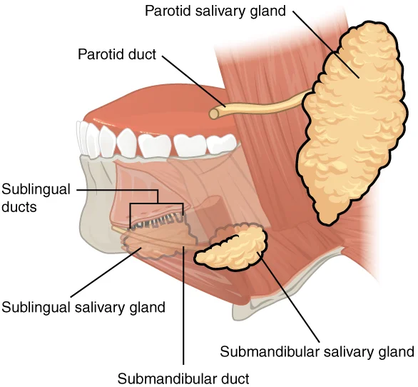
The parotid glands are the largest of the three major salivary glands. They secrete a watery solution rich in salivary amylase. Histologically, these glands are composed primarily of serous acini. The parotid duct, or Stensen’s duct, traverses the masseter muscle to open into the oral cavity opposite the second maxillary molar. The gland is located superficial to the masseter and ramus of the mandible. It is enclosed by the parotid sheath, a continuation of the deep cervical fascia. The facial nerve exits the stylomastoid foramen and divides within the gland. [1]
The submandibular glands produce saliva that is thicker than that of the parotid due to the presence of mucus. These glands contain both serous and mucous cells. The secretion contains amylase but is thickened with mucus. The submandibular duct, or Wharton’s duct, runs along the floor of the mouth. It opens at the sublingual caruncle on either side of the frenulum of the tongue. The gland is situated in the submandibular triangle, bounded by the anterior and posterior bellies of the digastric muscle and the inferior border of the mandible. The hypoglossal nerve runs deep to the gland. [1]
The sublingual glands are the smallest of the major salivary glands. They contain mostly mucous cells. Consequently, they secrete the thickest saliva with the least amount of salivary amylase. The ducts of the sublingual gland, known as the ducts of Rivinus, open directly into the floor of the mouth. A larger duct, the Bartholin duct, may join the submandibular duct. The gland lies superficial to the mylohyoid muscle. Minor salivary glands are also distributed throughout the oral mucosa. These include labial, buccal, palatal, and lingual glands. They ensure adequate saliva supply to all areas of the mouth. [1]
Figure 12408 Salivary Glands
Long description
The major salivary glands are located outside the oral mucosa and deliver saliva into the mouth through ducts.

Figure 1: The major salivary glands are located outside the oral mucosa and deliver saliva into the mouth through ducts.
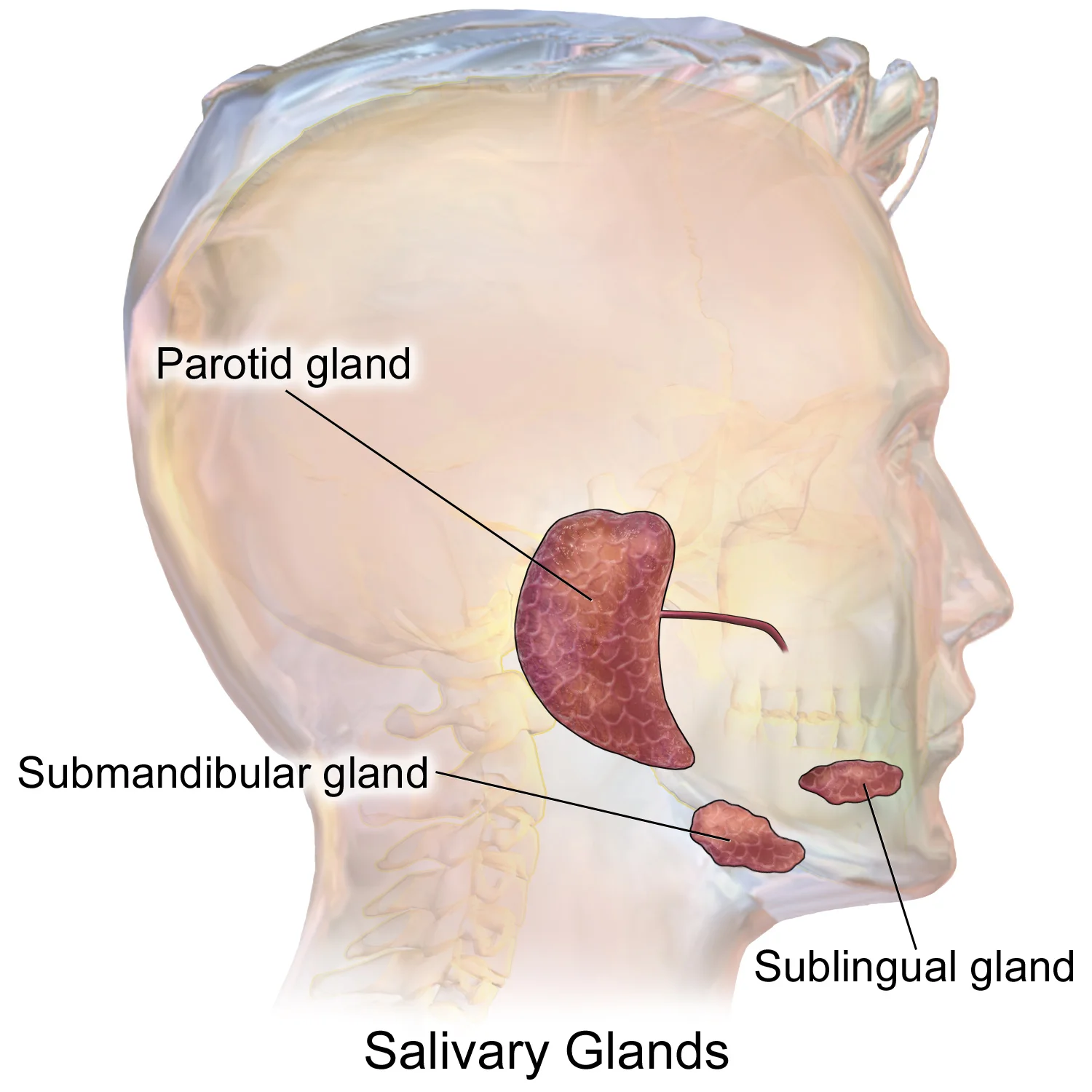
Figure 2Blausen 0780 SalivaryGlands
Long description
Salivary Glands. See a full animation of this medical topic.

Figure 2: Salivary Glands.
The hard palate forms the anterior roof of the mouth, acting as a rigid shelf created by the maxillary and palatine bones. Posterior to this lies the soft palate, composed mainly of skeletal muscle, which separates the oral and nasal cavities. The submandibular gland is situated in the submandibular triangle, bounded by the anterior and posterior bellies of the digastric muscle and the inferior border of the mandible. The parotid gland lies superficial to the masseter muscle and anterior to the ear. The ducts of these glands open into the oral cavity to facilitate digestion. [1]
The palate serves as a wall between the oral and nasal cavities. The anterior region is the hard palate, a bony structure against which the tongue pushes food. The posterior region is the soft palate, composed mainly of skeletal muscle. This arch allows simultaneous handling of digestion and respiration. [1]
The tooth consists of two main parts: the crown and the root. The crown projects above the gum line. The root is embedded within the maxilla and mandible. Both parts contain an inner pulp cavity with loose connective tissue, nerves, and blood vessels. The region of the pulp cavity running through the root is the root canal. Surrounding the pulp cavity is dentin, a bone-like tissue. In the root, dentin is covered by cementum, a layer of modified bone. In the crown, dentin is covered by enamel, the hardest substance in the body. [1]
This occurs at 24 days from the combination of the 3rd branchial arch arteries and the distal segments of the paired dorsal aortae. This portion becomes the ventral pharyngeal artery. Eventually, the ventral pharyngeal artery and the internal carotid artery fuse proximally to form the common carotid artery. At the 4 mm stage, which is 28 days, the internal carotid artery branches off into the anterior and the posterior division. [2]
DefinitionPalate
The palate is the roof of the mouth, consisting of the hard palate anteriorly and the soft palate posteriorly, separating the oral and nasal cavities.
| Structure | Location | Composition |
|---|---|---|
| Crown | Above gum line | Enamel covering dentin |
| Root | In maxilla/mandible | Cementum covering dentin |
| Pulp Cavity | Inner core | Loose connective tissue, nerves, vessels |
NoteEmbryology
The internal carotid artery develops from the 3rd branchial arch arteries and dorsal aortae at the 3 mm embryonic stage.
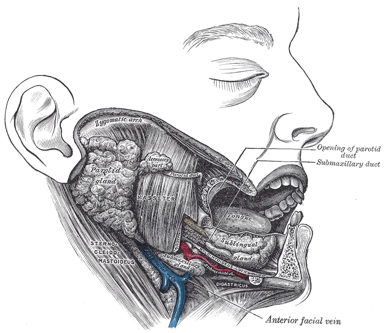
Figure 3Gray1024
Long description
Dissection, showing salivary glands of right side.

Figure 3: Dissection, showing salivary glands of right side.
The submandibular gland is situated in the submandibular triangle, bounded by the anterior and posterior bellies of the digastric muscle and the inferior border of the mandible. Deep to the gland lies the mylohyoid muscle, which forms the muscular floor of the mouth. The gland wraps around the posterior border of the mylohyoid, with a superficial part lying in the triangle and a deeper part extending into the sublingual space. [2]
The facial artery courses over the inferior border of the mandible, anterior to the masseter muscle, before entering the face. It lies superficial to the submandibular gland, often grooving its posterior aspect. The hypoglossal nerve (CN XII) runs deep to the gland, lying on the hyoglossus muscle. It is separated from the gland by the mylohyoid muscle and the lingual nerve in some regions, but generally lies inferior to the deep part of the gland. [1]
It runs anteriorly along the side of the tongue, deep to the mucosa of the floor of the mouth, opening at the sublingual caruncle on either side of the frenulum. The duct is closely related to the lingual nerve, which crosses over it laterally and then passes medial to it. This relationship is critical during surgical procedures to avoid injury to the nerve.
The sublingual gland lies superior to the mylohyoid muscle, in the sublingual space. It is smaller than the submandibular gland and drains via multiple small ducts (ducts of Rivinus) directly into the floor of the mouth, with one larger duct (Bartholin’s duct) sometimes joining the submandibular duct. The mylohyoid muscle separates the sublingual gland from the submandibular gland. The geniohyoid and genioglossus muscles lie superior to the mylohyoid, forming the muscular framework of the tongue base. [2]
The parotid gland, though not in the submandibular region, is related via the buccal space. The buccinator muscle forms the cheek, and the buccal nerve passes between the two heads of the lateral pterygoid muscle to supply sensation to the cheek. The masseter muscle covers the lateral aspect of the mandible. The temporalis tendon inserts on the coronoid process of the mandible.
DefinitionSubmandibular Triangle
A triangular area bounded by the anterior and posterior bellies of the digastric muscle and the inferior border of the mandible, containing the submandibular gland, facial artery, and hypoglossal nerve.
| Muscle | Origin | Insertion | Action | Nerve Supply |
|---|---|---|---|---|
| Mylohyoid | Mylohyoid line of mandible | Body of hyoid bone | Elevates hyoid, floor of mouth | Mylohyoid nerve (CN V3) |
| Digastric (Anterior) | Digastric fossa of mandible | Intermediate tendon | Depresses mandible, elevates hyoid | Mylohyoid nerve (CN V3) |
| Digastric (Posterior) | Mastoid notch of temporal bone | Intermediate tendon | Depresses mandible, elevates hyoid | Facial nerve (CN VII) |
The relations of the salivary glands are vital for understanding surgical approaches and potential complications. Injury to the lingual nerve can result in loss of sensation to the anterior two-thirds of the tongue and taste. Damage to the hypoglossal nerve causes ipsilateral tongue weakness and deviation towards the injured side. The facial artery can be ligated in this region for hemostasis, but care must be taken to avoid the nerve.
Exam trapLingual Nerve Course
The lingual nerve crosses lateral to the submandibular duct initially, then passes medial to it. Misidentifying this relationship can lead to nerve injury during duct repair.
It produces mixed serous and mucous secretions. The parotid gland is purely serous, while the sublingual gland is predominantly mucous. The autonomic nervous system regulates salivation, with parasympathetic stimulation increasing flow and sympathetic stimulation decreasing it. [2]
NoteClinical Relevance
The close proximity of the hypoglossal nerve to the submandibular gland necessitates careful dissection during gland excision to prevent tongue paralysis.
The internal architecture of the salivary glands and the submandibular region is defined by a precise arrangement of ducts, vessels, and nerves that ensure efficient secretion and drainage. Understanding this interior requires visualising the spatial relationships between the glandular parenchyma and the surrounding critical structures. This view presents a coronal section through the submandibular triangle, oriented with the anterior aspect to the left and the posterior aspect to the right. The image highlights the submandibular gland nestled within its fascial compartment, bordered superiorly by the mylohyoid muscle and inferiorly by the platysma.
DefinitionSubmandibular Gland
A compound tubuloalveolar gland located in the submandibular triangle, producing mixed serous and mucous secretions.
The gland is divided into superficial and deep lobes by the posterior edge of the mylohyoid muscle. The superficial lobe is the larger portion, lying in the submandibular fossa of the mandible. The deep lobe extends medially, wrapping around the posterior border of the mylohyoid to reach the floor of the mouth. Between these lobes, the facial artery and the submandibular ganglion are situated. It receives parasympathetic secretomotor fibres from the chorda tympani branch of the facial nerve, which hitchhikes along the lingual nerve to reach the ganglion. Postganglionic fibres then join the lingual nerve to innervate the gland.
| Structure | Relation to Submandibular Gland |
|---|---|
| Facial Artery | Courses over the superior border of the gland |
| Hypoglossal Nerve | Lies deep to the gland, on the hyoglossus muscle |
| Lingual Nerve | Lies superior and lateral to the submandibular duct |
| Submandibular Ganglion | Suspended from the lingual nerve, deep to the duct |
The internal ductal system begins with intercalated ducts within the acini, which merge into striated ducts and then into the main excretory duct. The main duct, approximately 5 cm long, travels anteriorly and medially. It is accompanied by the submandibular ganglion, which is connected to the duct by two roots. The duct opens into the oral cavity at the base of the plica sublingualis. Any obstruction or inflammation in this interior pathway can lead to sialadenitis or sialolithiasis, particularly given the upward course of the duct against gravity and the viscous nature of its mixed secretion.
The arterial supply to the salivary glands and the submandibular region is derived primarily from branches of the external carotid artery, with specific contributions ensuring adequate perfusion for secretion. Understanding this vascular architecture is essential for surgical planning and interpreting imaging in this anatomical zone.
The external carotid artery ascends in the neck, giving off the facial artery. The facial artery courses anteriorly, looping around the inferior border of the mandible. From this loop, the submental artery arises, running along the superior border of the hyoid bone. The facial artery then continues upwards, giving off the sublingual artery, which supplies the submandibular gland and adjacent structures. The submandibular gland itself is situated in the submandibular triangle, bounded by the mandible above and the anterior and posterior bellies of the digastric muscle below. The vascular supply ensures that the gland receives blood from both the facial and sublingual arteries, which anastomose within the glandular tissue. This arrangement allows for collateral circulation if one vessel is compromised. Understanding these relationships is crucial for avoiding vascular injury during surgery.
The primary source of blood for the submandibular gland is the facial artery. The facial artery is a branch of the external carotid artery. It arises in the neck, anterior to the internal carotid artery. The artery courses superiorly and anteriorly, passing deep to the posterior belly of the digastric muscle and the stylohyoid muscle. It then loops around the inferior border of the mandible at the anterior edge of the masseter muscle. This loop is a key landmark in surgery. After crossing the mandible, the artery enters the face, giving off branches to the submandibular gland. The submandibular branch arises just before the facial artery loops around the mandible. This branch enters the deep surface of the submandibular gland. The gland is drained by veins that accompany the arteries. The venous drainage is into the facial vein and the internal jugular vein. [2] [1]
DefinitionFacial Artery
A branch of the external carotid artery that supplies the face and submandibular region, looping around the mandible.
The sublingual artery, a branch of the lingual artery, also contributes to the blood supply. The lingual artery arises from the external carotid artery. It courses deep to the hyoglossus muscle. The sublingual artery arises from the lingual artery and supplies the sublingual gland and the floor of the mouth. It anastomoses with the submental artery and the facial artery. This anastomotic network ensures robust blood supply to the submandibular and sublingual regions.
| Artery | Origin | Key Relation |
|---|---|---|
| Facial Artery | External Carotid | Loops around mandible |
| Submental Artery | Facial Artery | Runs along hyoid |
| Sublingual Artery | Lingual Artery | Supplies floor of mouth |
The submandibular gland is located in the submandibular triangle. This triangle is bounded by the mandible superiorly, the anterior belly of the digastric muscle anteriorly, and the posterior belly of the digastric muscle posteriorly. The floor of the triangle is formed by the mylohyoid and hyoglossus muscles. The facial artery and vein pass through this triangle. The hypoglossal nerve runs along the superior surface of the hyoglossus muscle. The lingual nerve is also present, running superior to the submandibular duct. These structures are closely related to the gland and must be identified during surgery.
Exam trapSurgical Hazard
The hypoglossal nerve is at risk during submandibular gland excision. It runs on the hyoglossus muscle, deep to the gland.
The blood supply is critical for gland function. Ischemia can lead to glandular dysfunction. The anastomoses between the facial, submental, and sublingual arteries provide redundancy. This is important in cases of vascular disease or surgical ligation. The submandibular gland receives its blood from these multiple sources, ensuring viability even if one vessel is occluded.
NoteClinical Relevance
Understanding the vascular anatomy is vital for preventing complications during submandibular gland surgery.
The venous drainage of the salivary glands and the submandibular region follows a predictable pattern, mirroring the arterial supply but generally carrying deoxygenated blood towards the deep cervical veins. Understanding this drainage is crucial for appreciating how infections or malignancies in the head and neck spread.
DefinitionFacial Vein
The facial vein is a major tributary that drains the face and communicates with the cavernous sinus.
The facial vein originates near the medial angle of the eye, where it begins as the angular vein. It descends obliquely across the face, anterior to the masseter muscle and the submandibular gland. In the submandibular region, the facial vein lies superficial to the submandibular gland and the mylohyoid muscle. It receives tributaries from the submental vein and the deep facial vein. The deep facial vein connects the facial vein with the pterygoid plexus of veins, providing a potential route for [1]
Exam trapCavernous Sinus Connection
The facial vein lacks valves, allowing retrograde flow into the cavernous sinus, which can lead to cavernous sinus thrombosis. [1]
The common facial vein then descends vertically along the carotid sheath. It typically terminates by draining into the internal jugular vein, although it may also join the brachiocephalic vein directly in some variations. [1]
DefinitionCommon Facial Vein
The common facial vein is formed by the union of the facial vein and the anterior division of the retromandibular vein. [1]
The submandibular gland itself is drained by small venous channels that empty directly into the facial vein or the common facial vein. These veins run alongside the arterial supply, ensuring efficient removal of metabolic waste from the glandular tissue. [1]
| Structure | Drainage Destination | Clinical Significance |
|---|---|---|
| Facial Vein | Common Facial Vein | Valveless nature risks intracranial spread |
| Submental Vein | Facial Vein | Drains lower lip and chin |
| Deep Facial Vein | Pterygoid Plexus | Connects face to intracranial sinuses |
| Common Facial Vein | Internal Jugular Vein | Main outflow from anterior face |
The internal jugular vein serves as the final common pathway for venous blood from the head and neck. It descends through the neck and joins the subclavian vein to form the brachiocephalic vein. [1]
NoteAnatomical Landmark
The internal jugular vein is a key landmark in the carotid sheath. [1]
In summary, the venous drainage of the salivary glands and submandibular region is characterised by the facial vein and its tributaries, which ultimately empty into the internal jugular vein. The valveless nature of these veins is a critical anatomical feature with significant clinical implications for the spread of infection.
The lymphatic drainage of the salivary glands and the submandibular region follows a predictable pattern that is critical for understanding the spread of infection and malignancy.
This pathway is the most clinically significant for submandibular pathology.
The submandibular gland lies inferior to the mandible, with its efferent vessels passing deep to the gland to reach the deep cervical chain. The deep cervical nodes are arranged along the internal jugular vein, with the jugulodigastric node located at the angle of the mandible. This node is often enlarged in tonsillitis and submandibular gland infections.
| Structure | Primary Nodes | Secondary Nodes |
|---|---|---|
| Parotid Gland | Preauricular, Superficial Cervical | Deep Cervical |
| Submandibular Gland | Submandibular, Jugulodigastric | Deep Cervical |
| Sublingual Gland | Submandibular | Deep Cervical |
Lymphatic obstruction or inflammation can lead to significant clinical presentations. In cases of acute sialadenitis, the lymph nodes may become tender and enlarged, mimicking a neoplastic process. It is essential to distinguish between reactive lymphadenopathy and metastatic disease. The jugulodigastric node is particularly susceptible to enlargement due to its proximity to the oropharynx and submandibular gland.
Tumours of the salivary glands exhibit distinct patterns of lymphatic spread. Malignant tumours of the parotid gland, such as mucoepidermoid carcinoma, may metastasize to the preauricular nodes before involving the deep cervical chain. In contrast, malignant tumours of the submandibular gland often present with palpable submandibular lymphadenopathy. The presence of fixed, hard nodes suggests advanced disease with extracapsular extension.
Stones (sialolithiasis) typically affect the submandibular gland due to the anatomical characteristics of Wharton’s duct. The duct is longer, more tortuous, and the saliva is more alkaline and mucinous, promoting stone formation. Obstruction leads to glandular swelling, particularly during meals, known as mealtime syndrome. Chronic obstruction can result in fibrosis and atrophy of the gland.
Exam trapClinical Pitfall
Do not assume all submandibular swelling is due to stones; consider lymphadenopathy, cysts, and neoplasms in the differential diagnosis.
Understanding the lymphatic anatomy is crucial for surgical planning. Neck dissections must include the relevant nodal basins to ensure complete removal of metastatic disease. The level of nodes involved determines the extent of the dissection and the prognosis.
The nerve supply to the salivary glands involves both somatic and autonomic components. Somatic sensation is provided by branches of the trigeminal nerve. The parotid gland receives sensory innervation from the auriculotemporal nerve, a branch of the mandibular division of the trigeminal nerve. This nerve also carries postganglionic parasympathetic fibres to the gland. The submandibular and sublingual glands receive sensory innervation from the lingual nerve, another branch of the mandibular nerve. Pain from these glands may be referred to the ear or jaw due to shared sensory pathways.
Autonomic control is crucial for salivary secretion. The parasympathetic nervous system stimulates secretion. For the parotid gland, preganglionic parasympathetic fibres originate in the inferior salivatory nucleus. These fibres travel via the glossopharyngeal nerve. They branch off as the tympanic nerve and then the lesser petrosal nerve. The lesser petrosal nerve synapses in the otic ganglion. Postganglionic fibres then hitchhike on the auriculotemporal nerve to reach the parotid gland.
For the submandibular and sublingual glands, the pathway differs. Preganglionic fibres arise from the superior salivatory nucleus. They travel via the facial nerve. Specifically, they pass through the chorda tympani branch. The chorda tympani joins the lingual nerve. These fibres synapse in the submandibular ganglion. Postganglionic fibres then innervate the submandibular and sublingual glands. This secretomotor pathway ensures appropriate saliva production during digestion.
DefinitionSecretomotor Pathway
Parasympathetic stimulation increases saliva volume and watery content, essential for deglutition and digestion.
The autonomic nervous system controls the pharyngeal and esophageal phases of swallowing. This coordination ensures safe passage of food. The somatic nerves control the voluntary phase involving the tongue. Disruption of these nerves can lead to dysphagia. Understanding these pathways is vital for diagnosing lesions in the submandibular region.
| Gland | Parasympathetic Origin | Ganglion | Postganglionic Nerve |
|---|---|---|---|
| Parotid | Inferior salivatory nucleus | Otic ganglion | Auriculotemporal nerve |
| Submandibular | Superior salivatory nucleus | Submandibular ganglion | Lingual nerve |
| Sublingual | Superior salivatory nucleus | Submandibular ganglion | Lingual nerve |
Please refer to the dedicated histology page for detailed microscopic architecture of salivary tissue.
The submandibular gland is a mixed seromucous gland, meaning it produces both watery and viscous secretions. It is located in the submandibular triangle, bounded by the anterior and posterior bellies of the digastric muscle and the inferior border of the mandible. The gland wraps around the posterior border of the mylohyoid muscle, with a superficial lobe lying lateral to the muscle and a deep lobe extending medial to it.
The gland receives parasympathetic innervation from the facial nerve (CN VII). Fibres travel via the chorda tympani, join the lingual nerve, and synapse in the submandibular ganglion. Postganglionic fibres then innervate the glandular acini to stimulate secretion. Sympathetic innervation arises from the superior cervical ganglion, travelling along the external carotid artery branches to reach the gland, where it primarily causes vasoconstriction and modifies secretion consistency.
The main excretory duct is Wharton’s duct. It originates from the deep part of the gland and runs anteriorly along the side of the lingual nerve. It opens into the floor of the mouth at the sublingual caruncle, adjacent to the frenulum of the tongue. This anatomical position makes the duct susceptible to obstruction by sialoliths (salivary stones), which are more common in the submandibular gland than in other salivary glands due to the viscous nature of its secretion and the upward course of the duct against gravity.
Common pathologies include sialadenitis (inflammation), often bacterial in origin, and sialolithiasis (stone formation). Clinical presentation typically involves painful swelling of the gland, particularly during meals (mealtime syndrome), as saliva production increases but cannot escape the obstructed duct. Diagnosis is confirmed via ultrasound or CT imaging, which can visualise the calcified stones. Treatment ranges from conservative measures like hydration and gland massage to surgical removal of the stone or, in severe recurrent cases, excision of the gland.
Link to the embryology page.
The salivary glands and the submandibular region develop from the pharyngeal arches, with the submandibular gland arising from an ectodermal outgrowth of the floor of the mouth. This process begins early in gestation, establishing the ductal system that will eventually connect to the oral cavity. The parotid gland develops later from the ectoderm of the buccal mucosa, while the sublingual gland originates from multiple small buds.
Consequently, the full development of the ACA and the ACOMM mark the final realization of the adult CoW at the 6-7 weeks embryological stage. [2]
The parotid gland is the largest of the salivary glands. It lies superficial to the masseter muscle and extends deep into the parotid bed. On the axial section, it appears as a homogeneous, moderately dense structure lateral to the mandibular ramus. The gland is enclosed within the parotid sheath, a modification of the deep cervical fascia. Medially, it relates to the styloid process and the muscles of the pharynx. Laterally, it is covered by skin, subcutaneous tissue, and the platysma. The facial nerve emerges from the lateral aspect of the gland, dividing it into superficial and deep lobes. The retromandibular vein forms within the substance of the gland by the union of the superficial temporal and maxillary veins.
The submandibular gland occupies the submandibular triangle, bounded by the anterior and posterior bellies of the digastric muscle and the inferior border of the mandible. It is smaller than the parotid gland and has a more lobulated appearance on imaging. The gland wraps around the posterior border of the mylohyoid muscle. The anterior part lies superficial to the mylohyoid, while the posterior part lies deep to it. The hypoglossal nerve runs along the deep surface of the gland, medial to the mylohyoid. The submandibular duct arises from the anterior part of the gland and courses forward to open into the floor of the mouth at the sublingual caruncle.
The sublingual gland is the smallest of the major salivary glands. It lies in the floor of the mouth, medial to the body of the mandible and lateral to the genioglossus muscle. It is situated between the mylohyoid muscle and the mucosa of the floor of the mouth. The gland drains via multiple small ducts, known as the ducts of Rivinus, which open directly into the floor of the mouth. A larger duct, the Bartholin duct, may join the submandibular duct or open separately. On axial imaging, it appears as a small, triangular structure anterior to the submandibular gland.
The internal carotid artery and internal jugular vein lie deep to the parotid gland, separated by the styloid process and its attached muscles. The glossopharyngeal nerve passes between the internal and external carotid arteries. In the submandibular region, the lingual nerve loops under the submandibular duct. The mylohyoid nerve supplies the mylohyoid muscle and the anterior belly of the digastric. These relationships are critical for surgical planning and interpretation of cross-sectional imaging.
The submandibular gland is a mixed serous and mucous gland located in the submandibular triangle, bounded by the anterior and posterior bellies of the digastric muscle and the inferior border of the mandible. The duct of the submandibular gland, also known as Wharton’s duct, originates from the deep part of the gland. It courses anteriorly along the side of the tongue, deep to the lingual nerve, and opens into the floor of the mouth at the sublingual caruncle. This anatomical course makes the duct susceptible to obstruction by sialoliths, particularly at the mid-portion where the duct makes a sharp upward turn.
The parotid gland is the largest of the salivary glands, situated between the mastoid process, the sternocleidomastoid muscle, and the ramus of the mandible. It is enclosed within the parotid sheath, a modification of the deep cervical fascia. The facial nerve exits the stylomastoid foramen and enters the posterior aspect of the gland, dividing into its terminal branches within the substance of the gland. These branches, from superior to inferior, are temporal, zygomatic, buccal, marginal mandibular, and cervical. Understanding this intraglandular course is vital for surgical approaches to prevent iatrogenic facial nerve palsy. The retromandibular vein and external carotid artery also traverse the gland, lying deep to the facial nerve branches. [1]
Lesions in this region often present as swelling or pain. Sialolithiasis, the formation of calculi, most commonly affects the submandibular gland due to the alkaline, mucous-rich saliva and the uphill course of its duct. Imaging, such as ultrasound or CT, is used to identify these calcifications. The image also demonstrates the relationship of the sublingual gland, which is smaller and lies lateral to the genioglossus muscle, draining via multiple small ducts into the floor of the mouth. Injury to the marginal mandibular branch of the facial nerve during submandibular gland excision can result in asymmetry of the lower lip, highlighting the need for careful dissection below the mandible.
The uvula is a fleshy bead of tissue dropping from the center of the posterior edge of the soft palate. It moves upward during swallowing to prevent food and liquid from entering the nasal cavity. Two muscular folds extend downward from the soft palate on either side of the uvula. The palatoglossal arch lies next to the base of the tongue toward the front. The palatopharyngeal arch forms the superior and lateral margins of the fauces behind the palatoglossal arch. Between these two arches lie the palatine tonsils, which are clusters of lymphoid tissue protecting the pharynx. The lingual tonsils are located at the base of the tongue. Salivation is stimulated by taste receptors sending impulses to the superior and inferior salivatory nuclei in the brain stem. Parasympathetic impulses return through the glossopharyngeal and facial nerves to stimulate salivation. Dental caries develops when bacteria feeding on sugars release acids that degrade calcium crystals of the enamel. The pharynx runs from the posterior oral and nasal cavities to the opening of the esophagus and larynx. The voluntary phase of deglutition involves the tongue pushing the bolus into the oropharynx. [1]
Clinical pearlTonsil Location
Palatine tonsils sit between the palatoglossal and palatopharyngeal arches, while lingual tonsils are at the tongue base.
Exam trapSaliva Fate
Do not assume saliva is lost; most is swallowed and reabsorbed to maintain fluid balance.
DefinitionDental Caries
Acid degradation of enamel calcium crystals by bacteria feeding on oral sugars.
NotePharynx Subdivisions
The nasopharynx handles breathing and speech only; the oropharynx and laryngopharynx handle both breathing and digestion.
Clinical pearlVoluntary Swallowing
The tongue moves upward and backward against the palate to push the bolus into the oropharynx.
Exam trapUvula Function
It prevents nasal entry of food but can contribute to snoring sounds.
DefinitionFauces Margins
The palatopharyngeal arch forms the superior and lateral margins of the fauces.
NoteSalivary Nerves
Impulses travel via glossopharyngeal and facial nerves from brain stem nuclei to stimulate salivation.
Clinical pearlEnamel Vulnerability
Enamel is susceptible to mechanical and chemical erosion despite protecting dentin.
Exam trapPharynx Continuity
The oropharynx begins inferior to the nasopharynx and is continuous below with the laryngopharynx.
DefinitionDeglutition Phase
Chewing is completed and swallowing is set in motion during the voluntary oral phase.
NoteTonsil Protection
Palatine tonsils are lymphoid tissue clusters that protect the pharynx.
Students often confuse the origin and insertion of the extrinsic tongue muscles. The genioglossus originates from the mental spine of the mandible and inserts into the tongue, while the styloglossus originates from the styloid process and inserts into the tongue[1]. Another common mistake is misunderstanding the hyoid bone's unique articulation - it only distantly/indirectly articulates with other bones, unlike other bones in the skeletal system[1].
Many students incorrectly believe the tongue is the strongest muscle in the body proportionate to its size. While difficult to quantify, the tongue is indeed a workhorse facilitating ingestion, mechanical digestion, chemical digestion (lingual lipase), sensation, swallowing, and vocalization[1]. The lips are often misunderstood as simply red structures; their red color comes from being very vascular with a thin layer of keratin[1].
Exam trapLip structure
The lips have a huge representation on the cerebral cortex, which probably explains the human fascination with kissing!
Students frequently confuse the lower esophageal sphincter's function. It relaxes to let food pass into the stomach and then contracts to prevent stomach acids from backing up into the esophagus. When it doesn't completely close, it can cause gastroesophageal reflux disease (GERD)[1]. Another common error is misunderstanding the prevalence of vascular anomalies, such as a persistent stapedial artery which has a reported prevalence of 0.5%[1]. [2]
| Muscle | Origin | Insertion | Action | Nerve Supply |
|---|---|---|---|---|
| Genioglossus | Mental spine of mandible | Tongue | Protrudes tongue | Hypoglossal nerve |
| Styloglossus | Styloid process | Tongue | Elevates and retracts tongue | Hypoglossal nerve |
| Hyoglossus | Greater horn of hyoid | Tongue | Depresses tongue | Hypoglossal nerve |
| Palatoglossus | Palatine aponeurosis | Tongue | Elevates posterior tongue | Vagus nerve |
Knowledge of salivary glands and the submandibular region is essential at the bedside when examining patients for swelling or masses. Clinicians must understand the submandibular gland's location beneath the mandible and its duct's course forward to open at the submandibular caruncle[1]. This knowledge helps differentiate between salivary gland infections, stones, or tumors.
In the operating theatre, surgeons must be familiar with the submandibular triangle's boundaries and contents when performing procedures such as submandibular gland excision. The facial artery and vein are critical structures in this region that must be identified and preserved to prevent bleeding and preserve facial function[1]. The hypoglossal nerve's position deep to the submandibular gland is particularly important to avoid injury during surgery.
Submandibular Region Examination
- InspectLook for swelling, asymmetry, or scarring
- Question: Palpate for masses?
Yes
- Note location, size, mobility, tenderness
No
- Proceed to bimanual palpation
- Bimanual palpationOne finger intraorally, one extraorally
- Question: Express saliva?
Yes
- Note duct patency and saliva quality
No
- Consider imaging
On imaging studies, radiologists interpret CT and MRI scans to evaluate salivary gland pathology. Knowledge of normal anatomy helps identify abnormalities such as sialolithiasis, tumors, or inflammatory conditions. The reported prevalence of a persistent stapedial artery is 0.5%, and its absence can be an indirect sign on skull-base CT studies[1]. Vascular anomalies in the head and neck region, though rare, must be recognized to prevent complications during procedures.
In examinations, medical students are tested on their knowledge of salivary gland anatomy, including the parotid, submandibular, and sublingual glands. Students must correctly identify the submandibular gland as the second largest salivary gland and describe its duct's course. They should also understand that the submandibular gland's mixed serous and mucous secretions make it more prone to stone formation than the serous-dominated parotid gland[1].
- The parotid gland is the largest salivary gland and secretes a watery solution rich in salivary amylase through Stensen's duct.
- The submandibular gland is located in the submandibular triangle and produces a mixed serous and mucous secretion that drains via Wharton's duct.
- The sublingual gland is the smallest of the major salivary glands and primarily secretes thick, mucous saliva.
- The facial vein is valveless and communicates with the cavernous sinus via the deep facial vein, creating a potential route for intracranial infection.
- The submandibular gland's parasympathetic innervation originates in the superior salivatory nucleus and synapses in the submandibular ganglion.
- The lingual nerve has a critical relationship with the submandibular duct, crossing lateral to the duct before passing medial to it.
- Sialolithiasis is most common in the submandibular gland due to the viscous nature of its secretions and the upward course of its duct against gravity.
- The internal carotid artery develops from the 3rd branchial arch arteries and the distal segments of the paired dorsal aortae.
Answer each one in your head first, then open the answer.
Why is the relationship between the lingual nerve and the submandibular duct clinically significant during surgical procedures
Show answerHide answer
The lingual nerve crosses lateral to the duct and then passes medial to it, and misidentifying this relationship can lead to nerve injury during duct repair.How can an infection in the facial region potentially spread to the intracranial space
Show answerHide answer
The facial vein is valveless and connects to the pterygoid plexus via the deep facial vein, allowing retrograde flow into the cavernous sinus.Which anatomical and physiological factors make the submandibular gland more prone to stone formation than other salivary glands
Show answerHide answer
The gland produces more viscous, alkaline, and mucinous saliva, and its duct is longer, more tortuous, and courses upward against gravity.Contrast the parasympathetic pathways for the parotid gland versus the submandibular gland
Show answerHide answer
The parotid pathway involves the inferior salivatory nucleus, glossopharyngeal nerve, and otic ganglion, while the submandibular pathway involves the superior salivatory nucleus, facial nerve via the chorda tympani, and submandibular ganglion.What are the potential functional deficits resulting from injury to the hypoglossal nerve during submandibular gland excision
Show answerHide answer
Damage to the hypoglossal nerve causes ipsilateral tongue weakness and deviation of the tongue towards the injured side.
- Betts JG, Young KA, Wise JA, et al. The Mouth, Pharynx, and Esophagus. OpenStax, Anatomy and Physiology 2e (CC BY-NC-SA 4.0). Accessed .
- Menshawi K, Mohr JP, Gutierrez J. A Functional Perspective on the Embryology and Anatomy of the Cerebral Blood Supply. Journal of stroke 2015;17(2):144-158. doi:10.5853/jos.2015.17.2.144. Accessed .
Figure credits
- Figure 1: OpenStax College, via Wikimedia Commons, CC BY 3.0, CC BY 3.0.
- Figure 2: BruceBlaus. When using this image in external sources it can be cited as: Blausen.com staff (2014). "Medical gallery of Blausen Medical 2014". WikiJournal of Medicine 1 (2). DOI:10.15347/wjm/2014.010. ISSN 2002-4436., via Wikimedia Commons, CC BY 3.0, CC BY 3.0.
- Figure 3: Henry Vandyke Carter, via Wikimedia Commons, Public domain, Public domain.
Educational content for medical students and doctors. It is not a substitute for clinical judgement or local guidelines.