The pituitary gland and sella turcica
Anatomical and functional overview of the pituitary gland, its location in the sella turcica, and its connection to the hypothalamus.
ALIASEs: Hypophysis, Pituitary gland, Pituitary body
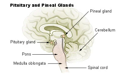
Figure 1Illu pituitary pineal glands
Long description
Illu pituitary pineal glands.jpg. Labelled illustration from Wikimedia Commons, used under its open licence.

Figure 1: Illu pituitary pineal glands.jpg.
The hypothalamus–pituitary complex can be thought of as the “command center” of the endocrine system[1]. This complex secretes several hormones that directly produce responses in target tissues, as well as hormones that regulate the synthesis and secretion of hormones of other glands[1]. In addition, the hypothalamus–pituitary complex coordinates the messages of the endocrine and nervous systems[1]. In many cases, a stimulus received by the nervous system must pass through the hypothalamus–pituitary complex to be translated into hormones that can initiate a response[1].
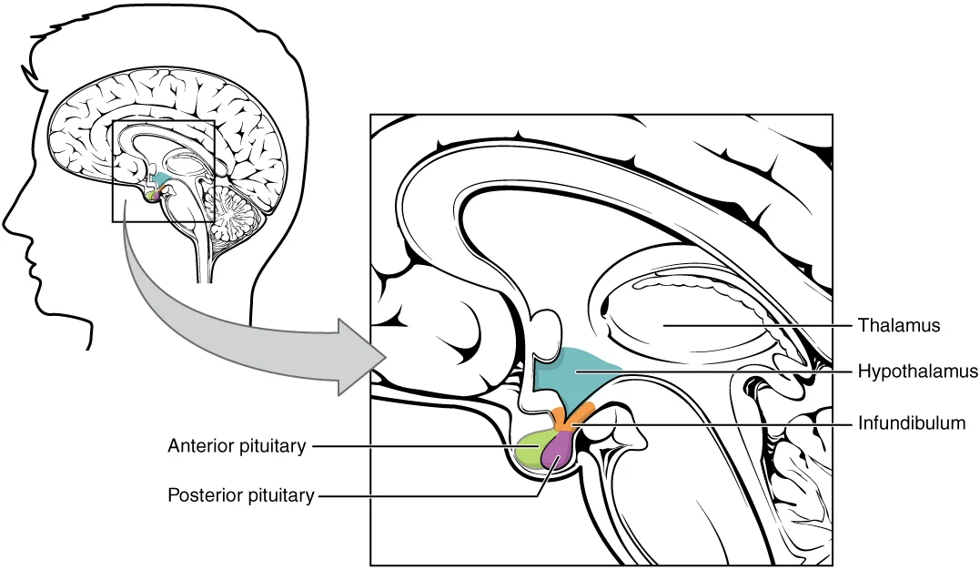
The hypothalamus is a structure of the diencephalon of the brain located anterior and inferior to the thalamus[1]. It has both neural and endocrine functions, producing and secreting many hormones[1]. In addition, the hypothalamus is anatomically and functionally related to the pituitary gland (or hypophysis), a bean-sized organ suspended from it by a stem called the infundibulum (or pituitary stalk)[1]. The pituitary gland is cradled within the sellaturcica of the
DefinitionSella Turcica
The saddle-shaped depression in the sphenoid bone that houses the pituitary gland.
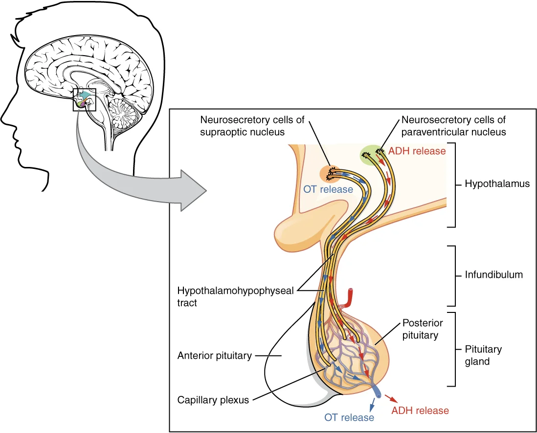
The posterior pituitary is actually an extension of the neurons of the paraventricular and supraoptic nuclei of the hypothalamus[1]. The cell bodies of these regions rest in the hypothalamus, but their axons descend as the hypothalamic–hypophyseal tract within the infundibulum, and end in axon terminals that comprise the posterior pituitary[1]. The posterior pituitary gland does not produce hormones, but rather stores and secretes hormones produced by the hypothalamus Recall that the posterior pituitary does not synthesize hormones, but merely stores them[1]. In contrast, the anterior pituitary does manufacture hormones[1]. However, the secretion of hormones from the anterior pituitary is regulated by two classes of hormones[1]. These hormones—secreted by the hypothalamus—are the releasing hormones that stimulate the secretion of hormones from the anterior pituitary and the inhibiting hormones that inhibit secretion[1].
Hypothalamic hormones are secreted by neurons, but enter the anterior pituitary through blood vessels[1]. Within the infundibulum is a bridge of capillaries that connects the hypothalamus to the anterior pituitary[1]. This network, called the hypophyseal portal system, allows hypothalamic hormones to be transported to the anterior pituitary without first entering the systemic circulation[1]. The hypophyseal portal system originates from the superior hypophyseal arte
Hormones produced by the anterior pituitary (in response to releasing hormones) enter a secondary capillary plexus, and from there drain into the circulation[1]. The hypothalamus–pituitary complex is located in the diencephalon of the brain[1]. The hypothalamus and the pituitary gland are connected by a structure called the infundibulum, which contains vasculature and nerve axons[1]. The pituitary gland is divided into two distinct structures with different embryonic origins[1]. The posterior lobe houses the axon terminals of hypothalamic neurons[1]. It stores and releases into the bloodstream two hypothalamic hormones: oxytocin and antidiuretic hormone (ADH)[1]. The anterior lobe is connected to the hypothalamus by vasculature in the infundibulum and produces and secretes six hormones[1]. Their secretion is regulated, however, by releasing and inhibiting hormones from the hypothalamus[1].
Anterior pituitary gland neoplasms have been designated as tumors more than a century ago[1]. The terms tumor and adenoma have been interchangeably used through the time[1]. Early on, both secretory and mass effects of the adenohypophysial tumors were recognized as it was “realized that the tumor need not necessarily be bulky but, quite to the contrary, striking clinical effects might be produced by minute, symptomatically predictable adenomas”[1]. Although typicall [3]
| Feature | Anterior Pituitary | Posterior Pituitary |
|---|---|---|
| Embryology | Primitive digestive tract | Neural tissue |
| Hormone Source | Manufactured locally | Stored from hypothalamus |
| Connection | Hypophyseal portal system | Hypothalamic-hypophyseal tract |
Figure 2Gray1181
Long description
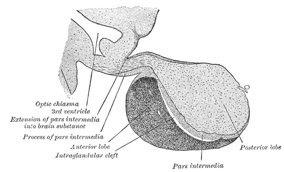
Pituitary gland. Labelled illustration from Wikimedia Commons, used under its open licence.

Figure 2: Pituitary gland.
Superior to the gland lies the diaphragma sellae, a dural fold with a central aperture for the infundibulum. Anteriorly, the tuberculum sellae forms the anterior boundary. Posteriorly, the dorsum sellae supports the posterior clinoid processes. The pituitary gland itself is divided into two main lobes. The anterior lobe, or adenohypophysis, is larger and lies anteriorly. The posterior lobe, or neurohypophysis, is smaller and lies posteriorly. Between them is the pars intermedia, a thin layer often indistinct in adults. The infundibulum, or pituitary stalk, connects the hypothalamus above to the posterior pituitary below. The pars tuberalis is a slender tube of anterior pituitary tissue that wraps around the infundibulum. The anterior pituitary originates from the digestive tract in the embryo and migrates toward the brain during fetal development. The three regions of the anterior pituitary are the pars distalis, the most anterior part; the pars intermedia, adjacent to the posterior pituitary; and the pars tuberalis. The posterior pituitary consists of neural tissue extending from the hypothalamus. It stores hormones produced in the hypothalamus. The anterior pituitary synthesizes its own hormones. These include thyroid-stimulating hormone (TSH), also called thyrotropin. TSH is released from the anterior pituitary in response to thyrotropin-releasing hormone (TRH) from the hypothalamus. It triggers the secretion of thyroid hormones by the thyroid gland. In a classic negative feedback loop, elevated levels of thyroid hormones in the bloodstream then trigger a drop in production of TRH and subsequently TSH. The cells in the zone between the pituitary lobes secrete a hormone known as melanocyte-stimulating hormone (MSH) that is formed by cleavage of the pro-opiomelanocortin (POMC) precursor protein. Local production of MSH in the skin is responsible for melanin production in response to UV light exposure. The role of MSH made by the pituitary is more complicated. For instance, people with lighter skin generally have the same amount of MSH as people with darker skin. Nevertheless, this hormone is capable of darkening of the skin by inducing melanin production in the skin’s melanocytes.
The pituitary gland, or hypophysis, sits within the sella turcica, a saddle-shaped depression in the body of the sphenoid bone. This anatomical position places it at the centre of the skull base, surrounded by critical neurovascular structures. Understanding these relations is fundamental for both clinical examination and surgical approaches, such as the transsphenoidal route, which accesses the gland through the sphenoid sinus. [1]
Figure 3Gray1180
Long description
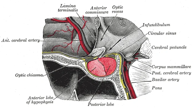
The hypophysis cerebri in position. Shown in sagittal section.

Figure 3: The hypophysis cerebri in position (shown in sagittal section).
Figure 4Gray721
Long description
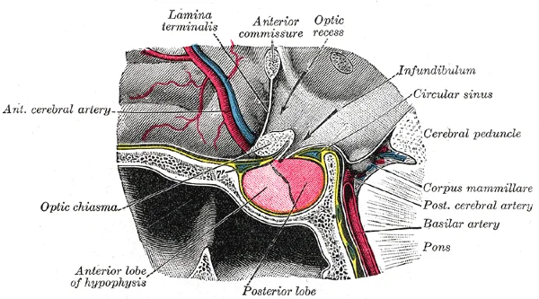
The hypophysis cerebri, in position. Shown in sagittal section.

Figure 4: The hypophysis cerebri, in position (shown in sagittal section).
Figure 51806 The Hypothalamus-Pituitary Complex
Long description
The hypothalamus region lies inferior and anterior to the thalamus. It connects to the pituitary gland by the stalk-like infundibulum. The pituitary gland consists of an anterior and posterior lobe, with each lobe secreting different hormones in response to signals from the hypothalamus.

Figure 5: The hypothalamus region lies inferior and anterior to the thalamus.
The anterior lobe (adenohypophysis) lies anteriorly, while the posterior lobe (neurohypophysis) is posterior. The sphenoid sinus lies inferior to the sella, separated only by a thin layer of bone. This relationship is crucial for the transsphenoidal surgical approach, where instruments pass through the nasal cavity and sphenoid sinus to reach the gland.
Superiorly, the pituitary gland is related to the optic chiasm. In most individuals, the chiasm lies above and anterior to the gland. This relation is clinically significant because enlargement of the pituitary, such as in a macroadenoma, can compress the chiasm, leading to bitemporal hemianopia. Laterally, the gland is bordered by the cavernous sinuses. These venous sinuses contain the internal carotid arteries and cranial nerves III, IV, V1, V2, and VI. Tumours extending laterally into the cavernous sinus can cause cranial nerve palsies, resulting in eye movement abnormalities or facial sensory loss.
The sella turcica itself is formed by the sphenoid bone. Anteriorly, it is bounded by the tuberculum sellae. Posteriorly, it is bounded by the dorsum sellae. The floor of the sella is formed by the body of the sphenoid. The sphenoid sinus, an air-filled cavity within the sphenoid bone, lies directly inferior to the sella. The thickness of the bone between the sinus and the sella varies between individuals, which is an important consideration for surgical planning.
The vascular supply to the region is complex. The internal carotid arteries run through the cavernous sinuses, lateral to the pituitary. Branches from the internal carotid, such as the meningo-hypophyseal trunk, contribute to the blood supply of the gland and its coverings. The venous drainage is primarily into the cavernous sinuses and the inferior petrosal sinuses.
Imaging of the pituitary gland is typically performed using magnetic resonance imaging (MRI), which provides excellent soft tissue contrast. MRI allows for detailed visualization of the gland, stalk, and surrounding structures, aiding in the diagnosis of tumours and other pathologies. The transsphenoidal route remains the most common surgical approach for pituitary tumours, leveraging the anatomical relationship between the sphenoid sinus and the sella turcica.
The pituitary gland, or hypophysis, sits within the sella turcica of the sphenoid bone. It comprises two distinct embryological origins and functional units: the anterior pituitary (adenohypophysis) and the posterior pituitary (neurohypophysis). This view displays the gland in sagittal section, revealing the anterior lobe occupying the anterior two-thirds of the sella and the posterior lobe occupying the posterior third. The infundibulum, or pituitary stalk, connects the posterior lobe superiorly to the hypothalamus. The anterior lobe is composed of glandular epithelial cells arranged in cords and clusters, separated by a rich network of fenestrated capillaries known as the hypophyseal portal system. These vessels transport releasing and inhibiting hormones from the hypothalamus directly to the anterior pituitary cells. The posterior lobe consists largely of unmyelinated axons terminating in Herring bodies, which store oxytocin and vasopressin produced in the hypothalamus.
The anterior pituitary produces seven hormones. These are the growth hormone (GH), thyroid-stimulating hormone (TSH), adrenocorticotropic hormone (ACTH), follicle-stimulating hormone (FSH), luteinizing hormone (LH), beta endorphin, and prolactin. Of the hormones of the anterior pituitary, TSH, ACTH, FSH, and LH are collectively referred to as tropic hormones (trope- = “turning”) because they turn on or off the function of other endocrine glands. The endocrine glands secrete a variety of hormones that control the development and regulation of the reproductive system (these glands include the anterior pituitary, the adrenal cortex, and the gonads—the testes and the ovaries). Much of the development of the reproductive system occurs during puberty and is marked by the development of sex-specific characteristics in adolescents. Puberty is initiated by gonadotropin-releasing hormone (GnRH), a hormone produced and secreted by the hypothalamus. GnRH stimulates the anterior pituitary to secrete gonadotropins—hormones that regulate the function of the gonads. The levels of GnRH are regulated through a negative feedback loop; high levels of reproductive hormones inhibit the release of GnRH. Throughout life, gonadotropins regulate reproductive function and, in the case of females, the onset and cessation of reproductive capacity. [1]
At 4 mm stage (28 days), the ICA branches off into the anterior and the posterior division. [1] [2]
DefinitionAdenohypophysis
The glandular anterior lobe of the pituitary, responsible for synthesising and secreting multiple peptide hormones under hypothalamic control.
DefinitionNeurohypophysis
The neural posterior lobe of the pituitary, storing and releasing hormones synthesised in the hypothalamus.
The blood supply to the pituitary gland is dual in nature, reflecting its embryological origin from both neural and mesodermal tissues. The anterior pituitary (adenohypophysis) is supplied by the hypophyseal portal system. This system begins with the superior hypophyseal arteries, which branch from the internal carotid artery and the posterior communicating artery. These arteries supply the median eminence and the infundibular stalk, forming a primary capillary network. Blood from this network drains into the hypophyseal portal veins, which descend along the stalk to the anterior pituitary. This arrangement allows hypothalamic hormones to reach the anterior pituitary in high concentrations without systemic dilution. [1]
The posterior pituitary (neurohypophysis) receives its blood supply directly from the systemic circulation. The inferior hypophyseal arteries, branches of the internal carotid artery, supply the posterior lobe. These arteries form a capillary network within the posterior pituitary, allowing for the direct release of antidiuretic hormone and oxytocin into the bloodstream. This direct vascular connection contrasts with the portal system of the anterior pituitary, ensuring rapid hormonal response to physiological changes. [1]
The sella turcica itself is surrounded by the cavernous sinuses, which contain the internal carotid arteries and cranial nerves. The arterial supply to the surrounding structures includes branches from the middle meningeal artery and the ethmoidal arteries. Understanding this vascular anatomy is crucial for interpreting imaging studies and planning surgical approaches to pituitary lesions. The close relationship between the pituitary gland and the internal carotid arteries means that pathology in one can affect the other, such as aneurysms compressing the gland or pituitary tumors invading the cavernous sinus.
The venous drainage of the pituitary gland and sella turcica is a specialized system that facilitates the hypothalamic-pituitary portal circulation. This arrangement allows hypothalamic releasing and inhibiting hormones to reach the anterior pituitary directly without entering the systemic circulation first. This unique vascular architecture ensures high concentrations of regulatory hormones are delivered precisely to the target cells. [1]
The pituitary gland is centrally located within the bony sella turcica. The cavernous sinuses are lateral to the gland, receiving venous drainage from the posterior pituitary via the inferior hypophyseal vein. The sphenoid sinus lies inferior to the sella turcica.
The cavernous sinuses drain anteriorly into the superior ophthalmic vein and posteriorly into the superior and inferior petrosal sinuses. The superior petrosal sinus drains into the transverse sinus, while the inferior petrosal sinus drains into the internal jugular vein. This drainage pathway is clinically significant because infections or thrombi in the facial veins can spread to the cavernous sinus via the ophthalmic veins, leading to cavernous sinus thrombosis.
The venous drainage of the pituitary gland is closely related to the surrounding structures. The diaphragma sellae, a dural fold, covers the sella turcica and has a central opening for the pituitary stalk. The cavernous sinuses contain the internal carotid artery and cranial nerves III, IV, V1, V2, and VI. Compression of these structures by a pituitary tumor can lead to specific neurological deficits. For example, compression of cranial nerve III can cause ptosis and a "down and out" eye position. The proximity of the venous drainage to the sphenoid sinus is also important for surgical access. Transsphenoidal surgery utilizes the sphenoid sinus to reach the pituitary gland, avoiding brain retraction. Understanding the venous anatomy is crucial for minimizing bleeding and complications during such procedures.
The lymphatic drainage of the pituitary gland and sella turcica follows a distinct pathway that bypasses the typical regional nodes, reflecting the gland’s unique embryological and anatomical position. Understanding this route is critical for appreciating how pathology in the sellar region may or may not present with lymphadenopathy.
The infundibulum, or pituitary stalk, extends upwards from the gland to connect with the hypothalamus. Instead, they follow the course of the internal carotid arteries. From the cavernous sinus, the lymphatic flow is directed towards the retropharyngeal nodes.
The primary route of drainage is towards the retropharyngeal lymph nodes. These nodes are located in the space between the buccopharyngeal fascia and the prevertebral fascia. They receive lymph from the nasopharynx, the posterior nasal cavity, and the paranasal sinuses. The pituitary gland’s proximity to the sphenoid sinus and the clivus facilitates this drainage pattern. The lymphatic vessels accompanying the internal carotid artery are a key feature. The cavernous sinus contains the internal carotid artery and several cranial nerves, including the oculomotor, trochlear, abducens, and the ophthalmic and maxillary divisions of the trigeminal nerve. The lymphatic vessels run alongside these structures, ensuring that any fluid or cells from the pituitary region are transported away from the central skull base.
It is important to note that the pituitary gland lacks a direct connection to the superficial cervical lymph nodes. This absence explains why primary pituitary tumours rarely present with palpable neck lumps. The drainage is deep and central, focusing on the retropharyngeal and paratracheal regions. This anatomical feature has clinical implications for the staging of pituitary lesions and the interpretation of imaging studies. When evaluating a patient with a sellar mass, radiologists look for signs of extension into the cavernous sinus or the sphenoid sinus, rather than lymph node enlargement. The lymphatic system in this region is sparse compared to other parts of the head and neck, reflecting the gland’s protected location within the bony sella turcica.
Clinical pearlClinical Relevance
The lack of direct lymphatic drainage to superficial nodes means pituitary adenomas do not typically cause cervical lymphadenopathy, distinguishing them from other head and neck malignancies.
The anatomical relationships described are consistent across standard anatomical texts. The sella turcica, named for its saddle-like shape, houses the pituitary gland. The anterior wall of the sella is formed by the tuberculum sellae, while the posterior wall is formed by the dorsum sellae. The floor of the sella is formed by the body of the sphenoid bone, which is pneumatized by the sphenoid sinus in most adults. This pneumatization varies among individuals, affecting the surgical approach to the pituitary gland via the transsphenoidal route. The lymphatic drainage pattern remains constant regardless of the degree of sinus pneumatization, as the vessels follow the vascular structures rather than the air-filled spaces.
Figure 61807 The Posterior Pituitary Complex
Long description
Neurosecretory cells in the hypothalamus release oxytocin (OT) or ADH into the posterior lobe of the pituitary gland. These hormones are stored or released into the blood via the capillary plexus.

Figure 6: Neurosecretory cells in the hypothalamus release oxytocin (OT) or ADH into the posterior lobe of the pituitary gland.
This dual supply explains the specific patterns of referred pain often reported in clinical practice. Understanding these connections requires an examination of the sympathetic, parasympathetic, and sensory contributions. [2]
Sympathetic fibres reach the pituitary via the superior hypophyseal arteries. These fibres originate from the internal carotid plexus. They travel along the arterial walls to the anterior lobe. This supply is primarily vasomotor, regulating blood flow rather than glandular secretion directly.
Parasympathetic innervation is derived from the facial nerve. Fibres travel via the greater petrosal nerve. They synapse in the sphenopalatine ganglion. Postganglionic fibres then join the maxillary nerve. These fibres eventually reach the pituitary via the lesser petrosal nerve and the otic ganglion. This pathway modulates local vascular tone.
Sensory innervation is critical for pain perception. The dura mater of the sella turcica is supplied by the ophthalmic division of the trigeminal nerve. Fibres from the cavernous sinus also contribute to this sensory network. Pain from pituitary lesions often refers to the frontal region or the eye. This referral pattern follows the trigeminal distribution.
The pituitary gland sits centrally within the sella turcica. The optic chiasm lies superior to the gland. The cavernous sinuses are lateral to the gland. The parasympathetic fibres from the sphenopalatine ganglion are traced towards the gland.
Clinical pearlReferred Pain
Pituitary pain often refers to the frontal region due to trigeminal innervation of the dura.
The integration of these nerve supplies ensures both vascular control and sensory feedback. Disruption of these pathways can lead to significant clinical symptoms. Recognition of the anatomical course is essential for surgical planning.
| Nerve Type | Origin | Target | Function |
|---|---|---|---|
| Sympathetic | Internal Carotid Plexus | Anterior Lobe | Vasomotor Control |
| Parasympathetic | Facial Nerve | Gland | Vascular Modulation |
| Somatic | Trigeminal Nerve | Dura Mater | Pain Sensation |
The pituitary gland, or hypophysis, sits within the sella turcica of the sphenoid bone. It comprises two distinct embryological origins: the adenohypophysis (anterior lobe) and the neurohypophysis (posterior lobe). The adenohypophysis arises from Rathke’s pouch, an ectodermal upgrowth of the stomodeum. The neurohypophysis develops from a downgrowth of the diencephalon.
The anterior pituitary contains three main cell types based on staining affinity. Acidophils stain with acidic dyes and include somatotrophs and lactotrophs. Somatotrophs secrete growth hormone. Lactotrophs secrete prolactin. Basophils stain with basic dyes and include corticotrophs, thyrotrophs, and gonadotrophs. Corticotrophs produce adrenocorticotropic hormone. Thyrotrophs produce thyroid-stimulating hormone. Gonadotrophs produce follicle-stimulating hormone and luteinising hormone. Chromophobes have few secretory granules and may represent degranulated acidophils or basophils.
The posterior pituitary consists mainly of unmyelinated axons and pituicytes. It stores hormones produced in the hypothalamus. Oxytocin and vasopressin travel down hypothalamo-hypophyseal tracts. These hormones are released from nerve terminals in the posterior lobe.
Pituitary neuroendocrine tumours (PitNETs) are classified by the WHO.
DefinitionPitNET
Pituitary neuroendocrine tumour.
| Cell Type | Staining | Hormone Secreted |
|---|---|---|
| Somatotroph | Acidophil | Growth hormone |
| Lactotroph | Acidophil | Prolactin |
| Corticotroph | Basophil | ACTH |
| Thyrotroph | Basophil | TSH |
| Gonadotroph | Basophil | FSH, LH |
| Chromophobe | Neutral | None (or degranulated) |
The pituitary gland, often referred to as the master gland, arises from two distinct embryological origins that converge to form the complex endocrine structure found within the sella turcica. Understanding its development requires linking to the embryology page for detailed histological lineage, but the gross anatomical formation is a classic example of ectodermal interaction.
The anterior pituitary, or adenohypophysis, develops from an upward growth of oral ectoderm known as Rathke’s pouch. This pouch originates from the roof of the primitive mouth (stomodeum). It migrates superiorly, passing between the developing forebrain and the nasal placode. As it ascends, it loses its connection to the oral cavity, though a remnant stalk, the infundibulum, persists. The anterior lobe consists of glandular tissue responsible for synthesizing and secreting multiple tropic hormones.
The posterior pituitary, or neurohypophysis, has a different origin. It develops as a downward extension of the diencephalon, specifically from the floor of the third ventricle. This neural tissue forms the infundibular stalk and the posterior lobe. Unlike the anterior lobe, the posterior pituitary does not synthesize hormones. Instead, it stores and releases oxytocin and vasopressin (antidiuretic hormone), which are produced by neuronal cell bodies in the hypothalamus. These hormones travel down axons through the infundibulum to be released into the systemic circulation from the posterior lobe.
The sella turcica, a saddle-shaped depression in the sphenoid bone, houses the pituitary gland. It is formed during cranial base development. The anterior part is the tuberculum sellae, and the posterior part is the dorsum sellae. The hypophyseal fossa lies between them. This bony enclosure protects the gland and provides attachment points for the diaphragma sellae, a dural fold that covers the superior aspect of the sella.
The relationship between the developing gland and surrounding structures is critical. The optic chiasm lies anterior and superior to the gland. The cavernous sinuses are located laterally. The sphenoid sinus is inferior. Any anomaly in the descent of Rathke’s pouch or the neural invagination can lead to congenital defects such as ectopic pituitary tissue or aplasia.
DefinitionRathke's Pouch
An ectodermal outpouching from the roof of the mouth that forms the anterior pituitary.
DefinitionInfundibulum
The stalk connecting the hypothalamus to the posterior pituitary, derived from neural tissue.
The final structure is a composite of glandular and neural tissues, reflecting its dual embryological origin. This unique development explains the distinct vascular supply and hormonal control mechanisms of the two lobes. The anterior lobe receives blood via the hypophyseal portal system from the hypothalamus, while the posterior lobe receives direct neural input.
The sella turcica is a saddle-shaped depression in the sphenoid bone that houses the pituitary gland. On axial imaging, the gland sits centrally within this bony enclosure. Superior to the gland lies the diaphragma sellae, a dural fold that forms the roof of the sella. This structure separates the pituitary fossa from the suprasellar cistern.
The pituitary gland itself is divided into two main lobes with distinct radiological appearances. The anterior lobe, or adenohypophysis, constitutes the majority of the gland's volume. It appears isointense to grey matter on T1-weighted images and slightly hyperintense on T2-weighted images. The posterior lobe, or neurohypophysis, is smaller and located posteriorly. It typically demonstrates high signal intensity on T1-weighted sequences due to the presence of neurosecretory granules. This 'bright spot' is a crucial landmark for identifying the posterior pituitary.
Lateral to the pituitary gland run the cavernous sinuses. These paired venous channels contain the internal carotid arteries and several cranial nerves. The internal carotid artery courses through the cavernous sinus, forming a concavity on the lateral wall of the sella turcica. This indentation is known as the carotid impression. The optic chiasm lies superior and anterior to the pituitary gland. It rests on the tuberculum sellae, which forms the anterior boundary of the sella. The optic nerves extend laterally from the chiasm towards the orbits.
Inferiorly, the pituitary gland rests on the sphenoid sinus. The floor of the sella turcica is thin and may be dehiscent in some individuals. This anatomical relationship is significant for transsphenoidal surgical approaches. The infundibulum, or pituitary stalk, connects the gland to the hypothalamus. It passes through the diaphragma sellae to reach the median eminence. The stalk is usually midline but can deviate slightly.
DefinitionSella Turcica
A saddle-shaped depression in the body of the sphenoid bone that houses the pituitary gland.
Clinical pearlPosterior Pituitary Signal
The posterior pituitary appears bright on T1-weighted MRI due to neurosecretory granules, serving as a key diagnostic landmark.
| Structure | Location | Radiological Feature |
|---|---|---|
| Anterior Lobe | Central sella | Isointense T1, Hyperintense T2 |
| Posterior Lobe | Posterior sella | Hyperintense T1 ('Bright Spot') |
| Cavernous Sinus | Lateral to sella | Contains ICA and CN III, IV, V1, V2, VI |
| Optic Chiasm | Superior/anterior | Crosses above tuberculum sellae |
| Sphenoid Sinus | Inferior to sella | Air-filled, surgical access route |
The surrounding bony structures provide context for normal anatomy. The clinoid processes anchor the diaphragma sellae. The anterior clinoid process is located anteriorly, while the posterior clinoid process is situated posteriorly. These landmarks help orient the viewer on axial and coronal slices. Understanding these relationships is essential for interpreting pituitary pathology.
The pituitary gland resides within the sella turcica, a bony depression in the sphenoid bone. This anatomical position places it in close proximity to critical neurovascular structures. Superiorly, the gland is related to the optic chiasm. Lateral relations include the cavernous sinuses, which contain the internal carotid arteries and cranial nerves III, IV, V1, V2, and VI. Inferiorly, the gland rests on the sphenoid sinus. Understanding these relations is essential for interpreting imaging and surgical approaches.
The pituitary gland is centrally located within the sella turcica. The cavernous sinus is lateral to the gland, containing the internal carotid artery. The sphenoid sinus is inferior.
Pituitary neuroendocrine tumours (PitNETs) are classified by behaviour. This classification aims to facilitate early recognition and aggressive treatment. [3]
Aggressive and metastatic PitNETs usually present as invasive “macroadenomas”, less frequently as giant tumors (largest diameter ≥4 cm), and display a highly variable clinical course. The most common subtypes are corticotroph and lactotroph tumors. Functioning corticotroph tumors (Cushing's disease) represent 40%–50% of the aggressive tumors, and 60% of the patients are men. This differs from the situation among “ordinary” non-aggressive PitNETs where only 5%–10% have Cushing's disease and the typical patient is a woman. [3]
Most aggressive tumors are identified only after multiple recurrences after surgery or upon progression after radiotherapy. Metastatic tumors behave similarly to the aggressive ones but are only identified after dissemination. [3]
Specific clinical characteristics should raise a suspicion of an aggressive tumor. These include a man with an invasive corticotroph macroadenoma, macro/giant lactotroph tumors initially not responding to, or losing sensitivity to dopamine agonists, somatotroph tumors demonstrating increasing GH levels /tumor progression while on somatostatin analogues, and patients with non-functioning invasive macroadenomas switching to clinically functioning, especially those becoming ACTH secreting. [3]
| Feature | Description |
|---|---|
| Presentation | Invasive macroadenomas or giant tumors (≥4 cm) |
| Common Subtypes | Corticotroph and lactotroph |
| Corticotroph Demographics | 40%–50% of aggressive tumors; 60% men |
| Definition | Large, invasive, rapid growth, or growth despite therapy |
| Identification | After recurrence or progression post-surgery/radiotherapy |
The pituitary gland sits within the sella turcica, a bony depression in the sphenoid bone. It is divided into the anterior pituitary (adenohypophysis) and posterior pituitary (neurohypophysis). The anterior pituitary secretes glycoprotein hormones including follicle-stimulating hormone (FSH) and luteinizing hormone (LH). FSH stimulates the production and maturation of sex cells, or gametes, including ova and sperm. LH triggers ovulation, as well as the production of estrogens and progesterone by the ovaries. LH stimulates production of testosterone by the testes. [1]
Clinical assessment often involves imaging the sella turcica. Congenital anomalies of cerebral arteries are relevant here. Fenestration is defined as the splitting of the arterial lumen into two different channels that eventually fuse during their course, restoring the primary vessel. [2]
Persistent carotid-vertebrobasilar anastomoses are embryological remnants.
Exam trapHypoplasia vs Absence
A hypoplastic artery has a patent lumen but is small (<1 mm), whereas an absent artery is completely missing. Do not confuse the two in imaging reports.
DefinitionFenestration
Fenestration is defined as the splitting of the arterial lumen into two different channels that eventually fuse during their course, restoring the primary vessel.
DefinitionGonadotropins
The gonadotropins include two glycoprotein hormones: follicle-stimulating hormone (FSH) stimulates the production and maturation of sex cells, or gametes, including ova and sperm. [1]
Clinical pearlLH Function
LH triggers ovulation, as well as the production of estrogens and progesterone by the ovaries.
Students frequently confuse the physiological effects of alcohol with pathological states of antidiuretic hormone. A common error is assuming that alcohol stimulates ADH release. In reality, alcohol consumption inhibits the release of ADH, resulting in increased urine production that can eventually lead to dehydration and a hangover. This mechanism is distinct from diabetes insipidus, a disease characterised by chronic underproduction of ADH that causes chronic dehydration. In diabetes insipidus [1]
Another significant pitfall involves the interpretation of skull-base imaging regarding the internal carotid artery. An aberrant ICA pathway occurs when the ICA deviates from its anatomical landmarks. The most commonly reported aberrant ICA involves the middle ear. In this variant, the ICA enters the floor of the middle ear and then turns forward, passing through the middle ear subsequently emerging through the foramen lacerum. Students often mistake this for a pathological anomaly, but the basic pathology is believed to be hypoplasia or agenesis of the ICA due to persistent collaterals. This condition substitutes the cervical and vertical petrous portions of the ICA through branches of the external carotid artery. An aberrant ICA pathway might be accompanied by a persistent stapedial artery. The persistent stapedial artery is a temporary embryonic artery that typically regresses by the third fetal month. Its reported prevalence is 0.5%. This artery supplies the middle meningeal artery territory, leading to the absence of the foramen spinosum. Absence of the foramen spinosum is seen in up to 3% of skull-base CT studies. This is an indirect sign of a persistent stapedial artery, but it can also be related to an ophthalmic origin of the middle meningeal artery. [2]
Students also misinterpret the significance of hypoplastic arteries. Hypoplastic arteries are probably the most common variants seen in adults. While their significance has not been fully validated in population-based studies, autopsy series have shown that hypoplastic arteries are more commonly found in patients who have had brain infarcts. The lumen of hypoplastic arteries is typically patent. In the absence of proximal large artery stenosis, the communicating arteries seem to affect the final flow volume to the brain only minimally. However, in the setting of large artery occlusion or severe stenosis, the communicating arteries become crucial. The caliber of the communicating arteries is inversely proportional to the risk of stroke. The smaller these communicating arteries are, the lesser the capacity to make up for the reduced or lost flow.
Azygous anterior cerebral artery is another frequent point of confusion. An azygous ACA, found in up to 2% of the population, consists of a single midline artery that arises from the confluence of both A1 segments and an absent anterior communicating artery. It supplies the medial aspects of both frontal and parietal lobes. This variant has been associated with saccular aneurysm in the same vessel. The hypothesis is that the increased flow resulting from the fused A2 leads to progressive dilatation of the wall. Additionally, as the azygous ACA can also be found in association with agenesis of corpus callosum, holoprosencephaly, and arteriovenous malformation, a more generalized abnormality of the tissues cannot be ruled out. [2]
This knowledge is applied at the bedside to manage fluid and electrolyte balance. Understanding the inhibition of ADH by alcohol explains the pathophysiology of dehydration and hangovers. Recognising diabetes insipidus allows clinicians to identify chronic dehydration despite high fluid intake. In the operating theatre, awareness of aberrant vascular pathways is critical. An aberrant ICA pathway involves the middle ear, where the ICA enters the floor and emerges through the foramen lacerum. Surgeons must recognise that this may be accompanied by a persistent stapedial artery. The persistent stapedial artery supplies the middle meningeal artery territory. On imaging, the absence of the foramen spinosum is a key indicator. This absence is seen in up to 3% of skull-base CT studies. It serves as an indirect sign of a persistent stapedial artery. True ICA agenesis, aplasia, or hypoplasia is reported in 0.01% of the population. [2]
In examinations, students must distinguish between normal and variant anatomy of the vertebral artery. The vertebral artery normally arises from the upper, posterior surface of the subclavian artery 0.5 to 2.0 cm medial to the thyrocervical trunk. Variants include a common trunk with the thyrocervical trunk (3.0%). An aortic origin of the left vertebral artery occurs in 2.5% of cases, arising between the left common carotid artery and the left subclavian artery. A right vertebral artery arising [2]
Imaging also reveals variations in the anterior cerebral artery. The A1 segment is among the most frequent sites of anatomic variation in the circle of Willis. An azygous ACA is found in up to 2% of the population. It consists of a single midline artery arising from the confluence of both A1 segments. A bihemispheric ACA exists when both A2 segments exist, but one is dominant. Occasionally, a small medial ACA arises from the anterior communicating artery. A persistent hyoid artery variant is occasionally seen, where the hyoid artery supplies the posterior inferior cerebellar artery directly. A higher origin is expected for the hyoid artery arising from the internal carotid artery at the level of the C1 vertebra or C1-C2 interspace. The origin of the proatlantal artery is typically at the C2 or C3 vertebra.
In clinical practice, the geometry of the carotid bifurcation influences disease. Intima thickening and atheromas rarely occur in the flow divider. They are more common in the outer wall of the bifurcation, where low or oscillating wall shear stress is found. Among young individuals (24±4 years), the inter-individual variability in the common carotid to internal carotid ratio is lower than among older individuals (63±10 years). This suggests that with aging, the geometry of the carotid bifurcation remodels into more heterogeneous configurations. This geometry is influenced by demographic variables such as sex and ethnicity, and by traditional vascular risk factors. The interplay between geometry and risk factors explains the broader spectrum of disease among older individuals.
Clinical pearlImaging Sign
Absence of the foramen spinosum on skull-base CT is an indirect sign of a persistent stapedial artery, seen in up to 3% of studies.
In examinations, students must distinguish between normal and variant anatomy of the vertebral artery. The vertebral artery normally arises from the upper, posterior surface of the subclavian artery 0.5 to 2.0 cm medial to the thyrocervical trunk. Variants include a common trunk with the thyrocervical trunk (3.0%). An aortic origin of the left vertebral artery occurs in 2.5% of cases, arising between the left common carotid artery and the left subclavian artery. A right vertebral artery arising at the site of bifurcation of the innominate artery occurs in 1.1% of cases. [2]
Alcohol inhibits ADH release, causing increased urine production and dehydration. Diabetes insipidus involves chronic underproduction of ADH, leading to chronic dehydration. An aberrant ICA may enter the middle ear floor and emerge through the foramen lacerum. A persistent stapedial artery is present in 0.5% of the population. Absence of the foramen spinosum is seen in up to 3% of skull-base CT studies. True ICA agenesis or hypoplasia is reported in 0.01% of the population. An azygous ACA is found in up to 2% of the population. The V1 segment of the vertebral artery ends at the 6th vertebra in 87.5% of cases. [2]
Answer each one in your head first, then open the answer.
What effect does alcohol consumption have on ADH release?
Show answerHide answer
It inhibits the release of ADH, resulting in increased urine production that can eventually lead to dehydration and a hangover.What is the reported prevalence of a persistent stapedial artery?
Show answerHide answer
0.5%.In what percentage of skull-base CT studies is absence of the foramen spinosum seen?
Show answerHide answer
Up to 3%.What percentage of the population has an azygous ACA?
Show answerHide answer
Up to 2%.At which vertebra does the V1 segment of the vertebral artery end in the majority of cases?
Show answerHide answer
The 6th vertebra (87.5%).
- Betts JG, Young KA, Wise JA, et al. The Pituitary Gland and Hypothalamus. OpenStax, Anatomy and Physiology 2e (CC BY-NC-SA 4.0). Accessed .
- Menshawi K, Mohr JP, Gutierrez J. A Functional Perspective on the Embryology and Anatomy of the Cerebral Blood Supply. Journal of stroke 2015;17(2):144-158. doi:10.5853/jos.2015.17.2.144. Accessed .
- Casar-Borota O, Burman P, Lopes MB. The 2022 WHO classification of tumors of the pituitary gland: An update on aggressive and metastatic pituitary neuroendocrine tumors. Brain pathology (Zurich, Switzerland) 2025;35(1):e13302. doi:10.1111/bpa.13302. Accessed .
Figure credits
- Figure 1: Unknown author (see the source page), via Wikimedia Commons, Public domain, Public domain.
- Figure 2: Henry Vandyke Carter, via Wikimedia Commons, Public domain, Public domain.
- Figure 3: Henry Vandyke Carter, via Wikimedia Commons, Public domain, Public domain.
- Figure 4: Henry Vandyke Carter, via Wikimedia Commons, Public domain, Public domain.
- Figure 5: OpenStax College, via Wikimedia Commons, CC BY 3.0, CC BY 3.0.
- Figure 6: OpenStax College, via Wikimedia Commons, CC BY 3.0, CC BY 3.0.
Educational content for medical students and doctors. It is not a substitute for clinical judgement or local guidelines.