Acid-base chemistry, buffers and blood gases
Comprehensive medical student guide to acid-base chemistry, buffers, blood gas physiology and buffering mechanisms.
Figure 1Concentration acide faible pH diag log
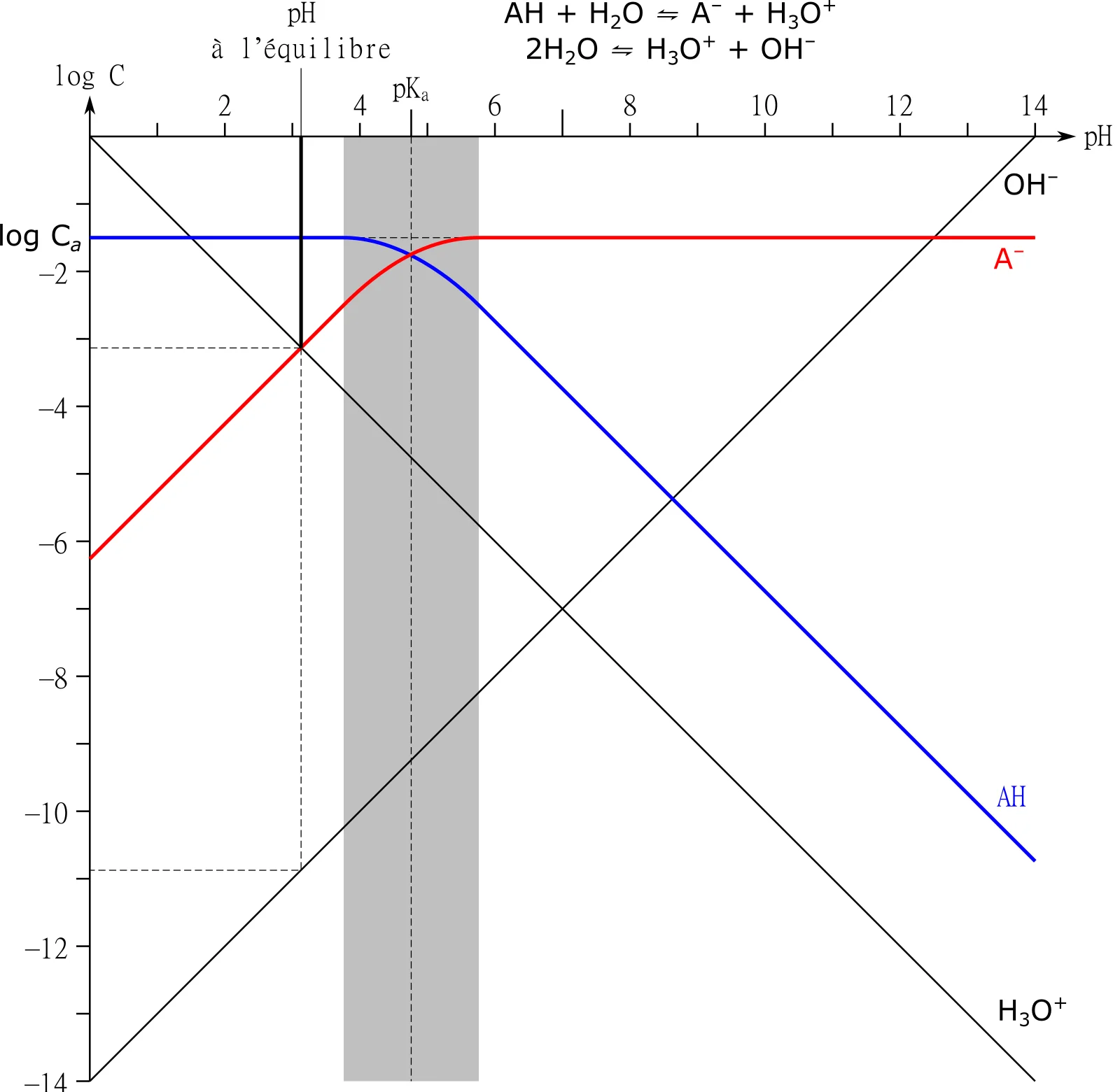
Long description
Log-log diagram of the concentration of chemical species versus the pH in the case of a weak acid, which dissociation constant is Ka, when a concentration Ca of salt of this acid is put in pure water.

Acid-base chemistry forms the bedrock of physiological homeostasis, governing every biochemical reaction within the human body. From first principles, an acid is defined as a chemical entity capable of donating a hydrogen ion, whilst a base is defined as a chemical entity capable of accepting a hydrogen ion. In clinical medicine and physiological contexts, hydrogen ion concentration is conventionally expressed using the negative logarithm scale, known as pH. The extracellular fluid pH must be maintained within an extremely narrow, critical range to ensure optimal enzymatic function, structural integrity of proteins, and uninterrupted cellular operations. Even very small changes in pH may significantly affect the function of various critical organ systems, such as the central nervous system and the cardiovascular system. [1]
To appreciate how the body maintains this stability, one must examine the physiological sources of acid production. Normal fetal and adult metabolism results in the continuous production of both carbonic acid and various organic acids. Carbonic acid is generated through the hydration of metabolically produced carbon dioxide, which can be rapidly eliminated via the respiratory system. Organic acids, such as lactic acid and ketoacids, are generated through intermediary cellular metabolism and must be buffered and eventually excreted by the renal and metabolic apparatus. Despite this continuous influx of acid loads, the extracellular pH is kept within a critical range due to the efficient buffer mechanisms of the blood system. [1]
When assessing acid-base status, clinicians rely on blood gas analysis to measure key parameters including arterial oxygen tension, partial pressure of carbon dioxide, and actual pH. During normal physiological states such as pregnancy, the maternal respiratory component of the acid-base system shows a progressive fall in arterial carbon dioxide tension alongside a parallel reduction in plasma bicarbonate within the metabolic element. This unique state results in a primary respiratory alkalosis paired with a secondary and compensatory metabolic acidosis, yielding a normal pH that usually sits comfortably on the alkaline side of the normal range. There is no true acidosis of pregnancy, but rather a coordinated reduction of total acid and total base, where the primary fall in the partial pressure of carbon dioxide is buffered by the action of the renal mechanism, which controls loss or conservation of bicarbonate. [1]
DefinitionPhysiological Acid-Base Balance
The dynamic equilibrium between acid production and elimination maintained by chemical buffers, respiratory ventilation, and renal excretion.
In specialized clinical settings, such as intrapartum obstetric care, evaluating acid-base balance becomes critical for detecting fetal compromise. The introduction of fetal blood sampling, whereby arterialized capillary blood could be obtained from the fetus during labor, revolutionized the diagnosis of intrapartum fetal asphyxia. Landmark work showed that a scalp blood pH of less than 7.20 was associated with poor clinical condition of the infant, leading investigators to designate values below 7.20 as acidosis. Furthermore, it was suggested that a fetal scalp blood pH of between 7.20 and 7.25 was indicative of early fetal deterioration, a range designated as preacidosis. Scalp blood pH is therefore useful to increase the specificity of cardiotocography by verifying readings, whilst cord blood pH is useful to differentiate a depressed baby due to hypoxemia and acidosis from other causes. [1]
| Category | pH Threshold | Clinical Interpretation |
|---|---|---|
| Normal | Above 7.25 | Reassuring fetal acid-base status |
| Preacidosis | 7.20 to 7.25 | Indicative of early fetal deterioration |
| Acidosis | Less than 7.20 | Associated with poor infant clinical condition |
Underlying these clinical measurements are complex physiological shifts. Fetal partial pressure of carbon dioxide is high, with approximately 60 percent being carried as bicarbonate. As fetal blood is oxygenated within the placental circulation, it releases hydrogen ions, which combine with bicarbonate and push the chemical equilibrium towards carbon dioxide and water, which then diffuse across the placenta down the concentration gradient for carbon dioxide. In the maternal circulation, the reaction proceeds in the opposite direction. Maternal pH in the placental blood thus falls slightly while that in the fetal placenta rises slightly. This small rise increases the oxygen affinity of fetal hemoglobin, shifting the oxyhemoglobin dissociation curve further to the left while the maternal curve is shifted right, a phenomenon known as the double Bohr shift. This sophisticated interaction enhances maternal to fetal oxygen transfer. [1]
Placental Gas Exchange and Buffering
- Fetal blood oxygenationOxygen binds fetal hemoglobin and releases hydrogen ions.
- Question: Hydrogen ions react with bicarbonate?
Yes
- Equilibrium shiftsForms CO2 and H2O which cross the placenta.
No
- Unbuffered stateLeads to localized acidosis if perfusion fails.
The Henderson-Hasselbalch equation is a mathematical derivation of the law of mass action that describes how the pH of a buffer solution is linked to the concentration of its constituent acid and conjugate base. From first principles, consider a weak acid, designated as HA, which dissociates in aqueous solution into a hydrogen ion and a conjugate base, designated as A minus. The dissociation constant, denoted as Ka, is expressed by multiplying the concentration of hydrogen ions by the concentration of conjugate base, divided by the undissociated acid concentration. By taking the negative logarithm of both sides of this expression and applying logarithmic rules, one arrives at the Henderson-Hasselbalch equation. In the context of the primary extracellular bicarbonate buffer system, the equation relates the blood pH to the pKa of the carbonic acid-bicarbonate system, the concentration of dissolved carbon dioxide, and the concentration of plasma bicarbonate.
In clinical practice, the Henderson-Hasselbalch equation provides the quantitative framework for interpreting arterial blood gas results. The equation is expressed as pH equals pKa plus the logarithm of the ratio of bicarbonate concentration to the concentration of dissolved carbon dioxide. For the carbonic acid-bicarbonate system in human plasma at normal body temperature, the pKa is approximately 6.1. The concentration of dissolved carbon dioxide is directly proportional to the partial pressure of carbon dioxide in the blood, multiplied by the solubility coefficient for carbon dioxide in plasma. This relationship demonstrates that blood pH is not determined by the absolute amounts of acid or base alone, but strictly by the ratio between the bicarbonate concentration and the partial pressure of carbon dioxide.
When analyzing disturbances using this chemical framework, primary alterations can originate from either the metabolic or the respiratory component. For example, during normal human pregnancy, a chronic reduction in the partial pressure of carbon dioxide occurs alongside a compensatory decrease in circulating bicarbonate concentrations, resulting in a state of compensated respiratory alkalosis. The diffusion of blood gases between the fetal and maternal circulations is thought to be driven by this maternal change in partial pressure. However, because of the adequate buffering that occurs within the blood system, there is a relatively marginal change in maternal arterial pH. The physiological consequence of this chronic reduction is a well-regulated adaptation that protects systemic enzyme function while maintaining efficient gas exchange across the placental interface. [1]
Clinical pearlRatio over Absolute Value
Blood pH is determined by the numerical ratio of bicarbonate to dissolved carbon dioxide, rather than the absolute concentration of either component alone.
To further explore how these variables interact, consider situations where pathological processes disrupt the normal homeostatic balance. Chronic clinical conditions associated with fetal and maternal acid-base derangement can originate from maternal, placental, or fetal sources. Maternal causes of chronic fetal acidosis include reduced oxygenation of maternal blood, such as in severe respiratory or cardiac disease, or reduced blood flow to the placenta, as seen in connective tissue diseases like systemic lupus erythematosus and preeclampsia. Furthermore, chronic placental factors usually result from impairment of placental transfer of oxygen. This is thought to result from inadequate trophoblast invasion of the myometrium in early pregnancy, leading to reduced perfusion of the intervillous spaces. In experimental models, such as acute cord compression studies, uteroplacental blood flow needs to be reduced by at least 50 percent to produce significant fetal hypoxia and subsequent acid-base derangement. [1]
Exam trapTransient Abnormalities
An abnormal fetal acid-base balance is not uncommon in a low-risk population during labor, and many cases are transient and of an innocuous respiratory type. [1]
Understanding the quantitative limits predicted by the Henderson-Hasselbalch equation helps clinicians differentiate between acute uncompensated states and chronic compensated disorders. When carbon dioxide is eliminated efficiently through hyperventilation, the numerator-to-denominator ratio in the equation shifts, preventing dramatic drops in pH despite heavy metabolic acid production. Conversely, when renal mechanisms adapt over days, bicarbonate reabsorption is modulated to restore the physiological ratio towards the normal baseline. This mathematical and physiological harmony ensures that cellular metabolism proceeds without the catastrophic inhibition that would otherwise result from uncontrolled hydrogen ion accumulation.
Buffer systems are chemical defenses that minimize changes in hydrogen ion concentration when an acid or base is added to a biological fluid. A buffer typically consists of a weak acid and its conjugate base, which together act as a chemical sponge that can absorb or release hydrogen ions. Within the human body, several distinct buffer systems operate across the intracellular and extracellular compartments. The major buffers utilized by the fetus and adult for neutralizing hydrogen ion production are plasma bicarbonate and hemoglobin, while inorganic phosphates and erythrocyte bicarbonate also serve as potential buffers that play a lesser role in fetal acid-base hemostasis. Each of these systems possesses unique chemical properties and distribution characteristics that dictate its specific physiological contribution. [1]
The bicarbonate buffer system is the most important extracellular buffer in the body, primarily because its components are regulated independently by two major organ systems: the lungs regulate the gaseous carbon dioxide component, while the kidneys regulate the non-volatile plasma bicarbonate component. Although plasma bicarbonate is remarkably potent in adults, fetal and neonatal plasma bicarbonate concentrations are low because of the immaturity of proximal tubular bicarbonate reabsorption. Consequently, buffering via the regulation of bicarbonate is slower in the neonate than in the adult. Despite this developmental immaturity, bicarbonate remains a cornerstone of extracellular defense, rapidly neutralizing metabolic acids as they diffuse through the vascular network. [1]
DefinitionIntracellular Hemoglobin Buffering
Intracellular acidosis in erythrocytes is mitigated predominantly by hemoglobin, which acts as a powerful intracellular buffer for hydrogen ions generated during gas transport.
Inside the erythrocyte, hemoglobin functions as a premier intracellular buffer. The imidazole groups of histidine residues within the globin chains of hemoglobin have pKa values close to physiological intracellular pH, making them exceptionally effective at binding hydrogen ions. During states of chronic physiological adjustment, such as pregnancy-induced respiratory alkalosis, circulatory changes may possibly include intracellular acidosis in the erythrocytes, in which the predominant buffer is hemoglobin. As carbon dioxide diffuses into the red blood cell from metabolizing tissues, it is rapidly converted into carbonic acid by carbonic anhydrase. The resulting hydrogen ions are immediately buffered by reduced hemoglobin, facilitating the transport of carbon dioxide without causing unacceptably large drops in intracellular pH.
Inorganic phosphate plays a complementary role in acid-base regulation. The phosphate buffer system operates chiefly within the intracellular fluid and the renal tubular fluid, where the concentration of phosphate is considerably higher than in plasma. The dihydrogen phosphate ion acts as a weak acid, whilst the monohydrogen phosphate ion acts as a weak base. Because the pKa of the phosphate buffer system is approximately 6.8, which is close to the normal intracellular pH of 7.0, it operates near its maximum buffering capacity. Although inorganic phosphates play a lesser role in fetal acid-base hemostasis compared to plasma bicarbonate and hemoglobin, they are vital for buffering tubular urine, allowing large quantities of hydrogen ions to be excreted by the kidneys without causing the urine pH to drop to damagingly low levels.
| Buffer System | Primary Location | Key Chemical Species | Relative Physiological Role |
|---|---|---|---|
| Bicarbonate | Extracellular fluid, plasma | HCO3- and dissolved CO2 | Major extracellular buffer; regulated by lungs and kidneys |
| Hemoglobin | Intracellular, erythrocytes | Histidine residues on globin | Major intracellular buffer in red blood cells |
| Phosphate | Intracellular, renal tubules | H2PO4- and HPO4(2-) | Important in renal tubular urine and intracellular fluid |
| Plasma Proteins | Extracellular, plasma | Albumin and globulin amino acid residues | Contributes to non-bicarbonate extracellular buffering |
Finally, plasma proteins, notably albumin and various globulins, contribute significantly to non-bicarbonate buffering in the extracellular space. Proteins are amphoteric molecules containing numerous ionizable amino acid side chains, particularly the carboxyl groups of aspartate and glutamate and the amino groups of lysine. These side chains can accept or release hydrogen ions in response to pH fluctuations. Although individual protein molecules have varying pKa values across a broad spectrum, their collective presence in blood plasma provides a robust tertiary defense mechanism against acute acid-base disturbances. Together, bicarbonate, hemoglobin, phosphate, and protein buffers form an integrated network that safeguards physiological function across all organ systems.
Understanding acid-base chemistry requires a solid foundation in first principles regarding how the human body maintains physiological pH within the narrow, life-sustaining range of 7.35 to 7.45. This delicate balance is governed by the bicarbonate-carbon dioxide buffer system, described by the Henderson-Hasselbalch equation. When an acid-base disturbance occurs, the body initiates compensatory mechanisms to return the pH toward normal, though complete normalisation is rare. Respiratory compensation happens rapidly, within minutes to hours, through alterations in alveolar ventilation mediated by the central and peripheral chemoreceptors. For instance, primary metabolic acidosis stimulates hyperventilation, which blows off carbon dioxide and lowers the partial pressure of carbon dioxide in arterial blood. Conversely, primary metabolic alkalosis results in hypoventilation to retain carbon dioxide. Renal compensation is slower, taking three to five days to reach maximal capacity, and involves adjusting the reabsorption of filtered bicarbonate and the excretion of hydrogen ions in the form of titratable acids and ammonium.
To systematically dissect complex acid-base disorders, calculating the anion gap is an indispensable diagnostic step. The anion gap represents the difference between routinely measured unmeasured serum cations and unmeasured anions. The standard formula is Anion Gap = Sodium minus the sum of Chloride and Bicarbonate, expressed in plain text as AG = Na - (Cl + HCO3). The normal reference range for the anion gap typically spans from 8 to 12 millimoles per litre, though this can vary slightly depending on the laboratory assay used, particularly regarding the measurement of serum potassium. An elevated anion gap indicates the presence of unmeasured anions in the extracellular fluid, which consume bicarbonate ions as they are buffered. Common pathological unmeasured anions include lactate, ketones, uraemic toxins, and exogenous ingested poisons such as salicylates, ethylene glycol, or methanol.
DefinitionAnion Gap Definition
The anion gap is the calculated difference between major measured cations and anions in serum, used primarily to classify metabolic acidoses into high anion gap and normal anion gap categories.
When evaluating an elevated anion gap metabolic acidosis, clinicians must calculate the delta-delta or delta ratio to uncover co-existing primary acid-base disorders. The change in the anion gap from normal should theoretically equal the change in serum bicarbonate concentration from its normal value. If the change in anion gap is disproportionately large compared to the drop in bicarbonate, a concurrent metabolic alkalosis is likely present. Conversely, if the drop in bicarbonate is much greater than the increase in the anion gap, a co-existing normal anion gap metabolic acidosis, also known as a hyperchloraemic metabolic acidosis, is present.
| Feature | High Anion Gap Acidosis | Normal Anion Gap Acidosis |
|---|---|---|
| Serum Chloride | Normal | Increased |
| Bicarbonate Level | Decreased | Decreased |
| Common Causes | Lactate, ketoacids, toxins | Diarrhoea, renal tubular acidosis |
| Delta Ratio | Typically greater than 2 for ketoacidosis | Not applicable |
Metabolic acidosis itself frequently stems from tissue oxygen debt and anaerobic metabolism, where incomplete oxidative metabolism of carbohydrates leads to the accumulation of organic acids such as lactic acid. Under conditions of oxygen deficiency, lactic acid is predominantly produced by anaerobic glycogenolysis. Accumulation of lactic acid can deplete the buffer system and result in metabolic acidosis. In extreme clinical scenarios, such as fetal hypoxia, maternal acidosis is associated with lower fetal pH and poor Apgar score. Furthermore, accumulation of lactic acid together with hypoxia leads to tissue oedema and cellular damage. Lactate can be measured in a small blood sample using an electrochemical strip method with results available by the bedside in 60 seconds. It is easier to measure than a full blood gas analysis using maintenance-free, battery-operated pocket-size devices. Lactate is comparable to pH and base deficit with respect to sensitivity, specificity, and predictive values of various perinatal complications. Despite the fact that a wide pH range of 7.00 to 7.16 is quoted in the literature for acidemia, there is a general consensus that a cord artery pH less than 7.00 is more significantly correlated with adverse neonatal outcome. The babies with pH less than 7.00 are more likely to suffer complications in the short term, while long-term outcome is correlated to neonatal encephalopathy rather than pH. Also, base deficit is now used as a surrogate to assess metabolic academia, where the commonest cut-off values to indicate abnormality include a base deficit greater than 8 millimoles as moderate and greater than 12 millimoles as severe metabolic acidosis. [1]
Acid-Base Diagnostic Approach
- Check pHDetermine if acidaemia or alkalaemia is present.
- Question: Is the primary disorder respiratory or metabolic?
Respiratory
- Check pCO2 and evaluate acute versus chronic compensation.
Metabolic
- Calculate the anion gap and evaluate expected compensation.
Interpreting an arterial blood gas requires a disciplined, step-by-step methodology to avoid misdiagnosis and ensure appropriate patient management. The systematic approach begins with evaluating the patient's clinical context alongside the numerical values provided by the blood gas analyzer. Step one is to inspect the pH to establish the primary disorder. A pH below 7.35 indicates acidaemia, while a pH above 7.45 indicates alkalaemia, keeping in mind that a normal pH does not rule out a mixed acid-base disorder where two opposing processes cancel each other out. Step two involves examining the partial pressure of carbon dioxide, which represents the respiratory component. Step three evaluates the serum bicarbonate level or calculated base excess, representing the metabolic component. Step four applies the appropriate compensation rules to determine if the secondary response is appropriate or if a mixed disorder is present.
Consider an illustrative worked example. A patient presents with acute shortness of breath and confusion. The arterial blood gas results show a pH of 7.21, a partial pressure of carbon dioxide of 65 millimetres of mercury, and a bicarbonate concentration of 25 millimoles per litre. Step one reveals a clear acidaemia because the pH is well below 7.35. Step two shows an elevated partial pressure of carbon dioxide, indicating a respiratory acidosis. Step three shows a normal bicarbonate level of 25 millimoles per litre. Step four involves assessing compensation; for an acute respiratory acidosis, the bicarbonate should rise by 1 millimole per litre for every 10 millimetres of mercury increase in carbon dioxide above 40. Here, carbon dioxide is elevated by 25 millimetres of mercury, so the expected bicarbonate is approximately 27 millimoles per litre. Because the measured bicarbonate has not had time to elevate, this represents an acute respiratory acidosis without metabolic compensation, commonly seen in acute opioid overdose or sudden airway obstruction.
Clinical pearlStepwise Interpretation
Always evaluate the pH first to set the primary direction, then look to carbon dioxide and bicarbonate to identify the constituent disorders before calculating expected compensation.
Consider a second illustrative worked example featuring a metabolic disturbance. A patient with severe sepsis presents with rapid, deep breathing. The arterial blood gas reveals a pH of 7.28, a partial pressure of carbon dioxide of 28 millimetres of mercury, and a bicarbonate concentration of 13 millimoles per litre. Serum electrolytes show a sodium of 140, chloride of 100, and bicarbonate of 13, yielding an anion gap of 27 millimoles per litre. Step one identifies acidaemia. Step two shows a low partial pressure of carbon dioxide, indicating hyperventilation. Step three shows a markedly reduced bicarbonate concentration, confirming a primary metabolic acidosis. Step four calculates the anion gap, which is significantly elevated at 27, pointing toward lactic ketoacidosis or a toxic ingestion. Winters formula, which states that the expected carbon dioxide equals 1.5 times the bicarbonate plus 8 with a margin of plus or minus 2, predicts an expected carbon dioxide of 27.5. The measured carbon dioxide of 28 falls precisely within this range, confirming an appropriate respiratory compensation for a high anion gap metabolic acidosis.
It is vital to contrast these findings with specialised clinical settings where extreme physiological stresses occur. In labor, the mother may exhibit forced hyperventilation, and similarly under anesthesia for surgery, the lungs may be excessively inflated, which is akin to forced hyperventilation; both situations can cause fetal acidosis and depression of the newborn. However, it should be noted that moderate hyperventilation could be beneficial as it can improve feto-maternal gradients at placental sites, but may rapidly become associated with adverse maternal and fetal consequences as it gets more pronounced. Usually when a maternal partial pressure of carbon dioxide of 15 millimetres of mercury and a pH of 7.650 is approached, there is a fall in fetal blood oxygen saturation; maternal muscle cramps and fetal heart irregularities in rate and rhythm appear. Experimental studies in animals have shown that excessive hyperventilation causes spasm of placental vessels and slowing of the umbilical circulation with fetal acidosis. Furthermore, hypoxia in fetuses is potentially very hazardous with an adverse outcome for the neonate because the fetus normally has a very low partial pressure of oxygen, approximately 35 millimetres of mercury in the umbilical vein, and a high partial pressure of carbon dioxide, approximately 42 millimetres of mercury in the umbilical vein. The inherent response therefore is preferential redistribution of blood to vital organs, and hence in acute severe fetal hypoxemia, there is associated and sustained maintenance of blood flow to the brain, adrenal gland, and the heart at the expense of other tissues. This results in activation of peripheral chemoreceptors, with an increase in sympathetic tone, and in later gestation there is also an increase in concentrations of circulating cortisol, catecholamines, angiotensin II, and vasopressin, with a resultant increase in peripheral resistance. [1]
Interpreting acid-base disturbances is fraught with potential errors that can lead to inappropriate clinical interventions if clinicians rely on automated machine readouts without critical appraisal. One of the most common pitfalls is failing to assess the clinical picture alongside the blood gas values. A blood gas must never be interpreted in isolation from the patient's history, physical examination, and current medications. Another frequent error is misjudging the chronicity of a disorder. Clinicians often expect full metabolic compensation in an acute respiratory disturbance, forgetting that renal compensation requires several days to reach its full physiological potential. Assuming a normal pH guarantees the absence of acid-base pathology is another major trap; mixed disorders, such as a simultaneous high anion gap metabolic acidosis and a primary respiratory alkalosis, can yield a completely normal pH while the underlying derangements are severely life-threatening.
Exam trapNormal pH Trap
Do not assume a normal pH means acid-base homeostasis; simultaneous opposing primary disorders can normalise the pH while leaving severe underlying pathology unaddressed.
Technical errors in sample collection and handling also introduce significant pitfalls. Air bubbles in the syringe can cause oxygen to diffuse into the sample and carbon dioxide to diffuse out, resulting in a falsely elevated partial pressure of oxygen and a falsely lowered partial pressure of carbon dioxide, which mimics respiratory alkalosis. Excessive heparin volume acts as a strong acid and dilutes the blood sample, leading to artificial depression of bicarbonate and low pH values. Delayed sample processing without prompt refrigeration allows ongoing cellular metabolism by white and red blood cells to consume oxygen and produce carbon dioxide and lactic acid, severely distorting the true in vivo chemistry. Furthermore, failure to correct blood gas values for patient temperature can mislead clinicians, as fever increases metabolic rate and carbon dioxide production, whereas hypothermia decreases gas solubility and metabolic turnover.
Additional pitfalls arise in specialised environments such as obstetric and neonatal care, where interpreting fetal or neonatal compromise requires understanding unique physiological baselines. Decelerations occur in at least 50% of labors where electronic fetal monitoring is used. Even with the worst pattern of tachycardia, reduced variability, and decelerations, only 50 to 60% of fetuses are acidotic. Acidosis is a function of time with ongoing insult. Cardiotocography has high sensitivity but low specificity. Meta-analysis of three trials comparing intermittent auscultation with electronic fetal monitoring used without fetal blood sampling showed a fourfold increase in caesarean section rate. This increase was limited to twofold where fetal blood sampling was used in conjunction with cardiotocography, with no improvement in outcome measures in either group. Use of fetal blood sampling for pH and blood gas analysis is still poorly domesticated in these centers, this is in spite of the fact that abnormalities of the fetal heart rate pattern are only suggestive of fetal asphyxia, and it has become generally accepted that the pH and blood gas analysis of a fetal scalp blood sample offers the most reliable indicator of fetal asphyxia in present-day use. Also, only very few centers undertake Doppler studies due to non-availability of the equipment and expertise. [1]
| Error Type | Mechanism | Resulting Blood Gas Artifact |
|---|---|---|
| Air Bubble in Syringe | Gas diffusion between sample and air | Decreased pCO2, Increased pO2 |
| Excess Heparin | Acidic diluent effect | Decreased HCO3, Decreased pH |
| Delayed Processing | Continued cellular respiration | Increased pCO2, Decreased pO2, Decreased pH |
| Temperature Mismatch | Altered gas solubility and kinetics | Miscalculated pH and partial pressures |
Acid-base chemistry and blood gas analysis are cornerstones of clinical practice across numerous medical specialties, guiding acute resuscitation, critical care management, perioperative monitoring, and neonatal assessment. In emergency departments and intensive care units, blood gas analysis provides real-time data on ventilation, oxygenation, and acid-base status, enabling clinicians to titrate mechanical ventilation settings, administer targeted buffer therapy, and monitor the response to shock resuscitation. In nephrology, blood gas and electrolyte panels are essential for diagnosing complex tubular acidoses and managing patients with end-stage renal failure undergoing dialysis. In pulmonology, chronic blood gas monitoring helps evaluate patients with chronic obstructive pulmonary disease, assess the need for long-term oxygen therapy, and monitor hypercapnic respiratory failure.
In obstetric and neonatal practice, acid-base assessment is vital for evaluating fetal well-being during labor and managing neonatal depression immediately after delivery. Fetal hypoxia can occur when maternal oxygenation is compromised, maternal perfusion of the placenta is reduced, or delivery of oxygenated blood from the placenta to the fetus is impeded. When adequate fetal oxygenation does not occur, complete oxidative metabolism of carbohydrates to carbon dioxide and water is impaired, and metabolism proceeds along an anaerobic pathway with production of organic acids, such as lactic acid, which are not readily excreted or metabolized. Similarly, the blood flow from the placenta to the fetus is often affected during labor and delivery by umbilical cord compression, which can sometimes happen before labor if there is reduced liquor or a true knot in the cord. Animal experiments have shown that there is significant reserve because the fetus can compensate by increased oxygen extraction, meaning blood flow to the fetus must be reduced by at least 50% to cause hypoxia. [1]
Clinical pearlClinical Utility
Blood gas analysis extends far beyond pulmonary medicine, serving as an indispensable tool in emergency medicine, intensive care, nephrology, and intrapartum fetal surveillance.
To manage these complex scenarios effectively, clinical facilities employ various diagnostic and monitoring modalities. Clearly from the foregoing, it is imperative that appropriate diagnostic and management modalities for fetal hypoxia be available and accessible, as this is the only way the burden of perinatal morbidity and mortality can be reduced with a view to achieving the millennium development goal of improved perinatal outcome. The modalities that can be used for diagnosis include clinical observation of meconium staining of liquor in labor, fetal heart rate abnormalities on auscultation or from cardiotocography, as well as fetal blood sampling for pH assessment and blood gas analysis. Very recently, fetal pulse oximetry has gained importance in the intrapartum assessment of the fetus and seems to compare favourably with fetal blood gas analysis. Over the immediate past years, a lot of tertiary health facilities have obtained cardiotocography machines for fetal monitoring, which in addition to readily available ultrasonography machines has made it possible to undertake biophysical profile studies. However, fetal arterial and scalp blood gas analysis remains the definitive gold standard for confirming tissue hypoxia and metabolic acidosis in compromised neonates, bridging the gap between clinical suspicion and definitive diagnosis. [1]
- Blood pH is tightly regulated between 7.35 and 7.45 via the bicarbonate-carbon dioxide buffer system and compensatory mechanisms.
- The anion gap is calculated as Sodium minus Chloride plus Bicarbonate, with a normal range of 8 to 12 millimoles per litre.
- An elevated anion gap indicates the presence of unmeasured anions such as lactate, ketoacids, or toxins.
- Systematic blood gas interpretation requires evaluating pH, partial pressure of carbon dioxide, and bicarbonate in a step-by-step sequence.
- Pre-analytical errors, such as air bubbles or delayed sample processing, can severely distort blood gas results.
- Fetal hypoxia during labor leads to anaerobic metabolism and lactic acid accumulation, which can be assessed via scalp blood sampling or cord blood gas analysis.
- Mixed acid-base disorders can present with a normal pH, necessitating calculation of expected compensation and anion gaps.
Answer each one in your head first, then open the answer.
What is the normal physiological range for arterial blood pH?
Show answerHide answer
7.35 to 7.45What formula is used to calculate the serum anion gap?
Show answerHide answer
Sodium minus the sum of Chloride and BicarbonateWhat primary blood gas abnormality is caused by excessive ventilation blowing off carbon dioxide?
Show answerHide answer
Respiratory alkalosisWhat primary organic acid accumulates during tissue oxygen debt and anaerobic glycolysis?
Show answerHide answer
Lactic acidWhat is the effect of an air bubble in a heparinised blood gas syringe on the partial pressure of carbon dioxide?
Show answerHide answer
Decreased partial pressure of carbon dioxide
- Omo-Aghoja L. Maternal and fetal Acid-base chemistry: a major determinant of perinatal outcome. Annals of medical and health sciences research 2014;4(1):8-17. doi:10.4103/2141-9248.126602. Accessed .
Figure credits
Educational content for medical students and doctors. It is not a substitute for clinical judgement or local guidelines.