Amino acids, the urea cycle and hyperammonaemia
Comprehensive medical student guide to amino acids, the urea cycle, and hyperammonaemia pathways.
Figure 141581 2020 267 Fig1 HTML
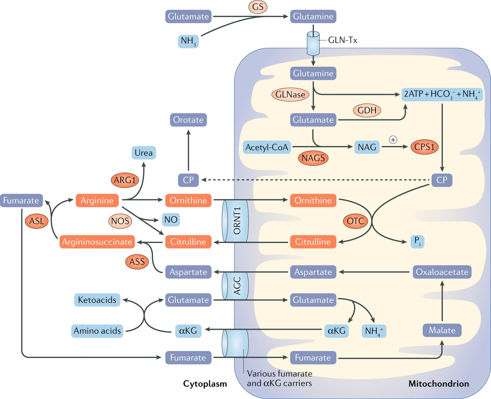
Long description
Hyperammonaemia in neonates and infants is typically caused by inborn errors of metabolism. Primary urea cycle disorders are caused by congenital deficiency of any of the six urea cycle enzymes: N-acetylglutamate synthas

Amino acids serve as foundational building blocks for proteins and act as critical metabolic intermediates across all human tissues. From first principles, every amino acid shares a common central carbon structure: an alpha-carbon bonded to a hydrogen atom, a carboxyl group, an amino group, and a variable side chain known as the R-group. This chemical diversity dictates protein folding, enzymatic catalysis, and intermediary metabolism. In human physiology, nitrogen homeostasis depends on the balance between amino acid consumption, protein synthesis, and the safe disposal of excess amino nitrogen.
When proteins are catabolized, amino acid carbon skeletons undergo oxidation to fuel energy production via the tricarboxylic acid cycle, while the released amino groups generate toxic ammonia. Because free ammonia is profoundly neurotoxic, human physiology converts it into urea through a specialized hepatic pathway. Urea formation mainly occurs in the urea cycle in the liver, and the products and substrates of urinary metabolites include arginine, citrulline and ornithine. In this cycle, several enzymes involved in removal of nitrogen from amino acids are regulated by glucagon and insulin. These enzymes are also involved in synthesis of nitric oxide. [2]
Hyperammonaemia arises when hepatic urea synthesis fails, overwhelming the brain's capacity to buffer ammonia through glutamine synthesis in astrocytes. This accumulation leads to cerebral oedema, astrocyte swelling, altered neurotransmission, and clinical encephalopathy. Understanding this pathway requires mastering the classification of amino acids, the precise enzymatic steps of ureagenesis, and the metabolic consequences of pathway blockades.
Amino acids are broadly categorized based on the synthetic capacity of human cells and the chemical properties of their side chains. Humans cannot synthesize a specific subset of canonical amino acids de novo because the requisite carbon skeleton pathways are absent from the human genome. These essential amino acids must be acquired directly from dietary protein intake to support normal cellular function, growth, and tissue repair. Conversely, non-essential amino acids can be synthesized within the body from simple metabolic intermediates, such as glucose metabolites and tricarboxylic acid cycle intermediates, often via transaminase reactions. [1]
The chemical properties of amino acid side chains govern protein architecture and metabolic channeling. Acidic amino acids, specifically aspartate and glutamate, possess negatively charged carboxylate side chains at physiological pH. These residues play pivotal roles in nitrogen shuttling, acting as direct amino donors for transamination reactions and serving as links between amino acid catabolism and the tricarboxylic acid cycle. Basic amino acids, which include arginine, lysine, and histidine, carry positively charged side chains at physiological pH, enabling them to bind nucleic acids, stabilize enzyme active sites, and participate directly in the urea cycle as intermediates or regulatory components.
DefinitionEssential vs Non-essential
Essential amino acids cannot be synthesized by human cells and must be ingested in the diet, whereas non-essential amino acids are produced internally via transamination and biosynthetic pathways.
To appreciate transport across cell membranes, clinicians must recognize that hydrophilic amino acids require specialized transmembrane carrier proteins. Transporters such as SLC6A14 exhibit broad substrate selectivity, encompassing all essential amino acids as well as glutamine. In pathological states such as cancer and certain metabolic syndromes, these transport mechanisms and biosynthetic circuits are heavily upregulated to sustain rapid cellular turnover and altered bioenergetics. [1]
| Category | Representative Amino Acids | Physiological Role |
|---|---|---|
| Essential | Valine, Leucine, Isoleucine, Lysine | Must be obtained from diet for protein synthesis and growth |
| Acidic | Glutamate, Aspartate | Serve as nitrogen donors and fuel metabolic intermediates |
| Basic | Arginine, Lysine, Histidine | Participate in enzyme catalysis, structural stability, and urea cycling |
The urea cycle is a five-step cyclic metabolic pathway localized across the mitochondrial matrix and the cytosolic compartment of hepatocytes. Its primary physiological purpose is the conversion of toxic waste nitrogen, derived from amino acid deamination, into highly water-soluble urea for renal excretion. In amino acid catabolism, most of the ammonia released is in the form of urea from a process of urea cycle of liver. The pathway begins in the mitochondrial matrix, where free ammonium ions combine with bicarbonate, driven by ATP consumption, to form carbamoyl phosphate. This initial reaction is catalyzed by carbamoyl phosphate synthetase I, an enzyme allosterically activated by N-acetylglutamate. [2]
Urea cycle pathway outline
- Mitochondria: NH4 + BicarbonateCarbamoyl phosphate synthetase I forms carbamoyl phosphate.
- Citrulline synthesisOrnithine transcarbamylase combines carbamoyl phosphate with ornithine.
- Question: Cytosol steps?
Yes
- Argininosuccinate formationCondensation with aspartate adds the second nitrogen.
- Cleavage to ArginineArgininosuccinase releases fumarate and yields arginine.
No
- Urea releaseArginase cleaves arginine to urea and regenerates ornithine.
Carbamoyl phosphate then condenses with ornithine, via ornithine transcarbamylase, to yield citrulline within the mitochondria. Citrulline is transported out into the cytosol, where it condenses with aspartate to form argininosuccinate. This step is critical because aspartate introduces the second nitrogen atom into the future urea molecule. Argininosuccinate is subsequently cleaved by argininosuccinase into fumarate and arginine. Finally, cytosolic arginase hydrolyzes arginine into urea and regenerates ornithine, which is transported back into the mitochondria to sustain the cycle.
Regulation of this pathway is tightly coupled to systemic nutritional status and hormonal signaling. Glucagon and insulin modulate the key enzymes responsible for the removal of nitrogen from amino acids. Insulin impacts the activity of arginase and nitric oxide synthase. Disruption of these regulatory steps or inherited deficiencies in any of the five urea cycle enzymes leads to severe hyperammonaemia, requiring immediate medical intervention to prevent irreversible neurological damage. [2]
Clinical pearlNitrogen Sources
The two nitrogen atoms in each urea molecule originate from free mitochondrial ammonium and the amino acid aspartate.
Ornithine transcarbamylase deficiency represents a key genetic error in the urea cycle, an essential metabolic pathway responsible for converting toxic nitrogenous waste into urea for safe renal excretion. When ornithine transcarbamylase is deficient or absent, the condensation of carbamoyl phosphate and ornithine to form citrulline within the mitochondrial matrix cannot proceed normally. Consequently, carbamoyl phosphate accumulates and spills over into extra-mitochondrial pathways, driving excessive pyrimidine synthesis and leading to profound systemic hyperammonaemia.
From first principles, the urea cycle operates to clear excess ammonia derived from amino acid catabolism. The liver serves as the primary site for this detoxifying process, utilizing specific enzymatic steps to bridge mitochondrial and cytosolic reactions. In ornithine transcarbamylase deficiency, the blockade prevents normal metabolic progression, causing nitrogen to back up in the form of ammonia and precursor molecules. This condition can present acutely in the neonatal period with life-threatening encephalopathy, or variably in late-onset forms triggered by catabolic stress, high protein intake, or intercurrent illness.
DefinitionOrnithine Transcarbamylase
Management strategies focus on lowering circulating ammonia levels, restricting dietary protein to minimize nitrogen loads, and using alternative pathway medications such as sodium phenylbutyrate or sodium benzoate to excrete waste nitrogen as phenylacetylglutamine or hippurate.
| Metabolic State | Biochemical Feature | Clinical Implication |
|---|---|---|
| Hyperammonaemia | Elevated blood ammonia | Encephalopathy and cerebral oedema |
| OTC Deficiency | Blocked citrulline synthesis | Accumulation of carbamoyl phosphate |
| Alternative Therapy | Nitrogen scavenging | Excretion via non-urea pathways |
Ammonia is a potent neurotoxin generated ubiquitously from amino acid deamination, amine catabolism, and gut microbial activity. Because free ammonia crosses biological membranes readily, safe transport between peripheral tissues and the central processing organs requires specific carrier and neutralisation mechanisms. Peripheral tissues, particularly skeletal muscle and the lungs, fix excess ammonia onto glutamate to generate glutamine via the action of glutamine synthetase. Glutamine serves as a neutral, non-toxic circulating vehicle for nitrogen, safely ferrying it through the bloodstream to the liver and kidneys.
Ammonia Transport and Processing
- Tissue Amino Acid CatabolismGenerates free ammonia in peripheral cells.
- Question: Is ammonia safely bound?
No (Free NH3)
- Cellular toxicity and cerebral diffusion
Yes (Glutamine synthesis)
- Transport via blood to liver and kidneysGlutamine acts as a neutral carrier.
- Question: Organ destination reached?
Liver
- Urea cycle conversion and excretion
Kidney
- Renal ammoniagenesis and acid-base regulation
Upon reaching the liver, glutamine is cleaved by hepatic glutaminase to release ammonia inside the portal hepatocyte, where it enters the urea cycle directly. Alternatively, when it reaches the renal proximal tubules, ammonia is liberated and secreted into the tubular lumen to buffer urinary hydrogen ions, thereby playing a vital role in systemic acid-base homeostasis.
Clinical pearlGlutamine as a Carrier
Failure of these transport and clearance mechanisms leads rapidly to hyperammonaemic encephalopathy. Excess ammonia within the central nervous system alters astrocyte function, depletes alpha-ketoglutarate from the tricarboxylic acid cycle through mass action, and impairs neurotransmission, underscoring why both efficient inter-organ transport and intact enzymatic pathways are mandatory for survival.
Amino acids serve as foundational precursors for an array of specialized nitrogenous derivatives with critical neurological, vascular, and metabolic functions. Catecholamines including dopamine, noradrenaline, and adrenaline are synthesized from the amino acid tyrosine through a well-defined enzymatic cascade. Tyrosine is first hydroxylated by tyrosine hydroxylase to L-DOPA, which is subsequently decarboxylated by aromatic L-amino acid decarboxylase to form dopamine. In noradrenergic and adrenergic neurons or the adrenal medulla, further enzymatic steps introduce a beta-hydroxyl group and a methyl group respectively.
Serotonin (5-hydroxytryptamine) and melatonin are derived from tryptophan. Tryptophan is hydroxylated and decarboxylated to yield serotonin, a key monoamine neurotransmitter involved in mood, gastrointestinal motility, and vasoconstriction. In the pineal gland, serotonin undergoes N-acetylation and O-methylation to produce melatonin, the principal regulator of circadian rhythms and the sleep-wake cycle. Gamma-aminobutyric acid (GABA), the primary inhibitory neurotransmitter in the central nervous system, is synthesized directly from L-glutamate via the action of glutamate decarboxylase, which requires pyridoxal phosphate (vitamin B6) as a coenzyme.
Nitric oxide (NO) is a potent gaseous signalling molecule generated from the metabolism of arginine via the citrulline-NO pathway. Nitric oxide synthase enzymes catalyze the oxidation of L-arginine to L-citrulline and NO. Nitric oxide supports vascular smooth muscle relaxation, inhibits platelet aggregation, and acts as an endogenous AMP-activated protein kinase activator that can indirectly repress hepatic lipogenesis and gluconeogenesis. Furthermore, nitric oxide supports tumor growth by promoting angiogenesis, though it can also function as a tumor suppressor by upregulating the p53 tumor suppressor pathway. [1] [2]
Haem, the prosthetic group of haemoglobin, myoglobin, and cytochromes, is synthesized starting in the mitochondria through the condensation of succinyl-CoA and glycine by the rate-limiting enzyme delta-aminolaevulinic acid synthase. Creatine, essential for rapid ATP buffering in skeletal muscle and brain tissue, is synthesized from arginine and glycine to form guanidinoacetate, which is subsequently methylated using S-adenosylmethionine.
Medical students often encounter several persistent misconceptions regarding amino acid derivatives and nitrogen disposal pathways. A frequent pitfall is confusing the nitrogen sources and metabolic destinations of purine versus pyrimidine nucleotide biosynthesis. Purine biosynthesis requires formate, bicarbonate, and three specific amino acids: aspartate, glycine, and glutamine. Glutamine and aspartate act as nitrogen sources for both nucleobases, while glycine contributes directly to the purine backbone at positions C4, C5, and N7, or generates one-carbon units via 5,10-methylenetetrahydrofolate. Students must remember that glycine incorporation builds the core framework, unlike in pyrimidine synthesis which follows a simpler biochemical route. [1]
Specifically, mitochondrial serine hydroxymethyltransferase 2 (SHMT2) is critical for redox regulation under hypoxic conditions, SHMT2 is induced by hypoxic stress through HIF1α and is involved in maintaining the cellular NADPH/NADP+ ratio [P23], [1]
Exam trapSerine depletion and glutathione
Ignoring the intersection between serine metabolism and the methionine cycle leads to the misconception that amino acids only build proteins. Serine depletion directly results in glutathione reduction, impairing cellular antioxidant defences. [1]
A final pitfall in clinical reasoning is misinterpreting alterations in amino acid profiles, such as those involving arginine, citrulline, and ornithine, in metabolic syndromes like type 2 diabetes mellitus. Although protein metabolism disorder is a typical symptom of type 2 diabetes, sensitivity analyses suggest that specific amino acid shifts are likely a cause rather than merely a consequence of diabetic complications such as diabetic nephropathy. [2]
Understanding amino acid pathways and their derivatives is essential for managing inherited metabolic disorders, nutritional deficiencies, and targeted oncologic therapies. In clinical biochemistry, automated ion-exchange chromatography systems, such as the Biochrom 30 plus amino acid analysis system, are employed to quantify physiological metabolites, including urea production from supernatant samples. This analytical capability is vital for diagnosing urea cycle defects and monitoring patients [3]
In oncology, the dependence of cancer cells on specific amino acid transport systems has become a major therapeutic target. Leucine acts as a well-known activator of mechanistic target of rapamycin (mTOR) signalling, promoting tumor growth. Leucine influx is mediated by L-type amino acid transporter 1 (LAT1), which functions in mandatory exchange with the sodium-coupled neutral amino acid transporter 2 (ASCT2, encoded by SLC1A5). ASCT2 mediates the influx of neutral amino acids like glutamine into cancer cells, which then efflux via LAT1 coupled to the entry of leucine. Pharmacological or genetic inhibition of ASCT2 impedes LAT1-mediated leucine uptake, leading to the inactivation of mTOR signalling in cancer cells. Inhibiting LAT1 suppresses tumor growth in conditions such as non-small cell lung cancer and oral cancer. [1]
Amino Acid Transport and mTOR Signalling
- ASCT2 (SLC1A5)Mediates Na+-coupled influx of glutamine into cancer cells.
- Question: Coupled transport active?
Yes
- LAT1 AntiporterGlutamine effluxes out as leucine enters the cancer cell.
- mTOR ActivationIntracellular leucine activates mTOR signalling and tumor growth.
No
- InhibitionLAT1/ASCT2 blockade impedes leucine uptake and inactivates mTOR.
Furthermore, investigating the citrulline-nitric oxide pathway provides clinical insights into vascular health and insulin resistance. Diabetic patients often exhibit a lower conversion of arginine to citrulline, reflecting impaired nitric oxide synthase activity and reduced nitric oxide bioactivity. Restoring nitric oxide synthesis can augment blood flow to insulin-sensitive tissues and improve metabolic regulation. [2]
| Derivative | Precursor Amino Acid | Primary Physiological Function |
|---|---|---|
| Catecholamines | Tyrosine | Neurotransmission and sympathetic response |
| Serotonin and Melatonin | Tryptophan | Mood regulation and circadian rhythm |
| GABA | Glutamate | Central nervous system inhibition |
| Nitric Oxide | Arginine | Vasodilation and AMPK activation |
| Haem | Glycine | Oxygen transport and electron transfer |
- Tyrosine is the foundational precursor for catecholamine neurotransmitters including dopamine, noradrenaline, and adrenaline.
- Tryptophan gives rise to serotonin for neurotransmission and melatonin for regulating circadian sleep cycles.
- GABA is synthesized directly from glutamate via glutamate decarboxylase using pyridoxal phosphate.
- Arginine is metabolized by nitric oxide synthase to produce nitric oxide, supporting vascular tone and AMPK activation.
- Purine biosynthesis requires aspartate, glycine, and glutamine as critical nitrogen and carbon sources.
- Serine-driven one-carbon metabolism via the folate cycle contributes substantially to cellular NADPH production and glutathione synthesis. [1]
- LAT1 and ASCT2 amino acid transporters couple glutamine efflux with leucine influx to activate mTOR signalling in cancer.
Answer each one in your head first, then open the answer.
Which amino acid is the direct precursor for the inhibitory neurotransmitter GABA?
Show answerHide answer
GlutamateName the rate-limiting enzyme that converts tyrosine to L-DOPA in catecholamine synthesis.
Show answerHide answer
Tyrosine hydroxylaseWhich amino acid serves as the nitrogen and carbon donor in the citrulline-NO pathway to generate nitric oxide?
Show answerHide answer
ArginineWhat cofactor is required by glutamate decarboxylase to convert glutamate into GABA?
Show answerHide answer
Pyridoxal phosphate (vitamin B6)Which amino acid transporter couples its influx with leucine entry to activate mTOR signalling in cancer cells?
Show answerHide answer
LAT1
- Lieu EL, Nguyen T, Rhyne S, Kim J. Amino acids in cancer. Experimental & molecular medicine 2020;52(1):15-30. doi:10.1038/s12276-020-0375-3. Accessed .
- Cao YF, Li J, Zhang Z, et al. Plasma Levels of Amino Acids Related to Urea Cycle and Risk of Type 2 Diabetes Mellitus in Chinese Adults. Frontiers in endocrinology 2019;10:50. doi:10.3389/fendo.2019.00050. Accessed .
- Laemmle A, Gallagher RC, Keogh A, et al. Frequency and Pathophysiology of Acute Liver Failure in Ornithine Transcarbamylase Deficiency (OTCD). PloS one 2016;11(4):e0153358. doi:10.1371/journal.pone.0153358. Accessed .
Figure credits
- Figure 1: Rupesh Raina, Jirair K. Bedoyan, Uta Lichter-Konecki, Philippe Jouvet, Stefano Picca, Nicholas Ah Mew, Marcel C. Machado, Ronith Chakraborty, Meghana Vemuganti, Manpreet K. Grewal, Timothy Bunchman, Sidharth Kumar Sethi, Vinod Krishnappa, Mignon McCulloch, Khalid Alhasan, Arvind Bagga, Rajit K. Basu, Franz Schaefer, Guido Filler & Bradley A. Warady, via Wikimedia Commons, CC BY 4.0, CC BY 4.0. resized.
Educational content for medical students and doctors. It is not a substitute for clinical judgement or local guidelines.