Cell cycle, cyclins and tumour suppressors
Regulation of cell division by cyclins, CDKs and tumour suppressors ensures orderly growth and prevents malignancy.
Figure 1Control Mechanism of Cell Cycle Progression
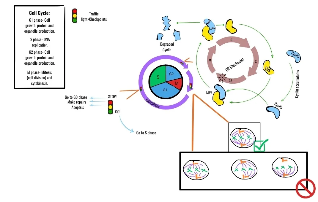
Long description
Regulation of the cell progression through the stages of division in a controlled and orderly manner. Checkpoints are where the cell assesses whether key processes have been properly completed. If there is an issue, the

The cell cycle is a series of ordered events that duplicate cellular components and divide a cell into two daughters. Central to its control are cyclins, which bind and activate cyclin‑dependent kinases (CDKs), and tumour‑suppressor proteins that can halt progression when DNA is damaged or growth signals are inappropriate.
DefinitionCyclin‑CDK complex
A heterodimer of a cyclin protein and a CDK that phosphorylates downstream substrates to drive the cell through a specific phase.
DefinitionTumour suppressor
A protein that restrains cell proliferation, often by enforcing checkpoints or inducing cell‑cycle arrest.
The cycle is divided into G1, S, G2 and M phases, each governed by distinct cyclin‑CDK pairs.
| Phase | Principal cyclin(s) | Partner CDK(s) | Main checkpoint |
|---|---|---|---|
| G1 | Cyclin D | CDK4/6 | RB‑E2F checkpoint (hypophosphorylated RB blocks E2F) |
| G1‑S transition | Cyclin E | CDK2 | DNA‑damage checkpoint (p53‑p21 axis) |
| S | Cyclin A | CDK2 | Replication stress checkpoint |
| G2 | Cyclin A/B | CDK1 | G2‑M checkpoint (CHK1/CHK2‑CDC25 regulation) |
| M | Cyclin B | CDK1 | Spindle assembly checkpoint |
During early G1, cyclin D‑CDK4/6 phosphorylates the retinoblastoma protein (RB), converting it from a hypophosphorylated repressor of E2F transcription factors to an inactive, hyperphosphorylated form. This releases E2F‑driven transcription of S‑phase genes. Hyperphosphorylation of RB is initiated by cyclin D‑CDK4/6 and reinforced later by cyclin E‑CDK2 and cyclin A‑CDK2, keeping RB inactive throughout S phase and beyond【P163】. [2]
Checkpoint kinases CHK1 and CHK2 act downstream of DNA damage sensors. CHK2 phosphorylates p53 on Ser‑20, reducing MDM2 binding and stabilising p53; CHK1 phosphorylates CDC25 on Ser‑216, preventing CDC25‑mediated activation of CDK1 and enforcing a G2 arrest【P96】. [1]
The CDK inhibitor p21^WAF1/CIP1 is a pivotal downstream effector of p53. High p21 levels inhibit cyclin D‑CDK4/6 activity, reinforcing G1 arrest, whereas low p21 can act as an assembly factor for the same complex, promoting progression【P26】. p21 is a direct transcriptional target of p53【P26】【P128】, but can also be induced independently of p53 by nuclear receptors such as androgen, vitamin D and retinoid receptors【P26】. [1]
Transforming growth factor‑β (TGF‑β) can maintain RB in a hypophosphorylated state, thereby inducing G1 arrest in lung epithelial cells【P41】. The INK4 family (p16^INK4A, p15^INK4B) specifically inhibits CDK4/6, mirroring the effect of high p21 on cyclin D‑CDK4/6 complexes【P41】. [1]
Exam trapCommon misconception
Only p21 can halt the cell cycle at any point; in fact, the INK4 family restricts arrest to G0/G1, while other CDKIs act at later phases.
p53 is a master regulator of genome integrity. Upon stress such as DNA damage, p53 levels rise, leading to transcription of genes that mediate cell‑cycle arrest, DNA repair or apoptosis【P132】. One of its most important targets is p21, which, when induced, can block cyclin‑D/CDK4/6 activity and thus keep RB hypophosphorylated, enforcing a G1 checkpoint【P26】. [1] [2]
RB functions as a gatekeeper at the G1/S transition by binding E2F transcription factors and repressing genes required for DNA synthesis. Hypophosphorylated RB exerts this repression; hyperphosphorylation by cyclin‑CDK complexes releases E2F, allowing S‑phase entry【P163】. In addition to the classic G1 role, RB‑dependent repression of E2F targets extends into later phases through cooperation with the DREAM complex, which silences a broad set of cell‑cycle genes, including those involved in mitosis【P48】.
The interplay between p53, p21 and RB creates a robust barrier to uncontrolled proliferation. p53‑mediated p21 induction maintains RB in a hypophosphorylated state, while RB‑DREAM cooperation reinforces repression of E2F‑driven transcription. Loss of either RB or p53 compromises this barrier; tumours frequently harbour mutations in both genes, suggesting that RB loss alone is insufficient for transformation and that p53‑dependent pathways provide compensatory control【P175】. [2]
During senescence, RB‑dependent chromatin reorganisation forms senescence‑associated heterochromatin foci (SAHF), which further silence E2F targets and stabilise arrest【P48】. This illustrates how tumour‑suppressor pathways can enforce long‑term growth arrest beyond transient checkpoint activation.
NoteClinical relevance
Mutations in TP53 or RB1 are hallmarks of many cancers; understanding their normal regulatory network guides the development of CDK4/6 inhibitors and p53‑reactivating agents.
Cell‑cycle control overview
- Growth signalCyclin D synthesis
- Question: Is p53 activated?
Yes
- p53 ↑ → p21 ↑Inhibit cyclin D‑CDK4/6
- RB remains hypophosphorylatedE2F repression
No
- Cyclin D‑CDK4/6 activeRB hyperphosphorylated → E2F active
- Progress to S phaseCyclin E‑CDK2, Cyclin A‑CDK2
Labile cells are continuously proliferating under normal physiological conditions. Examples include epithelial cells of the gastrointestinal tract, bone‑marrow haematopoietic precursors and skin keratinocytes. Because they are constantly cycling, they are highly dependent on intact cyclin‑CDK activity and are the primary targets of many antimitotic drugs. [1]
Stable cells divide only in response to a specific stimulus. Hepatocytes and fibroblasts belong to this group; they remain quiescent (G0) until growth factors such as hepatocyte growth factor or platelet‑derived growth factor trigger re‑entry into the cell‑cycle. The transition from quiescence to proliferation is tightly regulated by CDK inhibitors (CDKIs) such as p21^WAF1/CIP1, which can block cyclin‑CDK complexes when expressed at sufficient levels. [1]
Permanent cells have exited the cell‑cycle irreversibly. Mature neurons, cardiac myocytes and skeletal‑muscle fibres are classic examples. They are terminally differentiated and express lineage‑specific transcription factors that lock the cell‑cycle machinery in an off‑state. Although they no longer divide, they can still undergo a senescence‑like growth arrest if exposed to severe stress, but this does not restore proliferative capacity.
The distinction among these three categories is clinically important because the susceptibility to cytotoxic agents, the capacity for tissue regeneration, and the risk of tumourigenesis differ markedly.
Anticancer and immunosuppressive agents exploit the dependence of proliferating cells on specific phases of the cell‑cycle.
| Phase targeted | Representative drug(s) | Primary mechanism |
|---|---|---|
| G1 → S transition | CDK4/6 inhibitors (palbociclib, ribociclib) | Prevent phosphorylation of RB, keeping E2F bound and halting transcription of S‑phase genes |
| S‑phase DNA synthesis | Antimetabolites (methotrexate, 5‑fluorouracil) | Inhibit thymidylate synthase or dihydrofolate reductase, depleting nucleotides |
| G2 → M transition | Wee1 inhibitors (adavosertib) | Force premature CDK1 activation, leading to mitotic catastrophe |
| M‑phase (spindle) | Vinca alkaloids, taxanes | Disrupt microtubule dynamics, preventing chromosome segregation |
| Non‑phase‑specific | Alkylating agents (cyclophosphamide) | Cross‑link DNA, triggering damage‑induced checkpoints |
classical DDR mainly involves the p53/p21WAF1/CIP1 tumor suppressor pathway【P94】.
Illustrative example – A patient with hormone‑receptor‑positive breast cancer receives a CDK4/6 inhibitor. The drug binds CDK4/6, preventing RB phosphorylation; consequently, E2F‑dependent transcription of S‑phase genes is suppressed, leading to tumour‑cell arrest in G1.
Exam trapAssuming all non‑dividing cells are the same
Quiescent, senescent and terminally differentiated cells all lack proliferation, but their molecular signatures differ; mis‑identifying them can lead to erroneous conclusions about drug efficacy.
- Reliance on a single biomarker – p21^WAF1/CIP1 expression alone does not prove senescence because it is also up‑regulated transiently during DNA repair.
- Overlooking dose‑dependent effects – Low concentrations of microtubule‑targeting agents may arrest cells in mitosis, whereas higher doses can cause apoptosis; the therapeutic window is narrow.
- Ignoring tissue‑specific cell‑type – Drugs that are effective against labile epithelial tumours may be ineffective in cancers arising from stable cells (e.g., hepatocellular carcinoma) because the latter require a proliferative stimulus to become drug‑sensitive.
NoteClinical relevance
Understanding cell‑type proliferative status guides chemotherapy selection, dosing schedules, and predicts tissue‑specific toxicities.
- Oncology – CDK4/6 inhibitors are standard for hormone‑responsive breast cancer, exploiting the reliance of labile tumour cells on the G1‑S checkpoint.
- Transplant medicine – Antimetabolites such as mycophenolate mofetil suppress the proliferation of stable lymphocytes, preventing graft rejection.
- Regenerative medicine – Strategies to coax stable hepatocytes into the cell‑cycle (e.g., growth‑factor cocktails) aim to enhance liver regeneration after resection.
The following flowchart summarises a typical decision pathway when selecting a cell‑cycle‑targeted therapy.
Therapeutic decision pathway
- Identify tumour cell typeLabile, stable or permanent?
- Question: Is the tumour proliferating rapidly?
Yes
- Consider phase‑specific agentse.g., CDK4/6 inhibitors for G1 arrest
No
- Assess need for proliferative stimulusUse growth factors or combine with DNA‑damage agents
- Evaluate toxicity profileMatch drug to patient comorbidities
- Labile cells continuously cycle; stable cells divide only after stimulation; permanent cells are irreversibly post‑mitotic.
- p21^WAF1/CIP1 can inhibit all CDKs, enforcing G1 arrest when up‑regulated by p53.
- CDK4/6 inhibitors block RB phosphorylation, a first‑line therapy for hormone‑responsive breast cancer. [1]
- Single biomarkers (e.g., p21) are insufficient to distinguish senescence from other non‑proliferative states.
- Drug efficacy depends on the proliferative status of the target cell population.
- Microtubule‑targeting agents disrupt mitosis but have a narrow therapeutic window.
Answer each one in your head first, then open the answer.
Which CDKI can inactivate all cyclin‑CDK complexes?
Show answerHide answer
p21^WAF1/CIP1What is the primary effect of CDK4/6 inhibition on the RB pathway?
Show answerHide answer
Prevention of RB phosphorylation, keeping E2F boundName a stable cell type that requires a growth stimulus to divide.
Show answerHide answer
HepatocyteWhy is p21 alone not a definitive marker of senescence?
Show answerHide answer
It is also up‑regulated during DNA repair and transient arrestWhich class of drugs is first‑line for hormone‑receptor‑positive breast cancer?
Show answerHide answer
- Kumari R, Jat P. Mechanisms of Cellular Senescence: Cell Cycle Arrest and Senescence Associated Secretory Phenotype. Frontiers in cell and developmental biology 2021;9:645593. doi:10.3389/fcell.2021.645593. Accessed .
- Engeland K. Cell cycle regulation: p53-p21-RB signaling. Cell death and differentiation 2022;29(5):946-960. doi:10.1038/s41418-022-00988-z. Accessed .
Figure credits
Educational content for medical students and doctors. It is not a substitute for clinical judgement or local guidelines.