Cell organelles, trafficking and the cytoskeleton
Cellular pathways that move proteins and lipids between organelles and maintain cell shape.
Figure 1Cell Drawing
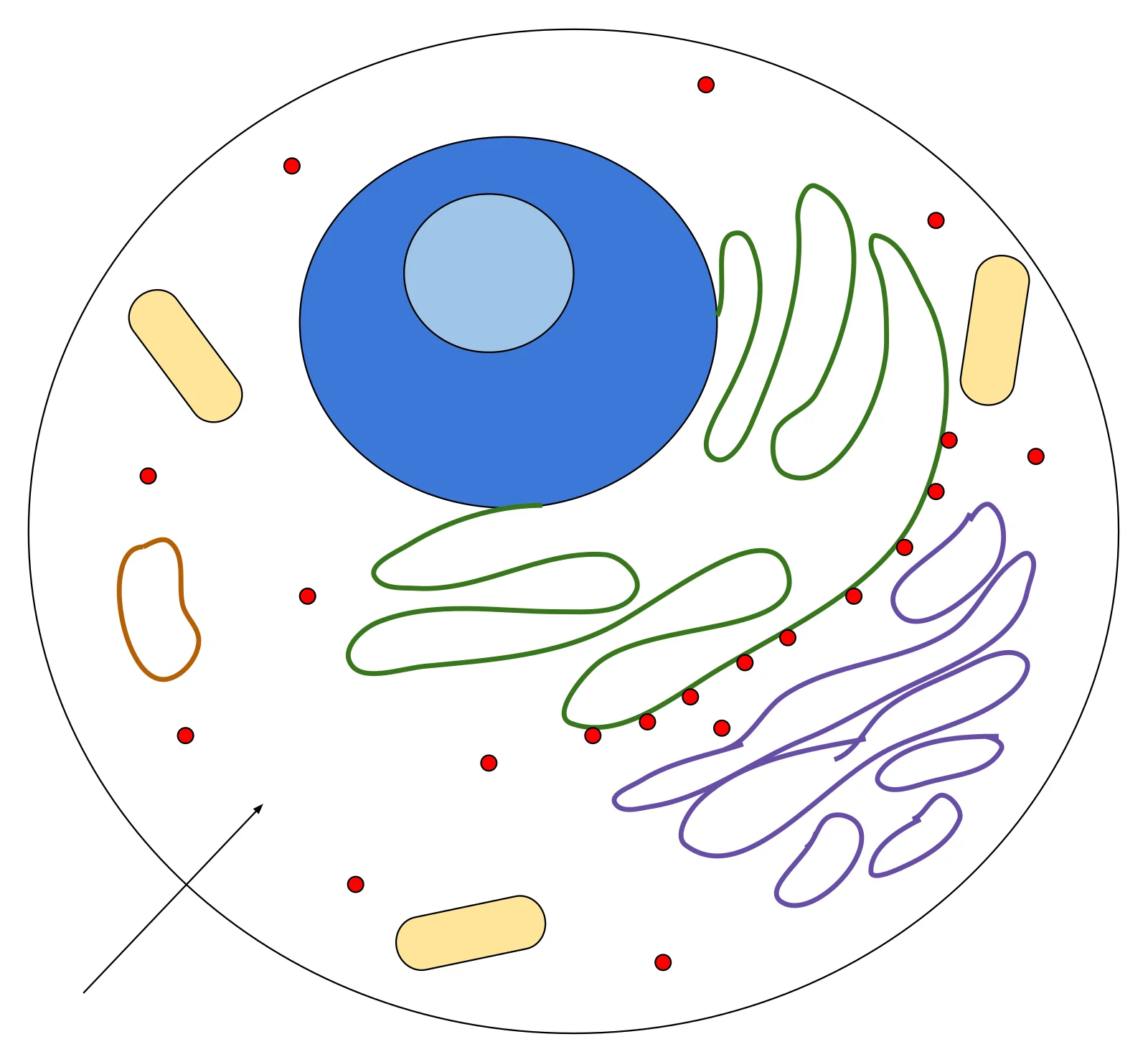
Long description
Illustration for biochemistry/cell organelles trafficking and cytoskeleton from Wikimedia Commons, titled 'Cell Drawing': Inside the Cell. The author and licence are given in the credit line.

DefinitionOrganelle trafficking
The eukaryotic cell contains specialised compartments – the endoplasmic reticulum (ER), Golgi apparatus, peroxisomes, lysosomes and mitochondria – each with distinct enzymatic activities. To function as an integrated system, newly synthesised proteins must be sorted, modified and delivered to their correct destination. This sorting relies on signal sequences, coat protein complexes (COPI, COPII, clathrin) and motor proteins that walk along actin filaments or microtubules. Disruption of any step can produce characteristic metabolic or structural disease phenotypes, making these pathways core teaching for future clinicians.
The rough ER (RER) is studded with ribosomes on its cytosolic face, giving it a “rough” appearance. It is the primary site of co‑translational insertion of secretory and membrane proteins. Nascent polypeptides enter the lumen where they fold, acquire N‑linked glycans and form disulphide bonds. Quality‑control mechanisms such as the unfolded protein response (UPR) monitor folding status; prolonged stress can trigger apoptosis via PERK‑eIF2α‑ATF4 signalling【P141】.
The smooth ER (SER) lacks ribosomes and is enriched in enzymes for lipid synthesis, steroidogenesis and detoxification. It also houses calcium‑binding proteins that regulate intracellular calcium stores. The transition between rough and smooth domains is fluid – a segment of ER can acquire ribosomes when demand for secretory protein synthesis rises, and conversely lose them when lipid production predominates.
Key points for students:
- RER → protein synthesis, N‑glycosylation, quality control.
- SER → lipid metabolism, steroid synthesis, calcium handling.
- Both share a continuous membrane; vesicles bud from ER exit sites (ERES) to deliver cargo to the Golgi.
The Golgi apparatus receives vesicles from the ER, further modifies glycoproteins (trimming and adding sugars), and sorts them into distinct transport carriers. Cis‑Golgi networks act as entry portals, while trans‑Golgi networks (TGN) serve as exit stations, directing cargo to the plasma membrane, lysosomes or secretory granules.
A classic example of disrupted Golgi trafficking is I‑cell disease (mucolipidosis II), caused by deficiency of the enzyme N‑acetylglucosamine‑1‑phosphotransferase. This enzyme normally tags lysosomal hydrolases with mannose‑6‑phosphate in the Golgi. Without the tag, hydrolases are missorted and secreted extracellularly, leading to accumulation of undegraded substrates within lysosomes. Clinically, patients present with severe developmental delay, skeletal dysplasia and coarse facial features. The disease illustrates how a single Golgi‑resident enzyme can have system‑wide consequences.
- Elevated plasma lysosomal enzymes (e.g., β‑hexosaminidase) despite normal urinary glycosaminoglycans.
- Absence of mannose‑6‑phosphate on lysosomal enzymes confirmed by lectin blotting.
Peroxisomes are small, single‑membrane organelles that perform β‑oxidation of very‑long‑chain fatty acids, α‑oxidation of phytanic acid and synthesis of plasmalogens – essential phospholipids for myelin and cardiac membranes. Peroxisomal diseases fall into two broad categories: peroxisome biogenesis disorders (PBD) and single‑enzyme deficiencies (SED)【P60】. [2]
Biochemical screening of blood and urine remains the first step, followed by functional studies in patient fibroblasts when results are equivocal. Important laboratory markers include:
- Pipecolic acid: Accumulates in Zellweger spectrum disorders (ZSDs) because L‑pipecolic acid oxidase, a peroxisomal enzyme, is deficient. Measurement by ESI‑MS/MS in plasma or urine is a useful diagnostic parameter【P9】.
- Immunoblotting of peroxisomal proteins: Detects the presence and processing of import‑competent peroxisomes. ACOX1 and the 3‑ketoacyl‑CoA thiolase are routinely examined; lack of processing of 3‑ketoacyl‑CoA thiolase distinguishes RCDP type 1 and the newly described type 5 from other RCDP forms【P28】. [1]
- Immunofluorescence microscopy: Visualises peroxisomal staining patterns. Aberrant staining that worsens at 40 °C suggests a subtle ZSD such as Heimler syndrome, where conventional metabolite screens may be normal【P43】. Conversely, normal fibroblast studies do not fully exclude a peroxisomal disorder【P44】. [1]
- Microscopy of peroxisomal morphology: Required for disorders of peroxisomal fission (e.g., PEX11β, DLP/DRP1, MFF defects). These present with enlarged, elongated peroxisomes or strings of peroxisomes, while biochemical parameters are often normal【P45】.
| Test | What it measures | Typical finding in PBD | Typical finding in SED |
|---|---|---|---|
| Plasma/urine pipecolic acid (ESI‑MS/MS) | Accumulation of a peroxisomal substrate | Elevated in ZSDs, normal in single‑enzyme defects【P9】 | Normal [1] |
| ACOX1 immunoblot | Presence of the first enzyme of β‑oxidation | Reduced or absent in severe PBD | Usually normal |
| 3‑ketoacyl‑CoA thiolase processing | Proteolytic maturation inside peroxisomes | Absent in RCDP type 1 and type 5【P28】 | Normal |
| Immunofluorescence of catalase | Peroxisomal matrix protein localisation | Aberrant staining, temperature‑sensitive in mild ZSDs【P43】 | Normal |
| Morphology by fluorescence microscopy | Size/number of peroxisomes | Variable; may be reduced in severe PBD | Normal, unless fission defect【P45】 |
- Zellweger spectrum disorders (ZSDs) represent the severe end of PBD, with multi‑system involvement: craniofacial dysmorphism, neuronal migration defects, hepatic dysfunction and sensorineural hearing loss.
- Rhizomelic chondrodysplasia punctata (RCDP) results from defects in peroxisomal 3‑ketoacyl‑CoA thiolase or related enzymes, leading to shortened limbs, cataracts and severe intellectual disability. The processing status of 3‑ketoacyl‑CoA thiolase helps differentiate subtypes【P28】.
- Heimler syndrome illustrates that very mild peroxisomal dysfunction can present with only sensorineural hearing loss and enamel defects; fibroblast studies remain essential for diagnosis【P43】.
Even when genetic sequencing identifies a pathogenic variant, biochemical confirmation is still required for prognosis, treatment decisions (e.g., dietary restriction of phytanic acid) and family counselling【P53】.
Peroxisomal diagnostic pathway
- Clinical suspicionNeurological, hepatic, or auditory signs
- Question: Blood/urine metabolites abnormal?
Yes
- Measure pipecolic acid (ESI‑MS/MS)Elevated suggests ZSD【P9】
- Proceed to fibroblast studies
No
- Perform fibroblast immunofluorescenceTemperature‑sensitive staining may reveal mild ZSD【P43】
- Consider peroxisomal fission disorder microscopyLook for elongated strings【P45】
- Immunoblot for ACOX1 & 3‑ketoacyl‑CoA thiolaseProcessing status informs severity【P28】
- Integrate biochemical, cellular and genetic dataConfirm diagnosis, guide management
DefinitionProteasome
The proteasome works in concert with the cytoskeletal network to maintain cellular homeostasis. When mis‑folded or damaged proteins accumulate, the ubiquitin‑proteasome system tags them with ubiquitin chains and delivers them to the 26S proteasome for degradation. This process prevents aggregation that could otherwise disrupt the architecture of microfilaments (actin filaments) and intermediate filaments (IFs).
Microfilaments are thin (≈7 nm) filaments composed of actin subunits. They provide mechanical support, drive cell motility, and generate contractile forces during cytokinesis. Actin dynamics are tightly regulated by ATP binding and hydrolysis, and by a host of actin‑binding proteins that cap, sever, or cross‑link filaments.
Intermediate filaments are rope‑like fibres (≈10 nm) formed from a diverse family of proteins such as keratins, vimentin, desmin and neurofilaments. Unlike actin or tubulin, IFs are not polar and do not undergo rapid polymerisation‑depolymerisation cycles. Their primary role is to bear tensile stress, anchor organelles, and maintain cell shape, especially in mechanically stressed tissues (epithelial, muscle, neuronal).
The proteasome indirectly influences both filament systems:
- Degradation of actin‑regulating proteins (e.g., cofilin, profilin) modulates filament turnover.
- Turnover of IF‑associated proteins (e.g., plectin, desmoplakin) controls filament network remodeling.
- Failure of proteasomal clearance can lead to protein aggregates that sequester actin or IF components, impairing cytoskeletal integrity and contributing to disease.
DefinitionMicrotubules
Microtubules display dynamic instability: the stochastic switching between growth (polymerisation) and shrinkage (depolymerisation). This behaviour is regulated by GTP‑bound tubulin at the plus end, microtubule‑associated proteins (MAPs), and the cellular concentration of free tubulin.
| Drug class | Mechanism | Clinical use |
|---|---|---|
| Vinca alkaloids (e.g., vincristine) | Bind β‑tubulin, suppress polymerisation, promote depolymerisation | Antineoplastic agents for leukaemia, lymphoma |
| Taxanes (e.g., paclitaxel) | Stabilise microtubules, prevent depolymerisation | Breast, ovarian, lung cancer chemotherapy |
| Colchicine | Binds tubulin, inhibits polymerisation | Acute gout flares, familial Mediterranean fever |
| Eribulin | Caps microtubule plus ends, induces non‑functional aggregates | Metastatic breast cancer |
These drugs exploit the essential role of microtubules in cell division; by disrupting spindle formation they trigger mitotic arrest and apoptosis in rapidly proliferating tumour cells. However, because microtubules are also vital for neuronal transport and ciliary function, toxicity (peripheral neuropathy, hair loss, ciliary dyskinesia) is common.
DefinitionMotile cilia
A typical motile cilium consists of:
- Basal body – a modified centriole anchoring the cilium.
- Transition zone – a diffusion barrier regulating protein entry.
- Axoneme – the core scaffold, usually a “9+2” arrangement of nine outer microtubule doublets (MTDs) surrounding two central singlet microtubules. Each doublet is decorated with inner and outer dynein arms (IDA, ODA) that generate sliding forces, radial spokes (RS) that coordinate activity, and numerous structural proteins.
PCD is a rare genetic disorder, affecting an estimated 1 in every 7,500 individuals. The disease is characterised by immotile or dyskinetic motile cilia, leading to chronic respiratory infections, subfertility and laterality defects. [3]
Recent proteomic studies have identified mutation‑specific alterations in axonemal composition. For example, patients with different DNAH5 (dynein heavy chain) mutations show variable loss of proteins such as VWA3B, KIAA1430/CFAP97, and DTHD1, which are normally present in wild‑type cilia but absent in patient samples. These proteins are not part of the classic dynein‑arm repertoire but appear to contribute to axonemal stability and dynein regulation.
- Diagnostic tools: high‑speed video microscopy, immunofluorescence, electron microscopy and genetic screening are employed to detect structural defects, protein loss, or pathogenic variants.
- Phenotypic variability: despite similar findings on conventional diagnostics, the specific protein deficits correlate with the severity of ciliary dysfunction, suggesting that genotype‑phenotype relationships are more nuanced than previously thought.
Understanding the precise composition of the axoneme and how individual mutations perturb it is essential for developing targeted therapies and improving diagnostic accuracy.
Ciliary dysfunction pathway
- Genetic mutation in ciliary gene (e.g., DNAH5)
- Question: Does mutation affect dynein arm assembly?
Yes
- Loss of dynein arms → reduced sliding force
- Impaired beat frequency and pattern
No
- Altered accessory proteins (VWA3B, CFAP97, DTHD1)
- Disrupted axonemal stability → dyskinetic beat
- Clinical phenotype: recurrent infections, infertility, laterality defects
DefinitionNa‑K ATPase
The Na‑K ATPase is a heterodimeric protein complex composed of an α‑subunit that carries the catalytic activity and a β‑subunit that assists in proper folding and membrane localisation. In cardiac myocytes the α1 isoform predominates, but the α2 and α3 isoforms are also present in vascular smooth muscle and neuronal tissue, respectively. By continuously extruding Na⁺, the pump creates a low intracellular sodium concentration (≈10 mmol L⁻¹) and a high intracellular potassium concentration (≈140 mmol L⁻¹). This gradient drives secondary active transporters, most notably the Na⁺/Ca²⁺ exchanger (NCX), which expels one Ca²⁺ ion in exchange for three Na⁺ ions entering the cell.
Digoxin, a cardiac glycoside derived from the foxglove plant, binds with high affinity to the extracellular domain of the α‑subunit. This interaction reduces the pump’s turnover rate, leading to a modest rise in intracellular Na⁺. The elevated Na⁺ diminishes the driving force for NCX, causing a secondary accumulation of intracellular Ca²⁺. The increased Ca²⁺ is taken up by the sarcoplasmic reticulum and released during each contraction, augmenting stroke volume – the classic positive inotropic effect of digoxin.
Beyond inotropy, digoxin exerts a vagomimetic action on the atrioventricular (AV) node by enhancing parasympathetic tone, which slows conduction and can be useful in controlling ventricular rate in atrial fibrillation. The dual mechanism – enhanced contractility and AV‑node slowing – underpins its historic role in heart‑failure management.
- Digoxin binds Na‑K ATPase → reduced Na⁺ extrusion.
- Intracellular Na⁺ rises → decreased Na⁺ gradient for NCX.
- NCX activity falls → less Ca²⁺ extrusion, net Ca²⁺ gain.
- Ca²⁺ load in sarcoplasmic reticulum increases → larger Ca²⁺ release on depolarisation.
- Myofilament activation is amplified → stronger systolic contraction.
The therapeutic window of digoxin is narrow because the same pump inhibition that benefits the heart also predisposes to arrhythmias when intracellular Na⁺ and Ca²⁺ become excessive. Toxicity manifests as nausea, visual disturbances (yellow‑green halos), and a spectrum of cardiac rhythm disturbances ranging from premature ventricular contractions to high‑grade AV block.
Exam trapAssuming all Na‑K ATPase isoforms behave alike
- Mis‑interpreting serum digoxin levels – Levels are measured as total digoxin; however, only the free (unbound) fraction is pharmacologically active. Renal impairment reduces clearance and raises the free fraction, increasing toxicity risk.
- Neglecting drug‑drug interactions – P‑glycoprotein inhibitors (e.g., amiodarone, quinidine) raise digoxin concentrations, while inducers (e.g., rifampicin) lower them. Failure to adjust the dose can precipitate either toxicity or therapeutic failure.
- Over‑reliance on the inotropic effect – Modern heart‑failure therapy (ACE inhibitors, β‑blockers, mineralocorticoid antagonists) provides mortality benefit, whereas digoxin improves symptoms without proven survival advantage. Using digoxin as a first‑line agent in contemporary practice is a common error.
- Ignoring electrolyte disturbances – Hypokalaemia, hypomagnesaemia, and hypercalcaemia potentiate digoxin toxicity because they enhance Na‑K ATPase binding of the drug. Routine monitoring of electrolytes is essential, especially in patients on diuretics.
- Confusing AV‑node slowing with anti‑arrhythmic efficacy – While digoxin slows AV conduction, it does not prevent atrial fibrillation recurrence and may even promote ectopic beats if concentrations rise.
NoteCurrent clinical niche
In many cardiology units, digoxin is prescribed after optimisation of guideline‑directed medical therapy (GDMT). Typical indications include:
- Heart failure with reduced ejection fraction (HFrEF) – added to ACE‑I/ARB/ARNI, β‑blocker, and mineralocorticoid antagonist when patients remain symptomatic despite optimal dosing.
- Atrial fibrillation with rapid ventricular response – particularly in sedentary or elderly patients where negative‑inotropic effects of β‑blockers are undesirable.
- Palliative care – where the goal is to improve quality of life rather than prolong survival.
Digoxin initiation pathway
- Assess eligibilityConfirm HFrEF or AF with rapid ventricular response; ensure GDMT is maximised.
- Question: Renal function adequate?
Yes
- Check electrolytesCorrect K⁺ >4 mmol/L, Mg²⁺ >0.8 mmol/L.
- Calculate doseStandard adult loading: 0.5 mg PO once, then 0.125–0.25 mg daily maintenance.
- Obtain baseline digoxin levelDraw 6–8 h post‑dose for trough.
No
- Adjust doseReduce maintenance dose by 50 % or avoid if CrCl <30 mL/min.
- Educate patientExplain signs of toxicity and need for regular monitoring.
- Follow‑upRe‑check level and electrolytes after 1 week, then periodically.
In acute decompensated heart failure, digoxin is generally withheld because its onset of action is slow (hours) and the risk of arrhythmia is heightened by the electrolyte shifts that accompany diuretic therapy. Conversely, in chronic stable disease, the modest increase in cardiac output (≈5–10 %) can translate into perceptible improvement in exercise tolerance and reduction in hospital admissions.
Understanding the quantitative distribution of inherited organellar and cytoskeletal disorders requires looking closely at specialized genetic panels and diagnostic metrics. For instance, when evaluating conditions like primary ciliary dyskinesia, comprehensive commercial genetic panel tests examine 19 distinct genes through next generation sequencing. This testing strategy comes at a specific financial cost of 1,990 and successfully detects approximately 50% of cases. [4]
Further expanding the economic and diagnostic scope, genetic testing costs for other commercial next generation sequencing panels range from 1,500 to 4,500 and frequently incorporate full cystic fibrosis transmembrane regulator protein analysis. Because these investigations often yield genetic variants of unknown significance where a definitive genetic diagnosis may remain unestablished, formal genetic counselling is strongly recommended. [4]
DefinitionDiagnostic Uncertainty
Patients with EM studies consistent with PCD should be referred to a PCD Foundation Clinical Center for confirmation.
| Panel Type | Target Scope | Cost Range | Diagnostic Yield |
|---|---|---|---|
| Standard NGS Panel | 19 specific genes | 1,990 | Approximately 50 percent |
| Expanded NGS Panel | Variable ciliary and CFTR | 1,500 to 4,500 | Variable with variants of unknown significance |
These diagnostic modalities form the basis of tracking regional epidemiology, allowing clinicians to map disease prevalence accurately despite the presence of complex genetic heterogeneity.
The underlying aetiology of organellar, trafficking, and cytoskeletal disorders is heavily rooted in distinct hereditary transmission patterns and genetic mechanisms. Almost all of these genes follow autosomal recessive inheritance [4]
Inheritance and Diagnostic Pathway
- Identify clinical presentation of organellar or ciliary disorderReview family history and phenotype.
- Question: Are genetic variants detected?
Yes
- Evaluate via next generation sequencing panel costing between 1,500 and 4,500Check for autosomal recessive patterns.
- Question: Is the result unclear?
Yes
- Refer to specialized clinical center for further investigationsManage variants of unknown significance.
No
- Establish genetic diagnosis and provide genetic counsellingConfirm two disease-causing mutations.
No
- Consider rare X-linked syndromic inheritanceInvestigate genes such as RPGR and OFD1.
Notable exceptions to this autosomal recessive rule include rare X-linked syndromic genes, specifically RPGR and OFD1. To date, no documented cases of digenic inheritance, defined as heterozygous mutations occurring in two different disease genes simultaneously and unequivocally associated with human disease, exist thus far. [4]
Clinical pearlDigenic Inheritance Rule
It is therefore important that emergency materials and drugs are always available and readily accessible when stronger drugs are used for analgesia and sedation.
NoteProcedural Safety
For absence of inner dynein arms in isolation, repeat biopsy and EM studies are always required to verify that this pathologic change persists and therefore is more likely genetic (primary) and not from secondary causes. [6]
Mitochondria function as the primary cellular powerhouse, orchestrating cellular respiration through the electron transport chain embedded in the inner mitochondrial membrane and executing oxidative phosphorylation to generate adenosine triphosphate. Mitochondrial genetics exhibit distinct maternal inheritance due to the unique presence of circular mitochondrial DNA encoding essential polypeptide subunits of the respiratory complexes. Intracellular transport systems rely heavily on molecular motors moving along cytoskeletal tracks; kinesins predominantly drive anterograde transport toward the plus end of microtubules, whereas dyneins mediate retrograde transport toward the minus end. Actin networks similarly support specialized motor protein interactions during cellular migration and local trafficking. Endocytosis encompasses several distinct physiological mechanisms, including clathrin-mediated endocytosis for specific receptor internalization, caveolae-dependent uptake utilizing cholesterol-rich membrane invaginations, phagocytosis for the engulfment of large particulate matter, pinocytosis for fluid-phase uptake, and receptor-mediated endocytosis tailored to macro-molecules. The nucleus and nucleolus coordinate genomic storage and ribosomal biogenesis, utilizing the nuclear pore complex to regulate macromolecular traffic via importins, exportins, and the regulatory GTPase Ran-GTP gradient.
For example, patients with biallelic DNAH11 mutations present with a classic clinical phenotype and low nasal nitric oxide levels, yet demonstrate normal electron microscopy ultrastructure coupl [P142]
DefinitionCiliary Biogenesis Defect
| Defect Type | Ultrastructural Finding | Clinical Implication |
|---|---|---|
| Dynein Arm Defect | Outer or inner arm loss | Confirms diagnosis on electron microscopy |
| Microtubule Disorganization | Inner dynein with disorganization | Requires repeat biopsy verification |
| DNAH11 Mutation | Normal ultrastructure | Low nasal nitric oxide with subtle waveform changes |
Primary ciliary dyskinesia and peroxisomal disorders require rigorous clinical staging and severity grading due to multisystem involvement. Prim
Primary Ciliary Dyskinesia Diagnostic Strategy
- Evaluate Clinical PhenotypeAssess for chronic oto-sino-pulmonary disease and associated syndromes.
- Question: Patient Age?
Under 5 Years
- Regional SelectionNorth America: Electron microscopy or genetics. Europe: High-speed videomicroscopy.
Older Patient
- Comprehensive Testing PanelIncorporate nasal nitric oxide, electron microscopy, immunofluorescence, and genetics.
Additionally, IF diagnostic results do not seem to be affected by secondary insults [P155].
Clinical pearlImmunofluorescence Utility
Additionally, repeat biopsies that fail to demonstrate any respiratory cilia could represent an oligociliary defect causing PCD.
Clinical pearlClassic presentations
The classic triad of Kartagener syndrome consists of situs inversus, chronic sinusitis or bronchiectasis, and subfertility, reflecting impaired motility of both nodal and respiratory cilia.
Most patients demonstrate three or more distinct clinical features, and the combination of multiple manifestations markedly increases the likelihood of a correct diagnosis. These hallmark features include neonatal respiratory distress, a chronic wet cough accompanied by recurrent lower respiratory infections and bronchiectasis, chronic nasal drainage associated with pansinusitis, recurrent otitis media particularly during childhood, laterality defects, and male infertility. Furthermore, genetic disorders affecting non-motile cilia can lead to distinct multisystem manifestations, including cystic kidneys, cystic or cholestatic liver disease, skeletal malformations, developmental delay, hydrocephalus, blindness, and deafness as seen in conditions such as Joubert syndrome, Bardet-Biedl syndrome, Usher syndrome, Jeune syndrome, and polycystic kidney disease. [4]
The clinical symptoms associated with primary ciliary dyskinesia frequently overlap with other prevalent pediatric and adult respiratory diseases, meaning each alternative condition must be carefully evaluated when patients present with chronic oto-sino-pulmonary symptoms. Diagnostic investigations should only be pursued when the specific clinical picture suggests the presence of those alternative conditions, establishing that primary ciliary dyskinesia is not merely a diagnosis of exclusion. Physicians treating primary ciliary dyskinesia often adapt therapeutic approaches utilized for other chronic respiratory conditions, such as cystic fibrosis and non-cystic fibrosis bronchiectasis. However, differences in various phenotypic parameters among primary ciliary dyskinesia, cystic fibrosis, and non-cystic fibrosis bronchiectasis indicate that simply extrapolating therapies may be inappropriate for management in certain clinical circumstances.
| Feature | Primary Ciliary Dyskinesia | Cystic Fibrosis | Severe Hypogammaglobulinemia |
|---|---|---|---|
| Onset of symptoms | Neonatal / Early infancy | Early childhood / Infancy | Variable (often later childhood / adult) |
| Sinus disease | Severe pansinusitis | Common | Recurrent sinopulmonary infections |
| Laterality defects | Present in about 50 percent | Absent | Absent |
| Diagnostic modality | Nasal nitric oxide / biopsy | Sweat chloride test / CFTR gene | Serum immunoglobulin levels |
Diagnostic pathway for suspected ciliary disorders
- Evaluate chronic oto-sino-pulmonary symptomsAssess for neonatal distress, wet cough, and pansinusitis.
- Question: Is the patient over 5 years old and cooperative?
Yes
- Measure nasal nitric oxide levelsLow values coupled with phenotype support diagnosis.
- Perform ciliary biopsy or genetic testingUse electron microscopy or high speed videomicroscopy.
No
- Proceed directly to specialized testingRefer to a specialized center for alternative diagnostics.
For children over 5 years of age and adults who can successfully cooperate with the required maneuvers, a low nasal nitric oxide value combined with an appropriate clinical phenotype may be adequate for establishing a clinical diagnosis, which should then be followed by ciliary biopsy for electron microscopy or high-speed videomicroscopy and genetic studies as needed. Minimal diagnostic criteria have been proposed by the Genetic Disorders of Mucociliary Clearance Consortium. Consultation with a geneticist or other subspecialists is strongly recommended when patients evaluated for possible primary ciliary dyskinesia exhibit features characteristic of non-motile ciliary dysfunction. For all patients diagnosed outside of specialized centers, at least one visit to an official clinical center is recommended to confirm the diagnosis. [4]
Establishing formal diagnostic criteria requires consensus recommendations from specialized physicians, such as those in North America who have been engaged in a primary ciliary dyskinesia centered research consortium for the last ten years, which have been adopted by the governing board of the Primary Ciliary Dyskinesia Foundation to provide guidance for clinical centers. Because primary ciliary dyskinesia is a genetically heterogeneous rare lung disease resulting in chronic oto-sino-pulmonary disease in both children and adults, many physicians incorrectly diagnose the condition or eliminate it from their differential diagnosis due to inexperience with diagnostic testing methods. [4]
| Parameter | Finding | Clinical Implication |
|---|---|---|
| Nasal NO cutoff | Less than 77 nl/min | High sensitivity (98%) and specificity (>99%) for detection |
| EM requirement | 20-50 clear cross-sections | Minimum amount for a diagnostic electron microscopy study |
| RSPH1 mutations | Borderline or normal nNO | Associated with later onset symptoms and subtle defects |
Thus, biopsies should be delayed until at least 2 weeks after full recovery from an illness.
Diagnostic Pathway
- Evaluate clinical phenotype of chronic oto-sino-pulmonary diseaseAssess patient suitability for cooperation and rule out cystic fibrosis.
- Question: Patient age 5 years or older?
Yes
- Measure nasal nitric oxide via chemiluminescence
- Question: nNO less than 77 nl/min?
Yes
- Proceed to confirmatory electron microscopy and high-speed videomicroscopy
No
- Consider PCD if phenotype is strong; evaluate multimodality test panel
No
- Utilize specialist referral and specialized tidal breathing or culture techniques
Biopsies should only be performed when patients are in their baseline state of health [P148]
Laboratory evaluation of complex cellular defects requires targeted assays depending on the suspected organelle dysfunction. For lysosomal storage disorders, definitive diagnosis relies upon specific enzyme activity assays measured in leukocytes, cultured fibroblasts, or plasma, identifying functional deficiencies in acid hydrolases such as glucocerebrosidase or hexosaminidase A. When investigating suspected mitochondrial cytopathies, multi-system dysfunction prompts functional and structural testing. Initial biochemical assessments include the lactate-to-pyruvate ratio from blood samples, while tissue diagnosis frequently utilizes open skeletal muscle biopsy. Histological examination of muscle specimens can reveal pathognomonic ragged red fibers on modified Gomori trichrome staining, reflecting subsarcolemmal mitochondrial accumulation. This is complemented by respiratory chain enzyme analysis to quantify individual electron transport chain complex activities.
Clinical pearlCilia screening technique
For primary ciliary dyskinesia screening, nasal nitric oxide measurement utilizing a chemiluminescence analyzer provides a sensitive, rapid, and non-invasive modality. Values are most reliable in school-aged children and adults who can reliably cooperate with blowing into an intentional resistor. Tidal breathing techniques for nasal nitric oxide measurement in children less than five years old are currently being investigated, but specific diagnostic cutoff values for tidal techniques are not currently available. Chemiluminescence devices are limited to research settings in North America, but they are gaining acceptance as a clinical tool in various countries across Europe through efforts by the BESTCILIA primary ciliary dyskinesia consortium. Extremely low nasal nitric oxide values are characteristic, and using a nasal nitric oxide cutoff value of less than 77 nl/min will detect primary ciliary dyskinesia resulting from ciliary axonemal defects or mutations in DNAH11, achieving a sensitivity of 98% and a specificity of greater than 99% once cystic fibrosis has been ruled out. [4]
Several older diagnostic tests are no longer recommended for PCD evaluation (Table ), including nasal saccharin testing, ciliary beat frequency calculation,, and visual assessment of ciliary motion without high speed recording devices.
Therapeutic strategies for disorders of cellular organelles target underlying metabolic blocks, protein accumulation defects, and impaired mucociliary clearance. For lysosomal storage diseases, management relies on enzyme replacement therapy to supply deficient hydrolytic enzymes and substrate reduction therapy to decrease the synthesis of complex glycolipids. In conditions driven by abnormal protein turnover, proteasome inhibitors such as bortezomib are deployed in multiple myeloma to overwhelm malignant plasma cells with toxic unfolded proteins. Mitochondrial disorders are managed through supportive mitochondrial cocktails and the strict avoidance of mitochondrial-toxic drugs that disrupt oxidative phosphorylation.
In Primary Ciliary Dyskinesia, management centers on rigorous airway clearance and infection control. Airway clearance through daily chest physiotherapy is highly recommended, as cough clearance is preserved. Daily cardiovascular exercise should also be strongly encouraged, because poor exercise capacity is linked to decreased pulmonary function, and exercise may improve mucus clearance. Antibiotics should be given for acute respiratory exacerbations, and oral antibiotics are recommended for mild exacerbations. Most physicians use a broad-spectrum oral antibiotic such as amoxicillin plus clavulinic acid or an equivalent cephalosporin. Typically, at least 2 to 3 weeks of oral antibiotics are recommended. Chronic suppressive inhaled antibiotics can be used on an individual basis, and chronic suppressive oral antibiotics including trimethoprim-sulfamethoxazole or macrolides can be used on a case by case basis. Inhaled corticosteroids are not routinely recommended and should be reserved for patients with associated asthma or airway reactivity. Similarly, intravenous immunoglobulin is not recommended for routine use, as immunodeficiency rarely exists with this condition. [4]
Clinical pearlAntibiotic Duration
Prescribe oral antibiotics for at least 2 to 3 weeks during exacerbations, mirroring regimens used in cystic fibrosis and non-cystic fibrosis bronchiectasis.
| Indication | First-line agent | Alternative or duration |
|---|---|---|
| Acute mild exacerbation | Amoxicillin plus clavulinic acid | Equivalent cephalosporin for 2 to 3 weeks |
| Chronic infection | Inhaled aminoglycosides or colistin | Trimethoprim-sulfamethoxazole or macrolides |
Defects in organelle trafficking, degradation, and energy production culminate in severe multiorgan failure. End-stage organ failure is a hallmark of advanced lysosomal, peroxisomal, and mitochondrial diseases, manifesting as progressive renal failure, restrictive or dilated cardiomyopathy, and hepatic failure driven by intracellular storage and metabolic exhaustion. In mitochondrial disorders, central nervous system involvement leads to profound neurodegeneration and recurrent stroke-like episodes that mimic vascular infarcts despite normal large vessel anatomy.
Primary ciliary dyskinesia presents with characteristic oto-sino-pulmonary manifestations that begin in early life and evolve over decades. At least 80% of patients with primary ciliary dyskinesia experience year-round daily nasal congestion, or chronic sinusitis in older children and adults, which appears during early infancy and does not resolve with seasonal changes or between viral infections. Nasal polyps may occur, and nearly all affected individuals demonstrate severe pansinusitis upon ev
Exam trapTiming of Distress
Do not mistake respiratory distress occurring at 12 to 24 hours of life for transient tachypnea of the newborn, which typically presents in the first few hours.
Primary Ciliary Dyskinesia Respiratory Complications
- Full-term birthWell initially.
- Question: Respiratory distress at 12 to 24 hours?
Yes
- Check for situs inversusInvestigate for PCD and provide supplemental oxygen.
- Long-term progressionMonitor for bronchiectasis and respiratory failure.
No
- Routine careWatch for delayed presentation within the first few weeks.
Life expectancy and overall prognostic outcomes vary widely across different organellar disease spectrums, reflecting the severity of the underlying defect in cellular trafficking, organelle biogenesis, or cytoskeletal maintenance. At the severe end of the peroxisomal biogenesis disorder spectrum, infantile Zellweger syndrome presents with profound neurological impairment, hypotonia, craniofacial dysmorphism, and early death typically within the first year of life. Conversely, milder allelic variants such as Heimler syndrome exhibit a much more attenuated phenotype, preserving developmental milestones and permitting long-term survival into adulthood, albeit often complicated by sensorineural hearing loss and enamel abnormalities.
Similar phenotypic variability is observed in lysosomal storage disorders and cytoskeletal channelopathies, where residual enzymatic or structural activity dictates the clinical trajectory. Patients with infantile-onset manifestations generally experience rapid multiorgan failure, whereas juvenile or adult-onset variants progress indolently. Monitoring disease progression often requires systematic investigations. For instance, in related conditions affecting respiratory clearance and organelle transport, chest radiography should be performed at diagnosis and during respiratory exacerbations, as indicated. Otherwise, chest radiography should be performed every 2 to 4 years in stable patients, in order to monitor disease progression. [4]
The decision to use serial computed tomography scans for monitoring disease progression should be decided on a case by case basis, and the lowest possible radiation doses should be used. However, a high-resolution chest computed tomography scan is generally recommended at least once after diagnosis to detect structural damage such as bronchiectasis, which may encourage better compliance to airway clearance in patients and parents who are aware of this finding. Prognostic counseling must therefore integrate genotype-phenotype correlations, longitudinal functional assessments, and multidisciplinary surveillance. [4]
Prevention and secondary identification of inherited metabolic, lysosomal, and organellar disorders rely heavily on robust population-level newborn screening initiatives, carrier testing, and advanced reproductive technologies. Newborn screening panels have transformed the clinical landscape by identifying affected neonates before irreversible tissue damage occurs, incorporating dried blood spot assays for conditions such as Krabbe disease, Pompe disease, Gaucher disease, and mucopolysaccharidosis type I (MPS-I). Early detection allows therapeutic interventions, such as enzyme replacement therapy or hematopoietic stem cell transplantation, to be initiated during the pre-symptomatic phase, significantly altering the natural history and long-term prognosis of these otherwise debilitating conditions.
For high-risk families with a known history of organellar, metabolic, or cytoskeletal genetic disorders, comprehensive carrier screening is an essential first-line preventative strategy. Identifying carrier status in prospective parents enables informed reproductive decision-making. When both parents are identified as carriers of an autosomal recessive condition, prenatal diagnosis via chorionic villus sampling or amniocentesis can be offered during early pregnancy.
Additionally, preimplantation genetic diagnosis performed in conjunction with in vitro fertilisation allows for the genetic profiling of embryos prior to uterine transfer, ensuring that only unaffected embryos are implanted. These integrated screening and diagnostic pathways drastically reduce the birth incidence of severe metabolic and organellar disorders, empowering families with actionable reproductive options while relieving the profound emotional and economic burdens associated with managing advanced systemic diseases.
Clinical pearlClinical pearl
| Modality | Primary Target / Condition | Frequency / Timing |
|---|---|---|
| Dried blood spot assay | Krabbe, Pompe, Gaucher, MPS-I | Newborn screening period |
| Chest radiography | Stable respiratory monitoring | Every 2 to 4 years |
| Computed tomography | Structural complications (e.g., bronchiectasis) | At least once after diagnosis |
Preventative and diagnostic pathway
- Carrier ScreeningIdentify at-risk parents prior to conception.
- Question: Carrier status confirmed?
Yes
- Prenatal / Preimplantation DiagnosisOffer chorionic villus sampling, amniocentesis, or preimplantation genetic testing.
No
- Routine CareStandard pregnancy and neonatal monitoring.
Multidisciplinary follow-up protocols for inherited organellar and metabolic disorders require coordinated management across multiple specialties. For primary ciliary dyskinesia (PCD), expert consensus panels comprising pediatric pulmonologists, adult pulmonologists, and otolaryngologists establish structured long-term monitoring schedules and clinical care guidelines. [4]
The monitoring routine for pulmonary function and bacterial colonization in PCD centers on regular physiological assessments. Spirometry using American Thoracic Society and European Respiratory Society criteria is suggested two to four times annually to follow disease progression in PCD. Although spirometry may not be the most sensitive test of pulmonary function in this population, it remains the most available testing method in both pediatric and adult centers. With further validation, alternative tests such as multiple breath washout may become useful adjuncts in clinical management. [4]
When evaluating patients for PCD, differential diagnoses must be thoroughly investigated. Sweat testing or cystic fibrosis genetic testing are recommended when evaluating patients for PCD because both conditions can present with similar clinical phenotypes and can produce nasal nitric oxide levels below the PCD diagnostic cutoff of 77 nl/min. Furthermore, immunodeficiency can present similarly to PCD, making laboratory studies investigating immunodeficiency necessary in suspected cases. Preliminary study of nasal nitric oxide in certain humoral immunodeficiencies has shown normal values well above 77 nl/min, though further study is required to determine if all forms of immunodeficiency consistently produce normal nasal nitric oxide levels. [4]
Therapeutic interventions during follow-up require careful consideration. Inhaled hyperosmolar agents can be used on a case-by-case basis to promote cough clearance and alter mucus rheology, although a recent meta-analysis reported unclear long-term benefits of hyperosmolar agents in non-cystic fibrosis bronchiectasis. Hypertonic saline at concentrations ranging from 3% to 7% has not been formally studied in PCD, and trials comparing inhaled hypertonic saline to isotonic saline show limited positive effects in non-cystic fibrosis bronchiectasis. When physicians prescribe inhaled hypertonic saline in PCD, proper equipment sterilization is essential. Inhaled dry powder mannitol has also been studied in non-cystic fibrosis bronchiectasis with inconclusive outcomes, and mannitol has not been studied in PCD. [4]
Similarly, dornase-alfa or Pulmozyme can be used on an individual basis in PCD. Although prospective trials are lacking in PCD, studies of dornase-alfa in adults with non-cystic fibrosis bronchiectasis show no clinical benefits in one study and an increased frequency of respiratory exacerbations with worsened lung function in another. Several case reports suggest possible benefit when used for short- and long-term periods, but larger prospective clinical studies in children and young adults are required before potential negative effects can be dismissed. Inhaled bronchodilators are also used on a case-by-case basis. Long-acting bronchodilators with inhaled corticosteroids do not show clinical efficacy in non-cystic fibrosis bronchiectasis, and PCD studies show mixed results: one study demonstrated significant improvement in lung function after a single bronchodilator dose, whereas another showed unchanged lung function after six weeks of regular use. [4]
Surgical interventions are approached with caution. Lobectomy is not routinely suggested as therapy in PCD and requires multidisciplinary discussion between pulmonologists, intensivists, and surgeons. In the post-operative period, airway clearance is limited by pain and immobility, placing PCD patients at risk of pulmonary deterioration. Although lobectomy may be beneficial in rare cases with severe, localized bronchiectasis, it must be considered with caution. Lung transplantation can be considered in advanced pulmonary disease, but situs anomalies may surgically complicate the procedure. [4]
Clinical pearlFollow-up Intensity
It is estimated that EM will detect approximately 70% of all PCD cases, but in centers inexperienced with EM processing and interpretation, this percentage will be notably less. [5]
| Parameter | Finding or Threshold |
|---|---|
| Nasal nitric oxide cutoff | Below 77 nl/min |
| Situs inversus totalis | Slightly less than 50% of PCD patients |
| Situs ambiguus | At least 12% of PCD patients |
| Recurrent otitis media | Affects at least 80% of children in first year |
Organellar biology encompasses intricate intracellular systems, beginning with lysosomes and lysosomal storage diseases. Conditions such as Gaucher, Tay-Sachs, Niemann-Pick, Fabry, and Hurler or Hunter syndromes result from specific enzymatic deficiencies leading to macromolecular accumulation. I-cell disease (mucolipidosis II) involves a fundamental defect in the Golgi apparatus: a deficiency of UDP-N-acetylglucosamine-1-phosphotransferase, which prevents the addition of mannose-6-phosphate tags to lysosomal enzymes. Without this crucial tagging mechanism, lysosomal enzymes are secreted constitutively into the extracellular space instead of being routed to endosomes and lysosomes, resulting in dense inclusion bodies within fibroblasts and severe clinical manifestations.
Mitochondrial biology relies on a specialized double-membrane structure comprising an outer membrane and an extensively folded inner membrane forming cristae, which house the electron transport chain complexes responsible for oxidative phosphorylation. Because mitochondria are inherited almost exclusively maternally, mitochondrial DNA mutations exhibit maternal inheritance patterns. Classic mitochondrial syndromes include mitochondrial encephalopathy, lactic acidosis, and stroke-like episodes (MELAS), myoclonic epilepsy with ragged-red fibers (MERRF), Leber hereditary optic neuropathy (LHON), and Leigh syndrome.
Intracellular transport depends heavily on cytoskeletal motor proteins that traffic cargo along microtubule tracks. Kinesins function as motor proteins that move toward the plus-end of microtubules, mediating anterograde transport away from the cell body toward the periphery. Conversely, dyneins move toward the minus-end of microtubules, driving retrograde transport toward the cell center.
Defects in cytoskeletal and trafficking machinery manifest in distinct monogenic conditions. Chédiak-Higashi syndrome is caused by a mutation in the LYST gene, which regulates lysosomal trafficking and results in impaired vesicular fusion, giant inclusion bodies in leukocytes, and associated microtubule defects. Disease causing EM defects in the outer dynein arms, outer and inner dynein arms, inner dynein arms with microtubule disorganization, radial spokes, or central apparatus, provide confirmation of PCD diagnosis
Beyond inherited organellar disorders, targeting intracellular degradation pathways has revolutionized therapeutics. Proteasome inhibitors, such as bortezomib and carfilzomib, are successfully employed in clinical therapeutics for multiple myeloma. By blocking the ubiquitin-proteasome system, these agents induce terminal endoplasmic reticulum stress and apoptosis in malignant plasma cells that synthesize high volumes of immunoglobulins.
Cytoskeletal Motor Traffic
- Microtubule TrackPolarized cellular cytoskeleton.
- Question: Direction of Transport?
Anterograde (Plus-end)
- KinesinMoves cargo toward the cell periphery.
Retrograde (Minus-end)
- DyneinMoves cargo toward the cell center.
- Na‑K ATPase maintains intracellular Na⁺/K⁺ gradients essential for excitability.
- Digoxin binds the α‑subunit, reducing pump activity and raising intracellular Na⁺.
- Elevated Na⁺ diminishes Na⁺/Ca²⁺ exchange, leading to increased intracellular Ca²⁺ and stronger cardiac contraction.
- The therapeutic window is narrow; toxicity is potentiated by hypokalaemia, renal impairment, and interacting drugs.
- Modern heart‑failure therapy provides mortality benefit; digoxin is used for symptom relief and rate control when other agents are unsuitable.
- Regular monitoring of digoxin levels, renal function, and electrolytes is mandatory.
- AV‑node slowing is useful in atrial fibrillation but does not prevent arrhythmia recurrence.
Answer each one in your head first, then open the answer.
What is the primary ion transport stoichiometry of the Na‑K ATPase?
Show answerHide answer
Three Na⁺ out, two K⁺ in per ATP hydrolysed.How does digoxin increase intracellular calcium in cardiac myocytes?
Show answerHide answer
By inhibiting Na‑K ATPase, raising intracellular Na⁺, which reduces Na⁺/Ca²⁺ exchanger activity, causing Ca²⁺ accumulation.Name two electrolyte abnormalities that increase digoxin toxicity.
Show answerHide answer
Hypokalaemia and hypomagnesaemia.Why is digoxin less useful as a first‑line therapy in contemporary heart‑failure management?
Show answerHide answer
It improves symptoms but does not reduce mortality, unlike ACE‑I/ARNI, β‑blockers, and mineralocorticoid antagonists.What is a key clinical sign of digoxin toxicity affecting vision?
Show answerHide answer
Yellow‑green halos around lights.
- Ferdinandusse S, Ebberink MS, Vaz FM, et al. The important role of biochemical and functional studies in the diagnostics of peroxisomal disorders. Journal of inherited metabolic disease 2016;39(4):531-543. doi:10.1007/s10545-016-9922-4. Accessed .
- Shimozawa N, Takashima S, Kawai H, et al. Advanced Diagnostic System and Introduction of Newborn Screening of Adrenoleukodystrophy and Peroxisomal Disorders in Japan. International journal of neonatal screening 2021;7(3):58. doi:10.3390/ijns7030058. Accessed .
- de Ceuninck van Capelle C, Luo L, Leitner A, et al. Proteomic and structural comparison between cilia from primary ciliary dyskinesia patients with a DNAH5 defect. Frontiers in molecular biosciences 2025;12:1593810. doi:10.3389/fmolb.2025.1593810. Accessed .
- Shapiro AJ, Zariwala MA, Ferkol T, et al. Diagnosis, monitoring, and treatment of primary ciliary dyskinesia: PCD foundation consensus recommendations based on state of the art review. Pediatric pulmonology 2016;51(2):115-132. doi:10.1002/ppul.23304. Accessed .
- Figueredo A, Rumble RB, Maroun J, et al. Follow-up of patients with curatively resected colorectal cancer: a practice guideline. BMC cancer 2003;3:26. doi:10.1186/1471-2407-3-26. Accessed .
- Lago P, Garetti E, Merazzi D, et al. Guidelines for procedural pain in the newborn. Acta paediatrica (Oslo, Norway : 1992) 2009;98(6):932-939. doi:10.1111/j.1651-2227.2009.01291.x. Accessed .
Figure credits
Educational content for medical students and doctors. It is not a substitute for clinical judgement or local guidelines.