Cell signalling: receptors, G proteins and second messengers
Mechanisms of cell guidance receptor signalling including oligomerisation states co-receptors and divergent functional outcomes
Figure 1Cell Signaling Reception
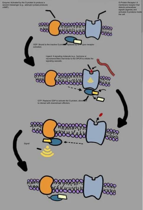
Long description
Illustration for biochemistry/cell signalling receptors g proteins and second messengers from Wikimedia Commons, titled 'Cell Signaling Reception': Diagram of G-Coupled Protein Reception. The author and licence are given in the credit line.

Cell signalling is the fundamental language by which cells communicate, coordinate their behaviour, and maintain tissue homeostasis across the life of an organism. During embryogenesis and throughout adult life, multicellular organisms rely on exquisitely tuned signalling systems to direct complex cellular events such as migration, differentiation, and survival. At the heart of these cascades are ligand-receptor interactions occurring at the plasma membrane. Extracellular signals, whether they are secreted molecules or cell-surface-attached ligands, must engage specific transmembrane receptors to provoke a precise intracellular response. [6]
From a first-principles perspective, understanding these pathways requires tracing the signal from the extracellular ligand through the plasma membrane to cytoplasmic second messengers and ultimate transcriptional or cytoskeletal outcomes. Guidance cues provide classic archetypes for these processes. Members of four major cell guidance molecule families—the netrins, slits, ephrins, and semaphorins—interact with their cognate cell surface receptors to guide cells and neuronal growth cones with ex Although ‘axon guidance’ molecules were first characterised by their role in the development of the nervous system these secreted or cell surface attached proteins are now known to perform cell guidance functions in a broad range of tissues [6]
Signalling fidelity is governed by multiple structural mechanisms operating at the cell surface, including receptor auto-inhibition, precise oligomeric states, and the assembly of multi-receptor complexes. Most guidance receptors are type 1 single membrane-spanning proteins with diverse cytoplasmic domains. The classical trigger for downstream signalling is ligand-induced receptor dimerisation. However, structural and cell-based analyses reveal that simple dimerisation is frequently insufficient for biological activation. For instance, ephrins are cell-surface-attached ligands that signal through Eph receptor tyrosine kinases, where higher-order oligomerisation beyond simple dimers is an absolute requirement to trigger downstream signalling. Visualisation of EphB2 receptors in living cells demonstrates that activation occurs through trimers and tetramers, whereas mere dimerisation fails to initiate the characteristic cellular collapse response associated with repulsive guidance. [6]
DefinitionReceptor oligomerisation
The process by which multiple receptor monomers aggregate into dimers, trimers, or higher-order clusters upon ligand binding, shifting the receptor from an inactive to an active enzymatic or conformational state.
Beyond clustering size, the spatial composition of the signalling assembly determines functional outcomes. Signalling complexes often incorporate co-receptors that drive context-dependent switching of cellular behaviour. For example, the semaphorin ligand Sema3E binds to its primary receptor PlexinD1, but the presence of the co-receptor neuropilin switches the signalling outcome from repulsive to attractive. Similarly, Netrin-1 interacts with DCC to generate an attractive response, whereas the inclusion of UNC5 in the receptor complex results in a repulsive guidance signal. These modular arrangements highlight how cell-surface interactions integrate multiple extracellular inputs into a unified intracellular command. [6]
Although guidance cues utilise receptor tyrosine kinases and single-pass transmembrane proteins, another monumental class of cell surface receptors relies on heterotrimeric GTP-binding proteins to transduce extracellular signals: the G-protein-coupled receptors. While the provided texts focus heavily on structural paradigms of guidance molecules such as netrins, slits, ephrins, and semaphorins—exemplified by molecules like Netrin-1 whose DCC ectodomain comprises four immunoglobulin-like domains followed by six fibronectin type 3 domains—the general principles of membrane receptor assembly, auto-inhibition, and subunit cooperation translate directly to G-protein-coupled signalling systems. [6]
Heterotrimeric G proteins consist of alpha, beta, and gamma subunits. In the basal, inactive state, the alpha subunit binds guanosine diphosphate (GDP) and associates tightly with the beta-gamma dimer. Upon agonist binding to the extracellular domain of the G-protein-coupled receptor, a conformational change is transmitted across the membrane, promoting the exchange of bound GDP for guanosine triphosphate (GTP) on the alpha subunit. This nucleotide exchange triggers the dissociation of the GTP-bound alpha subunit and the free beta-gamma dimer from the receptor and from each other, allowing both moieties to regulate downstream effector enzymes and ion channels.
G-protein alpha subunits are categorised into major families based on their primary downstream signalling effectors and physiological roles:
| G-Protein Family | Primary Effector | Second Messenger / Pathway |
|---|---|---|
| Gs (Stimulatory) | Adenylyl cyclase activation | Increases cyclic AMP (cAMP) |
| Gi (Inhibitory) | Adenylyl cyclase inhibition | Decreases cyclic AMP (cAMP) |
| Gq (Phospholipase C) | Phospholipase C-beta activation | Increases IP3, DAG, and intracellular calcium |
The stimulatory G-protein alpha subunit, Gs, activates adenylyl cyclase, an enzyme embedded in the plasma membrane that catalyses the conversion of cytosolic adenosine triphosphate into cyclic AMP. Conversely, the inhibitory G-protein alpha subunit, Gi, suppresses adenylyl cyclase activity, thereby lowering intracellular cyclic AMP levels and modulating downstream protein kinase A activity. The third major pathway, driven by Gq, stimulates membrane-bound phospholipase C-beta, initiating the cleavage of membrane phospholipids into potent intracellular messengers.
Clinical pearlReceptor cross-talk
Direct cross-talk frequently occurs between distinct membrane signalling systems at the cell surface, such as plexins mediating slit guidance functions, demonstrating that receptors rarely operate in complete isolation.
These systems are subject to rigorous negative and positive feedback mechanisms. Just as guidance cue receptors form intricate supercomplexes with diverse receptor families and proteoglycans to achieve spatial and temporal precision, G-protein-coupled receptors undergo rapid desensitisation, internalisation, and recycling. Structural studies examining how receptor complexes connect to downstream internalisation machinery underscore the importance of spatial control in preventing aberrant signalling. [6]
Second messengers act as intracellular relay molecules that amplify the initial extracellular signal manifold, translating membrane-level binding events into widespread biochemical changes across the cytoplasm and nucleus. Among the most prominent second messengers are cyclic AMP (cAMP), cyclic GMP (cGMP), inositol 1,4,5-trisphosphate (IP3), and diacylglycerol (DAG). Each is produced or released in response to specific upstream receptor and G-protein cascades, operating with exquisite temporal and spatial precision to regulate cellular metabolism, transcription, and cytoskeletal dynamics.
Cyclic AMP is synthesized by adenylyl cyclase following Gs activation and is degraded by phosphodiesterases. The primary intracellular target of cAMP is protein kinase A (PKA). When cAMP binds to the regulatory subunits of PKA, active catalytic subunits are released to phosphorylate specific serine and threonine residues on target transcription factors and metabolic enzymes. In neuronal growth cones and migrating cells, intracellular gradients of cAMP and cGMP act in concert with guidance cue receptors—such as DCC and UNC5—to dictate whether a cell is attracted toward or repelled away from a source of Netrin-1.
The Gq pathway diverges into two distinct second messenger arms through the action of phospholipase C-beta. Phospholipase C-beta cleaves phosphatidylinositol 4,5-bisphosphate, a minor lipid component of the inner plasma membrane leaflet, yielding two vital second messengers: IP3 and DAG.
Gq-Phospholipase C Signalling Pathway
- Gq-protein-coupled receptor activated by agonistGDP is exchanged for GTP on the G-alpha-q subunit.
- Question: Does Gq-GTP activate Phospholipase C-beta?
Yes
- Cleavage of PIP2Phosphatidylinositol 4,5-bisphosphate is split into IP3 and DAG.
Inositol 1,4,5-trisphosphate is a water-soluble molecule that diffuses rapidly through the cytosol to bind to ligand-gated calcium channels (IP3 receptors) located on the membrane of the endoplasmic reticulum. This triggers a rapid efflux of stored calcium ions into the cytoplasm, sharply elevating intracellular calcium concentration. Calcium itself acts as a ubiquitous second messenger, binding to proteins such as calmodulin to regulate diverse enzymatic cascades.
Concurrently, diacylglycerol remains lipophilic and confined within the plane of the plasma membrane. DAG binds to and activates conventional isoforms of protein kinase C. Complete activation of protein kinase C requires both the membrane tethering provided by DAG and the binding of calcium ions released by the IP3 pathway. Once active, protein kinase C phosphorylates downstream targets involved in cell proliferation, secretion, and cytoskeletal remodelling.
Exam trapOligomerisation requirements
Assuming simple dimerisation is sufficient for all receptors is a frequent error; higher-order oligomerisation into trimers or tetramers is frequently mandatory for productive signal transduction, as seen with EphB2 receptors.
These multi-level control mechanisms ensure that second messenger generation is tightly restricted in both time and space. Through the combinatorial interplay of G proteins, second messengers, and receptor clustering, cells achieve the remarkable accuracy required for embryonic development and tissue homeostasis. [6]
Cellular communication relies heavily on surface receptors that convert extracellular cues into intracellular signals. Receptor tyrosine kinases (RTKs) comprise a major family of single-pass transmembrane proteins expressed on various cell types, including those within the tumor microenvironment. Growth factors and chemokines bind to these receptors, activating diverse signaling cascades that regulate normal physiology but can also drive cancer progression when dysregulated. Structurally, an RTK spans the cell membrane once, featuring an extracellular ligand-binding domain, a hydrophobic transmembrane region, and an intracellular domain possessing intrinsic tyrosine kinase activity. [5]
Binding of an appropriate extracellular ligand induces conformational changes that typically promote receptor dimerization. For instance, EGFR is notable because it can bind seven different ligands, demonstrating ligand promiscuity within the family. The interactions between the receptor ectodomain, the plasma membrane, and the juxtamembrane and kinase regions are critical. Molecular dynamics studies suggest that a delicate balance of receptor-receptor, receptor-lipid, and receptor-ligand interactions controls the switch from inactive dimer-based states to active signaling clusters. [5]
Once dimerization occurs, the intrinsic kinase domains cross-phosphorylate each other on specific intracellular tyrosine residues. These newly phosphorylated tyrosine residues act as high-affinity docking sites for downstream effector molecules containing Src homology 2 (SH2) or phosphotyrosine-binding (PTB) domains. Effector proteins can bind directly to the activated RTKs or through intermediary docking proteins that become tyrosine phosphorylated. Well-known docking proteins that orchestrate the formation of large macromolecular signaling complexes include fibroblast growth factor receptor substrate 2 (FRS2), insulin receptor substrate 1 (IRS1), and Grb2-associated binder 1 (Gab1). These recruitment hubs then launch canonical cascades such as the MAPK/ERK, PI3K/Akt, and JAK/STAT pathways. [4]
DefinitionReceptor tyrosine kinase
Therapeutic targeting of RTKs is central to modern oncology, yet resistance remains a clinical hurdle. Impaired interaction between anti-RTK agents and their receptors can cause resistance, often driven by structural alterations or masking proteins. For example, mucin-4 and CD44 are cell surface proteins that can be overexpressed in trastuzumab-resistant breast cancer patients, and their physical proximity to the HER2 epitope masks the receptor and blocks trastuzumab binding while enhancing tumor growth. Alternatively, expression of a truncated version of HER2, known as p95HER2, overrides trastuzumab sensitivity by forming heterodimers with HER3 and activating downstream signaling in a ligand-independent manner.
| Docking protein | Abbreviation | Function |
|---|---|---|
| FGF receptor substrate 2 | FRS2 | Orchestrates large protein complexes |
| Insulin receptor substrate 1 | IRS1 | Mediates downstream signaling |
| Grb2-associated binder 1 | Gab1 | Recruits effector molecules |
RTK activation and signaling
- Ligand BindingGrowth factor binds to the extracellular domain of the single-pass RTK.
- Question: Does dimerization occur?
Yes
- AutophosphorylationIntracellular kinase domains cross-phosphorylate tyrosine residues.
- Effector RecruitmentProteins with SH2 or PTB domains bind phosphotyrosines, activating downstream cascades.
No
- Inactive StateReceptor remains monomeric and silent.
Beyond the cell surface, cells utilize intracellular transcription factors known as nuclear receptors to directly regulate gene expression in response to lipophilic molecules. Peroxisome proliferator-activated receptors (PPARs) belong to this superfamily of phylogenetically related proteins, which were identified in rodents in 1990 and comprise a nuclear hormone receptor superfamily containing 48 members[1]. Structurally, PPARs resemble steroid or thyroid hormone receptors and are stimulated by small lipophilic ligands[1]. PPARs act on specific DNA response elements as heterodimers with the retinoid X receptor (RXR)[1]. Their natural activating ligands are lipid-derived substrates[1], reflecting their primary roles in regulating lipid homeostasis, energy metabolism, and inflammation.
The family of PPARs is represented by three distinct isoforms: PPAR-alpha, PPAR-delta, and PPAR-gamma[1]. Other notable members of the nuclear receptor superfamily include retinoic acid, estrogen, thyroid, vitamin D, and glucocorticoid receptors, alongside several proteins involved in xenobiotic metabolism[1]. Nuclear receptors characteristically feature conserved domains for DNA binding and ligand binding, allowing them to translocate to the nucleus and modulate transcription upon ligand activation.
Clinical pearlNuclear receptor heterodimerization
Complex feedback mechanisms govern nuclear receptor signaling in metabolic pathways. A key repressive mechanism involves the activation of the farnesoid X receptor (FXR) by accumulating bile acids (BAs), which then induces the small heterodimer partner (SHP). SHP acts as a repressor of hepatic nuclear factor 1 alpha and 4 alpha (HNF-1 alpha and HNF-4 alpha), and also interferes with retinoid X receptor (RXR) and retinoic acid receptor (RAR) heterodimers in rats, or the glucocorticoid receptor in humans, all of which are required for normal sodium-taurocholate cotransporting polypeptide (NTCP) expression. [3]
Understanding nuclear receptors is also crucial for pharmacology and toxicology. Although rodent studies identified certain proliferative agents linked to peroxisomes—subcellular organelles performing H2O2-based respiration, fatty acid beta-oxidation, and cholesterol metabolism[1]—these exact agents are associated with no cellular proliferation in human beings[1], highlighting important species-specific differences in nuclear receptor biology.
| Receptor / Factor | Subtype / Partner | Key Feature |
|---|---|---|
| PPAR | Alpha, Delta, Gamma | Acts as heterodimer with RXR, binds lipid substrates |
| FXR | Farnesoid X receptor | Activated by accumulating bile acids, induces SHP |
| SHP | Small heterodimer partner | Acts as a transcriptional corepressor |
Nuclear receptor transcriptional control
- Ligand EntrySmall lipophilic molecules enter the cell and bind the nuclear receptor.
- Question: Does it heterodimerize?
Yes
- RXR PartnershipForms a heterodimer, such as PPAR-RXR.
- DNA BindingInteracts with response elements to alter gene transcription.
No
- Corepressor ActionInduction of factors like SHP to repress target genes.
The Janus kinase signal transducer and activator of transcription pathway represents an evolutionarily conserved signaling network. From first principles, cell surface receptors often lack intrinsic enzymatic activity and must couple to intracellular catalytic machinery to relay extracellular cues into transcriptional changes. The JAK-STAT cascade fulfills this requirement across multiple critical physiological domains, including hematopoiesis, cellular differentiation, metabolic control, and immune modulation. Structurally, the cascade relies on three main components: transmembrane receptors, receptor-associated cytosolic tyrosine kinases termed JAKs, and signal transducers and activators of transcription known as STATs. [4]
The mammalian protein architecture for this cascade features four distinct JAK family members—JAK1, JAK2, JAK3, and TYK2—alongside seven distinct STAT family proteins designated STAT1, STAT2, STAT3, STAT4, STAT5A, STAT5B, and STAT6. Historically, the entire signaling architecture was discovered through pioneering investigations centered on interferon-related transcriptional activation. Over a subsequent research period spanning about 20 years, the fundamental components and pathogenic outlines of the pathway were fully mapped. More than 50 distinct types of cytokines—including interferons, interleukins, and specific growth factors—utilize this machinery to execute regulatory tasks governing cell differentiation, survival, homeostasis, and immune responses. [4]
DefinitionCore Pathway Mechanics
When an extracellular ligand engages its cognate receptor, receptor-associated JAKs initiate direct tyrosine phosphorylation of the receptor complex. This enzymatic modification creates docking sites that recruit corresponding cytoplasmic STAT proteins. Once recruited, the phosphorylated STAT molecules undergo dimerization and transit directly into the nucleus, where they bind specific DNA sequences to regulate target gene transcription. This direct relay mechanism enables rapid transmission of extracellular signals directly to the nuclear machinery. [4]
Cytokines drive both humoral and cellular immune responses. A wide array of cytokines tied to autoimmune disorders—such as interferons, interleukins, and colony-stimulating factors—exert pleiotropic biological effects primarily through combinations of type I and type II cytokine receptors. The engagement of these specific receptor types activates the associated JAKs, which subsequently recruit STATs to transduce cytokine signals. Beyond immunology, upstream cytokines and their cognate receptors are pivotal for modulating overall pathway activity. Therapeutic agents designed to manipulate these upstream nodes include siltuximab and tocilizumab, which achieve classical blockade of interleukin-6 to inhibit downstream signal transduction. [4]
| Family Class | Component Members | Primary Functional Roles |
|---|---|---|
| Janus kinases (JAKs) | JAK1, JAK2, JAK3, TYK2 | Receptor-associated cytosolic tyrosine kinases |
| STAT proteins | STAT1, STAT2, STAT3, STAT4, STAT5A, STAT5B, STAT6 | Dimerization and nuclear transcription regulation |
| Upstream ligands | Interferons, interleukins, growth factors | Extracellular receptor activation |
Under physiological conditions, cytokine-induced activation dictates immune cell differentiation, development, and system homeostasis. Different cytokine stimulations prompt distinct functional outcomes: interferon signaling utilizes STAT1 and STAT2 to mediate cancer cell recognition, whereas interleukin-6 acting via STAT3 can drive immune escape. Furthermore, receptor tyrosine kinases such as epidermal growth factor receptors, vascular endothelial growth factor receptors, platelet-derived growth factor receptors, insulin-like growth factor receptors, and fibroblast growth factor receptors can also activate this pathway alongside the mitogen-activated protein kinase and phosphoinositide 3-kinase cascades. Elevated receptor tyrosine kinase levels are strongly linked with heightened breast cancer aggressiveness and decreased survival. [4] [5]
Dysregulation of these cascades contributes heavily to human pathology. Hyperactivation of the pathway is implicated in poor clinical outcomes across multiple malignancies, including melanomas, glioblastomas, and cancers of the head, neck, lung, pancreas, breast, rectum, and prostate. Conversely, favorable or suppressive regulatory roles have been documented in head and neck squamous cell carcinomas as well as prostate and colorectal cancers. Constitutively activated STAT3 specifically promotes constitutively activated STAT3 promotes lineage plasticity and associated cell migration and invasion [4]
JAK-STAT Signal Transduction Outline
- Ligand BindingExtracellular cytokine or growth factor engages transmembrane receptor.
- JAK ActivationReceptor-associated JAKs phosphorylate receptor tyrosine residues.
- Question: STAT Recruitment?
Yes
- Dimerization & Nuclear EntryPhosphorylated STATs dimerize, enter the nucleus, and alter gene transcription.
No
- Baseline StatePathway remains unactivated.
Cell signalling cascades, particularly those driven by receptors coupled to tyrosine kinase enzymes and intracellular second messengers, provide the mechanistic foundation for numerous targeted pharmacotherapies in clinical medicine. From a first-principles perspective, breaking or modulating these signalling webs allows clinicians to halt aberrant cell proliferation, dampen runaway immune responses, and reverse pathological gene transcription.
The Janus kinase-signal transduction and activation of transcription pathway, universally known as the JAK-STAT pathway, serves as a prime illustration of this principle. Type I and type II cytokine receptors lack intrinsic enzymatic activity; instead, they rely on non-covalently bound Janus kinases (JAKs) to phosphorylate the receptor tails upon ligand binding, creating docking sites for STAT proteins. These STAT transcription factors then dimerise, translocate to the nucleus, and switch on cytokine-specific genetic programmes.
When this pathway becomes hyperactive, it drives autoimmune, inflammatory, and myeloproliferative disorders. This pathophysiological insight led to the development of small-molecule inhibitors targeting JAKs, known as Jakinibs. For instance, ruxolitinib is a JAK1 and JAK2 inhibitor with moderate inhibitory activity against Tyk2. Ruxolitinib is FDA approved for the treatment of polycythemia vera and intermediate- and high-risk primary myelofibrosis, where inappropriate activation of JAK2 underlies disease pathogenesis. Furthermore, ruxolitinib is effective in the treatment of essential thrombocythemia and has been granted Breakthrough Therapy Designation for the treatment of graft versus host disease. [2]
Another targeted Jakinib is baricitinib, whose metabolism and pharmacokinetics show that renal clearance is the primary route of excretion; therefore, the role of cytochrome P450-mediated medication interactions is thought to be minimal for this drug. It is speculated that its half-life would be prolonged in disease states with reduced renal function. Filgotinib represents yet another compound in this class, investigated for the treatment of rheumatoid arthritis. Phase IIa studies in patients with active rheumatoid arthritis and an inadequate response to methotrexate showed efficacy of filgotinib over placebo at doses of 30 mg daily and higher. This was followed by two phase IIb trials, designated as DARWIN1 and DARWIN2. DARWIN1 is a study on 595 methotrexate-treated rheumatoid arthritis patients where filgotinib was added at a range of doses from 50 mg daily to 100 mg twice daily. The DARWIN2 study assessed filgotinib monotherapy in 280 rheumatoid arthritis patients with doses ranging from 50 to 200 mg daily. In both studies, filgotinib was found to be superior to placebo in controlling disease activity as measured by the American College of Rheumatology criteria ACR 20, ACR 50, Disease Activity Score using 28 joints and C-reactive protein (DAS28-CRP), Simple Disease Activity Index (SDAI), and clinical disease activity index. [6] [2]
| Agent | Primary Targets | Key Indications |
|---|---|---|
| Ruxolitinib | JAK1, JAK2, Tyk2 (moderate) | Polycythemia vera, myelofibrosis, essential thrombocythemia |
| Baricitinib | JAK1, JAK2 | Rheumatoid and inflammatory disorders (renal clearance) |
| Filgotinib | JAK family selectivity | Rheumatoid arthritis (assessed in DARWIN trials) |
Beyond haematological and rheumatologic pathways, receptor tyrosine kinases (RTKs) such as the epidermal growth factor receptor (EGFR) family play critical roles in solid tumours, particularly breast cancer. Increased expressions of EGFR1, HER2, or EGFR3 are associated with reduced survival, whereas elevated levels of EGFR4 are connected with better survival in breast cancer patients. Upon binding to the ligand, EGFR activates various downstream signaling molecules including Ras, PI3K, phospholipase C-γ (PLC-γ), and JAK leading to cell survival, cell growth, and tumor progression [5]
Therapeutically, this is exploited using monoclonal antibodies like trastuzumab (Herceptin), a humanized monoclonal antibody that targets the extracellular domain of HER2 in HER2-positive breast cancer, enhancing the survival of patients at early and late stages of the disease. Trastuzumab inhibits HER2-HER3 heterodimerization, which occurs in a ligand-independent manner in HER2-positive breast cancer, and may also induce HER2 degradation. [5]
Clinical pearlReceptor cross-talk
Receptor tyrosine kinases do not operate in isolation; FGFRs are mechanistically linked to the functions of other RTKs and drug resistance, making them potential targets for breast cancer treatment. [5]
When studying and applying cell signalling pathways in clinical pharmacology, students and clinicians frequently encounter several conceptual traps. The first major pitfall is assuming that a single cytokine receptor family uses only one linear signalling cascade with uniform physiological outcomes. In reality, pathways like JAK-STAT exhibit immense pleiotropy. The effects of the JAK-STAT pathway on disease progression are complex because individual JAKs pair with multiple distinct cytokine receptors, and downstream STAT dimers regulate vast arrays of genes that can either promote inflammation, drive neoplastic proliferation, or exert immunomodulatory feedback. [4]
Another common trap involves misunderstanding target specificity and toxicity profiles. Because JAK3 is highly expressed in immune cells, patients with autosomal recessive severe combined immunodeficiency are spared from extra-immune disease manifestations. This observation formed the basis for historical and ongoing interest in selective JAK3 blockade as a potential immunosuppressive therapy with limited off-target effects. Students often generalise this and assume all Jakinibs share clean safety profiles, ignoring the fact that broader inhibition of JAK1 and JAK2 leads to dose-limiting cytopenias, lipid abnormalities, and infection risks due to interference with erythropoietin, thrombopoietin, and interferons. [2]
Pharmacokinetic assumptions represent a third diagnostic and therapeutic trap. Clinicians may mistakenly manage all small-molecule kinase inhibitors using standard hepatic cytochrome clearance assumptions. However, clearance mechanisms vary profoundly. For instance, renal clearance is the primary route of excretion for baricitinib, meaning that CYP-mediated drug interactions are minimal, but dose adjustments or extensions in half-life are mandatory when encountering renal impairment. Assuming hepatic metabolism for a renally cleared drug risks severe accumulation and toxicity.
Exam trapAssuming uniform dosing strategies
Optimal dosing strategies of Jakinibs deserve further study; while currently approved regimens rely on a single dose for induction of remission and maintenance, it is not clear that this is optimal, and loading patients with high doses followed by a lower maintenance dose mirrors successful paradigms like corticosteroid therapy. [2]
At this time, the only definitive treatment for this disease is hematopoietic stem cell transplantation. [2]
Cell signalling pathways and second messenger cascades dictate clinical decision-making across rheumatology, clinical immunology, medical oncology, and personalised medicine. Understanding these pathways allows clinicians to interpret diagnostic assays, select targeted biological or small-molecule therapies, and navigate treatment resistance.
In rheumatology and inflammatory bowel disease, clinicians deploy Jakinibs when conventional synthetic disease-modifying antirheumatic drugs (such as methotrexate) fail. Therapeutic monitoring relies on validated composite scoring systems. For example, clinical trials such as DARWIN1 and DARWIN2 demonstrated efficacy by measuring improvements in ACR 20, ACR 50, DAS28-CRP, SDAI, and clinical disease activity index. Clinicians use these scores to titrate targeted therapies that block intracellular cytokine signal transduction.
Targeting Cell Signalling in Clinical Practice
- Identify Pathological PathwayDetermine if disease is driven by cytokine overexpression (JAK-STAT) or growth factor receptors (RTK/HER2).
- Question: Receptor Type?
Cytokine Receptor (Type I/II)
- Deploy Small Molecule JakinibSelect ruxolitinib, baricitinib, or filgotinib based on renal function and target isoform.
Tyrosine Kinase Receptor (RTK)
- Deploy Monoclonal AntibodyUse agents like trastuzumab to disrupt heterodimerization and trigger receptor degradation.
- Monitor Response and ResistanceAssess clinical indices (DAS28, ACR) and watch for primary or acquired drug resistance via pathway cross-talk.
In oncology, immunohistochemistry and molecular pathology reports dictate treatment by quantifying receptor expression levels. Pathologists evaluate oestrogen receptor (ER) status alongside EGFR and HER2 expression. Notably, increased expressions of EGFR1, HER2, and EGFR3 are coupled with reduced expression of oestrogen receptors, and various studies find that ER expression is inversely correlated with EGFR or cancer stem-like cell phenotypes. This inverse relationship is supported by data indicating higher expression of EGFR and the presence of stem cell populations in triple-negative breast cancers (TNBCs), which lack ER expression. Consequently, determining the precise receptor profile dictates whether an anti-HER2 monoclonal antibody like trastuzumab is indicated to enhance survival. [5]
Biomarker development and patient stratification represent the cutting edge of clinical practice. Complex rheumatologic and oncologic diseases are driven by JAK-dependent cytokines and RTK networks to a different degree in different patients. Ongoing biomarker research aims to identify subgroups most likely to respond to specific signalling inhibitors, moving medicine away from trial-and-error prescribing toward precision molecular alignment. Finally, non-oral formulations of Jakinibs are in preliminary stages of investigation for localized conditions including psoriasis, allergic dermatitis, and ocular disease, expanding the clinical utility of second-messenger pathway modulation beyond systemic administration. [2]
DefinitionPrecision biomarker utilisation
Biomarker development is ongoing to identify groups of patients with disease likely to respond to Jakinibs, as complex rheumatologic diseases may be driven by JAK-dependent cytokines to a different degree in different patients. [2]
- Type I and II cytokine receptors use the JAK-STAT pathway to phosphorylate transcription factors and activate specific genetic programmes.
- Ruxolitinib is a JAK1 and JAK2 inhibitor approved for polycythemia vera, myelofibrosis, and essential thrombocythemia.
- Renal clearance is the primary route of excretion for baricitinib, minimizing CYP-mediated interactions while prolonging half-life in renal impairment.
- Filgotinib showed superior efficacy over placebo in rheumatoid arthritis trials (DARWIN1 and DARWIN2) across multiple dosing schedules.
- Approximately 65 percent of HER2-positive breast cancer patients do not respond to primary trastuzumab treatment, highlighting clinical resistance barriers. [5]
- Oestrogen receptor expression is inversely correlated with EGFR expression and cancer stem cell phenotypes in triple-negative breast cancers.
- Biomarker development is essential because complex diseases are driven by JAK-dependent cytokines to varying degrees across different patients.
Answer each one in your head first, then open the answer.
Which kinase family is primarily employed by type I and II cytokine receptors to phosphorylate STAT proteins?
Show answerHide answer
Janus kinase or JAKWhat is the primary route of excretion and metabolism consideration for baricitinib?
Show answerHide answer
Renal clearanceWhat percentage of HER2-positive breast cancer patients do not respond to primary trastuzumab treatment?
Show answerHide answer
Approximately 65 percentWhich two phase IIb clinical trials assessed filgotinib monotherapy and combination therapy in rheumatoid arthritis?
Show answerHide answer
DARWIN1 and DARWIN2What is the functional relationship between estrogen receptor expression and EGFR in breast cancer?
Show answerHide answer
Inverse correlation
- Tyagi S, Gupta P, Saini AS, et al. The peroxisome proliferator-activated receptor: A family of nuclear receptors role in various diseases. Journal of advanced pharmaceutical technology & research 2011;2(4):236-240. doi:10.4103/2231-4040.90879. Accessed .
- Banerjee S, Biehl A, Gadina M, et al. JAK-STAT Signaling as a Target for Inflammatory and Autoimmune Diseases: Current and Future Prospects. Drugs 2017;77(5):521-546. doi:10.1007/s40265-017-0701-9. Accessed .
- Halilbasic E, Claudel T, Trauner M. Bile acid transporters and regulatory nuclear receptors in the liver and beyond. Journal of hepatology 2013;58(1):155-168. doi:10.1016/j.jhep.2012.08.002. Accessed .
- Xue C, Yao Q, Gu X, et al. Evolving cognition of the JAK-STAT signaling pathway: autoimmune disorders and cancer. Signal transduction and targeted therapy 2023;8(1):204. doi:10.1038/s41392-023-01468-7. Accessed .
- Butti R, Das S, Gunasekaran VP, et al. Receptor tyrosine kinases (RTKs) in breast cancer: signaling, therapeutic implications and challenges. Molecular cancer 2018;17(1):34. doi:10.1186/s12943-018-0797-x. Accessed .
- Rozbesky D, Jones EY. Cell guidance ligands, receptors and complexes - orchestrating signalling in time and space. Current opinion in structural biology 2020;61:79-85. doi:10.1016/j.sbi.2019.11.007. Accessed .
Figure credits
Educational content for medical students and doctors. It is not a substitute for clinical judgement or local guidelines.