Cholesterol, bile acid and steroid hormone biosynthesis
How cells make cholesterol, convert it to bile acids and steroid hormones, keep it in balance, and what happens when an enzyme in the pathway fails.
- Cholesterol is a sterol made mainly in the liver from acetyl-CoA through the mevalonate pathway; its rate-limiting enzyme is HMG-CoA reductase.[1]
- The body uses cholesterol as a membrane component, as the starting material for bile acids and for all steroid hormones, and as the precursor of vitamin D.
- Cholesterol balance is maintained by changing how much is made, how much is taken up from the blood by LDL receptors, and how much is excreted as bile acids.
- Statins, ezetimibe, bile-acid sequestrants and PCSK9 inhibitors all work on this pathway or on cholesterol absorption and clearance.
- Inherited defects in the enzymes of cholesterol synthesis or of steroidogenesis produce well-recognised syndromes, from Smith-Lemli-Opitz syndrome to congenital adrenal hyperplasia.
By the end of this page you should be able to:
- Trace cholesterol synthesis from acetyl-CoA to cholesterol, naming the rate-limiting step and its cofactors.
- Explain how HMG-CoA reductase is regulated and why statins lower LDL cholesterol.
- Describe bile-acid synthesis, conjugation and the enterohepatic circulation.
- Outline steroidogenesis in the adrenal cortex and gonads, including the role of StAR and the main cytochrome P450 enzymes.
- Recognise the commoner inherited defects of cholesterol and steroid-hormone synthesis and their clinical pictures.
- Interpret the laboratory tests used to investigate this pathway.
Figure 1Cholesterol metabolism
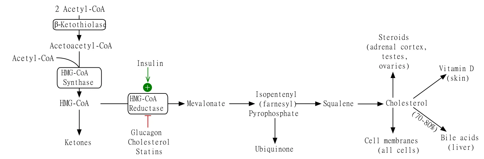
Long description
Illustration for biochemistry/cholesterol bile acid and steroid hormone biosynthesis from Wikimedia Commons, titled 'Cholesterol metabolism': Cholesterol metabolic pathway. The author and licence are given in the credit line.

Cholesterol is not just a number on a lipid profile. It is an essential structural lipid of cell membranes, the precursor of bile acids that digest fat, the precursor of every steroid hormone, and the molecule from which vitamin D is made in skin. Because humans can make all the cholesterol they need, dietary cholesterol is not essential, but the amount in blood matters: too much LDL cholesterol drives atherosclerosis, and too little, or a block in its synthesis, disrupts development.
This page covers the pathway as a whole: cholesterol synthesis, its regulation and homeostasis, bile-acid synthesis and recycling, steroid hormone synthesis, the disorders that arise when an enzyme fails, the drugs that act on the pathway, and the tests used to investigate it. It is written for a first encounter, so each enzyme is introduced with what it does and why it matters.
- Cholesterol synthesis occurs in the cytoplasm and on the endoplasmic reticulum of almost all nucleated cells, but most of it happens in the liver. The brain makes its own cholesterol because cholesterol does not cross the blood-brain barrier well.
- Bile-acid synthesis occurs in hepatocytes.
- Steroidogenesis occurs in the adrenal cortex, the gonads (testes and ovaries) and the placenta. These tissues take up cholesterol from lipoproteins rather than making it all themselves.
- The rate-limiting step of steroidogenesis is not a synthetic enzyme but the delivery of cholesterol into mitochondria by the steroidogenic acute regulatory protein (StAR).
The pathway has two halves. The first half, from acetyl-CoA to the sterol lanosterol, is called the mevalonate pathway. The second half trims and rearranges lanosterol into the 27-carbon cholesterol.
| Step | Reaction | Enzyme | Key points |
|---|---|---|---|
| 1 | 2 acetyl-CoA to acetoacetyl-CoA | Thiolase (acetyl-CoA acetyltransferase) | Reversible condensation |
| 2 | Acetoacetyl-CoA + acetyl-CoA to 3-hydroxy-3-methylglutaryl-CoA (HMG-CoA) | HMG-CoA synthase | Cytoplasmic isoform for sterol synthesis |
| 3 | HMG-CoA to mevalonate | HMG-CoA reductase | Rate-limiting; uses NADPH; target of statins |
| 4 | Mevalonate to mevalonate-5-phosphate | Mevalonate kinase | ATP-dependent; defects cause mevalonic aciduria |
| 5 | Mevalonate-5-phosphate to mevalonate-5-pyrophosphate | Phosphomevalonate kinase | ATP-dependent |
| 6 | Mevalonate-5-pyrophosphate to isopentenyl pyrophosphate (IPP) | Mevalonate pyrophosphate decarboxylase | Releases CO2; ATP-dependent |
| 7 | IPP to dimethylallyl pyrophosphate (DMAPP) | IPP isomerase | Reversible |
| 8 | DMAPP + IPP to geranyl pyrophosphate (GPP) | Farnesyl pyrophosphate synthase | First C10 intermediate |
| 9 | GPP + IPP to farnesyl pyrophosphate (FPP) | Farnesyl pyrophosphate synthase | C15; branch point for non-sterol isoprenoids |
| 10 | 2 FPP to squalene | Squalene synthase | C30; uses NADPH; first committed sterol step |
| 11 | Squalene to squalene-2,3-epoxide | Squalene epoxidase (monooxygenase) | Uses NADPH and O2 |
| 12 | Squalene-2,3-epoxide to lanosterol | Lanosterol synthase (oxidosqualene cyclase) | Cyclisation |
| 13 | Lanosterol to cholesterol | Many enzymes (CYP51A1, EBP, SC5D, DHCR7, DHCR24 and others) | Two routes: Bloch and Kandutsch-Russell |
Two terminal routes exist. In the Bloch pathway, the side chain is reduced last and the immediate precursor is desmosterol, converted to cholesterol by 24-dehydrocholesterol reductase (DHCR24). In the Kandutsch-Russell pathway, the side chain is reduced early and the immediate precursor is 7-dehydrocholesterol, converted to cholesterol by 7-dehydrocholesterol reductase (DHCR7). Both routes operate in different tissues and developmental stages.
A crucial point is that the mevalonate pathway is not only for cholesterol. Farnesyl pyrophosphate (FPP) and geranylgeranyl pyrophosphate feed protein prenylation, ubiquinone (coenzyme Q) synthesis, dolichol synthesis and heme A synthesis. This is why blocking the pathway early, as statins do, has effects beyond cholesterol.
HMG-CoA reductase catalyses the conversion of HMG-CoA to mevalonate. It is the rate-limiting enzyme of cholesterol synthesis and the target of the statin drugs. The reaction uses two molecules of NADPH and is essentially irreversible in vivo.
Statins (atorvastatin, simvastatin, rosuvastatin, pravastatin and others) are competitive inhibitors of HMG-CoA reductase. They mimic HMG-CoA and bind the active site. Lowering intracellular cholesterol in hepatocytes increases the activity of SREBP-2 (see below), which raises LDL-receptor expression, so more LDL is cleared from blood. This dual action, less synthesis plus more clearance, is why statins are effective LDL-lowering drugs.
A Cochrane systematic review of statins for primary prevention found reductions in all-cause mortality, major vascular events and revascularisations in people without established cardiovascular disease, with no excess of adverse events in the trials reviewed.[10] NICE (NG238, 2023) recommends statins for primary and secondary prevention of cardiovascular disease, with optimisation of treatment and alternatives when statins are not tolerated.[11] WHO's cardiovascular-risk guidelines likewise frame prevention around absolute cardiovascular risk rather than single risk factors.[12]
Cells keep cholesterol within a narrow range by balancing three processes: synthesis, uptake and efflux.
- Synthesis is controlled at HMG-CoA reductase (below).
- Uptake is controlled by the LDL receptor. When intracellular cholesterol is low, SREBP-2 activates transcription of the LDL receptor gene, so the cell imports more LDL. When cholesterol is abundant, LDL receptors are down-regulated and also degraded through PCSK9-mediated lysosomal targeting.
- Efflux moves cholesterol out of cells to HDL via ABCA1 and ABCG1, for return to the liver (reverse cholesterol transport). Excess intracellular cholesterol is esterified by ACAT and stored in lipid droplets.
The master transcriptional regulator is SREBP-2 (sterol regulatory element-binding protein 2).[2] SREBP-2 is made as an inactive precursor bound to SCAP in the endoplasmic reticulum membrane. When sterols are low, the SREBP-2-SCAP complex moves to the Golgi, where SREBP-2 is cleaved to its active form, which enters the nucleus and drives expression of HMG-CoA reductase, HMG-CoA synthase, squalene synthase and the LDL receptor. When sterols accumulate, INSIG proteins bind SCAP and trap the complex in the endoplasmic reticulum, shutting transcription down.
HMG-CoA reductase is also controlled after it is made:
- Sterol-accelerated degradation. Accumulation of sterols, particularly lanosterol and certain oxysterols, triggers INSIG-dependent ubiquitination and proteasomal degradation of HMG-CoA reductase.
- Phosphorylation. AMP-activated protein kinase (AMPK) phosphorylates and inactivates HMG-CoA reductase; insulin and thyroid hormone favour dephosphorylation and activation. This links sterol synthesis to cellular energy status.
- Feedback by non-sterol isoprenoids. Farnesyl and geranylgeranyl pyrophosphates also contribute to degradation of the reductase.
The net effect is that a hepatocyte making too much cholesterol slows its own synthesis and stops importing LDL; a hepatocyte making too little does the opposite.
- Acetyl-CoA is the starting material; it comes from glycolysis, fatty-acid oxidation and amino-acid catabolism, so cholesterol synthesis competes with other fates of acetyl-CoA.
- The mevalonate pathway also supplies isoprenoid precursors for ubiquinone, dolichol, heme A and protein prenylation, as noted above.
- Cholesterol is the precursor of bile acids, steroid hormones and vitamin D (a secosteroid made in skin from 7-dehydrocholesterol by ultraviolet light).
- Oxysterols derived from cholesterol are signalling molecules in their own right, for example through the liver X receptor (LXR).
Bile acids are amphipathic molecules made from cholesterol in hepatocytes.[3] They emulsify dietary fat in the intestine and are also signalling molecules through the farnesoid X receptor (FXR) and TGR5.
Two routes convert cholesterol to the primary bile acids, cholic acid and chenodeoxycholic acid:
- The classical (neutral) pathway, which begins with 7α-hydroxylation of cholesterol by CYP7A1 (cholesterol 7α-hydroxylase), the rate-limiting enzyme of bile-acid synthesis. CYP8B1 (12α-hydroxylase) then determines how much cholic acid is made relative to chenodeoxycholic acid.
- The alternative (acidic) pathway, initiated by CYP27A1 (sterol 27-hydroxylase) and continued by CYP7B1. It contributes less under normal conditions but becomes important when the classical pathway is impaired.
Newly made bile acids are conjugated in the hepatocyte with glycine or taurine, which makes them more water-soluble and less likely to precipitate. They are secreted into bile, stored in the gallbladder and released into the duodenum after meals.
Bile acids are not used once and discarded. Most are reabsorbed in the terminal ileum by the apical sodium-dependent bile acid transporter (ASBT) and returned to the liver in portal blood. This recycling means a small pool of bile acids can be used many times a day. A small fraction escapes reabsorption and is lost in stool; the liver replaces this loss by making new bile acids from cholesterol. Interrupting this loop, for example with bile-acid sequestrants or ileal disease, increases hepatic conversion of cholesterol to bile acids and lowers LDL cholesterol.
Bile acids regulate their own synthesis through FXR. In the ileum, bile acids activate FXR, which induces fibroblast growth factor 19 (FGF19, FGF15 in rodents). FGF19 travels to the liver and, together with the hepatic FXR-SHP pathway, suppresses CYP7A1. In the liver, FXR also induces the bile salt export pump (BSEP) and represses further synthesis. This negative feedback keeps the bile-acid pool stable.
Steroid hormones are made from cholesterol in the adrenal cortex, gonads and placenta. All begin the same way and then diverge.[4]
- Cholesterol is delivered from the outer to the inner mitochondrial membrane by StAR (steroidogenic acute regulatory protein). This is the true rate-limiting step of steroidogenesis.
- CYP11A1 (P450scc, cholesterol side-chain cleavage enzyme) converts cholesterol to pregnenolone. This reaction uses NADPH and requires ferredoxin and ferredoxin reductase.
- 3β-hydroxysteroid dehydrogenase type 2 (HSD3B2) converts pregnenolone to progesterone and 17-hydroxypregnenolone to 17-hydroxyprogesterone. This step is required for all three classes of steroid (mineralocorticoids, glucocorticoids and androgens).
- Mineralocorticoids (aldosterone). Progesterone is 21-hydroxylated by CYP21A2, then 11β-hydroxylated and oxidised by CYP11B2 (aldosterone synthase) in the zona glomerulosa. Aldosterone synthesis is driven mainly by angiotensin II and potassium.
- Glucocorticoids (cortisol). Progesterone (or 17-hydroxyprogesterone) is 17α-hydroxylated by CYP17A1, then 21-hydroxylated by CYP21A2, then 11β-hydroxylated by CYP11B1 in the zona fasciculata. Cortisol synthesis is driven by ACTH.
- Androgens. CYP17A1 also has 17,20-lyase activity, which converts 17-hydroxypregnenolone to dehydroepiandrosterone (DHEA). DHEA is converted to androstenedione by HSD3B2, then to testosterone by 17β-hydroxysteroid dehydrogenase. In target tissues, 5α-reductase converts testosterone to dihydrotestosterone (DHT). A backdoor pathway can also make DHT from progesterone without going through testosterone.
- Oestrogens. CYP19A1 (aromatase) converts androstenedione and testosterone to oestrone and oestradiol in the ovaries, placenta and peripheral tissues.
- DHEA sulphate. DHEA is sulphated by SULT2A1 in the adrenal zona reticularis and circulates as a major adrenal androgen precursor.
All cytochrome P450 enzymes in steroidogenesis need NADPH and, for the mitochondrial ones (CYP11A1, CYP11B1, CYP11B2), ferredoxin and ferredoxin reductase. The microsomal ones (CYP17A1, CYP21A2, CYP19A1) depend on cytochrome P450 oxidoreductase (POR).
Regulation is hormonal: ACTH for glucocorticoids and adrenal androgens, angiotensin II and potassium for aldosterone, and LH/FSH (and hCG in pregnancy) for gonadal steroids. Chronic ACTH stimulation, as in untreated congenital adrenal hyperplasia, enlarges the adrenal cortex.
| Enzyme (gene) | Location | Reaction | Deficiency syndrome |
|---|---|---|---|
| StAR (STAR) | Mitochondria (all steroidogenic tissues) | Moves cholesterol into mitochondria | Lipoid congenital adrenal hyperplasia |
| CYP11A1 (CYP11A1) | Inner mitochondrial membrane | Cholesterol to pregnenolone | Lipoid CAH / P450scc deficiency |
| HSD3B2 (HSD3B2) | Microsomes | Pregnenolone to progesterone; 17-OH-pregnenolone to 17-OH-progesterone; DHEA to androstenedione | 3-beta-HSD deficiency |
| CYP17A1 (CYP17A1) | Microsomes | 17-alpha-hydroxylation and 17,20-lyase | 17-hydroxylase/17,20-lyase deficiency |
| CYP21A2 (CYP21A2) | Microsomes | 21-hydroxylation of progesterone and 17-OH-progesterone | 21-hydroxylase deficiency (about 95% of CAH) |
| CYP11B1 (CYP11B1) | Inner mitochondrial membrane (zona fasciculata) | 11-beta-hydroxylation, cortisol synthesis | 11-beta-hydroxylase deficiency |
| CYP11B2 (CYP11B2) | Inner mitochondrial membrane (zona glomerulosa) | Aldosterone synthase | Aldosterone synthase deficiency |
| CYP19A1 (CYP19A1) | Microsomes | Aromatisation of androgens to oestrogens | Aromatase deficiency |
| SULT2A1 (SULT2A1) | Cytosol (zona reticularis) | Sulphation of DHEA | Not a common clinical syndrome |
| SRD5A2 (SRD5A2) | Microsomes (target tissues) | Testosterone to dihydrotestosterone | 5-alpha-reductase deficiency |
Cholesterol synthesis is expensive. It consumes many molecules of acetyl-CoA, ATP and NADPH, and several steps require oxygen. The cell therefore makes cholesterol mainly when it is needed, and otherwise takes it up from lipoproteins. The mevalonate pathway's early steps also supply isoprenoids for other essential products, so a complete block of the pathway is lethal in early development, whereas a partial block produces the malformation syndromes described below.
These are rare autosomal recessive conditions.[6] The biochemical hallmark is accumulation of a precursor sterol proximal to the blocked enzyme, with low or low-normal cholesterol.
- Smith-Lemli-Opitz syndrome (SLOS) is caused by mutations in DHCR7, blocking the final step of cholesterol synthesis.[9] It affects an estimated 1 in 20,000 to 60,000 newborns and is most common in people of European ancestry.[9] Features include distinctive facial features, microcephaly, intellectual disability, behavioural problems (often with autistic features), growth restriction, polydactyly and 2-3 toe syndactyly, and congenital heart defects. Diagnosis rests on an elevated plasma 7-dehydrocholesterol level and genetic testing.
- Desmosterolosis (DHCR24 deficiency) and lathosterolosis (SC5D deficiency) are rarer, with overlapping developmental phenotypes. They are identified by sterol profiling.
- Familial hypercholesterolaemia is not a synthesis defect but a clearance defect[7]: mutations in LDLR, APOB, LDLRAP1 or PCSK9 reduce LDL-receptor function, so LDL accumulates. It affects about 1 in 200 to 250 people in most countries and is the commonest inherited cause of premature coronary artery disease.[7] Tendon xanthomas and corneal arcus are clinical clues.
The commonest group is congenital adrenal hyperplasia (CAH).[5]
- 21-hydroxylase deficiency (CYP21A2) accounts for about 95% of CAH.[8] Classic forms occur in about 1 in 15,000 newborns[8] and non-classic forms in about 1 in 1,000 people.[8] Cortisol and aldosterone synthesis are impaired, androgens are overproduced, and ACTH rises. About 75% of classic cases are the salt-wasting type, which can be life-threatening in early infancy.[8] Females with classic disease have virilised external genitalia; males may present with salt-wasting or, in the non-classic form, with early puberty and reduced fertility.
- 3β-hydroxysteroid dehydrogenase type 2 deficiency (HSD3B2) impairs all three steroid classes and can present as salt-wasting CAH.
- 17α-hydroxylase/17,20-lyase deficiency (CYP17A1) impairs cortisol and sex-steroid synthesis; mineralocorticoid precursors accumulate, causing hypertension and hypokalaemia. Affected 46,XY individuals usually have undervirilised or female external genitalia; 46,XX individuals have primary amenorrhoea.
- Cholesterol side-chain cleavage deficiency (CYP11A1) and defects of StAR produce lipoid CAH, the most severe form, with near-absent steroidogenesis. These are rare.
- Cytochrome P450 oxidoreductase (POR) deficiency impairs several microsomal steroidogenic enzymes together and can be associated with skeletal abnormalities (Antley-Bixler syndrome).
Diagnosis of CAH combines clinical picture, electrolytes, 17-hydroxyprogesterone and other steroid measurements, and genetic testing. Newborn screening in many countries measures 17-hydroxyprogesterone.
Approach to a suspected disorder of steroid or cholesterol synthesis
- Clinical suspicionAmbiguous genitalia, salt-wasting crisis, hypertension with hypokalaemia, developmental delay with multiple anomalies, or very high LDL cholesterol
- First-line testsElectrolytes, 17-hydroxyprogesterone, cortisol/ACTH, steroid profile; lipid profile and sterol profile as indicated
- Question: Pattern of results?
High 17-OHP, low cortisol
- 21-hydroxylase deficiency likelyConfirm with genetic testing; start glucocorticoid and mineralocorticoid replacement
Hypertension and hypokalaemia
- Consider 17α-hydroxylase or 11β-hydroxylase deficiencyMeasure DOC, corticosterone and 11-deoxycortisol; genetic testing
Elevated 7-dehydrocholesterol
- Smith-Lemli-Opitz syndromeDHCR7 testing; multidisciplinary developmental and cardiac care
Very high LDL cholesterol, tendon xanthomas
- Familial hypercholesterolaemiaLDLR, APOB, LDLRAP1, PCSK9 testing; cascade screening of relatives
- Statins inhibit HMG-CoA reductase (above).
- Ezetimibe inhibits the intestinal cholesterol transporter NPC1L1, reducing cholesterol absorption. It is additive to statins.
- Bile-acid sequestrants (cholestyramine, colesevelam) bind bile acids in the gut, interrupt the enterohepatic circulation, and force the liver to convert more cholesterol into bile acids.
- PCSK9 inhibitors (evolocumab, alirocumab) prevent degradation of the LDL receptor and markedly lower LDL.
- Bempedoic acid inhibits ATP citrate lyase, upstream of HMG-CoA, and is used as an adjunct.
- Fibrates (PPARα agonists) mainly lower triglycerides; niacin has mixed effects on lipoproteins.
- Inhibitors of steroidogenesis are used therapeutically: ketoconazole and metyrapone reduce cortisol synthesis; abiraterone inhibits CYP17A1 in prostate cancer; aromatase inhibitors (anastrozole, letrozole) block oestrogen synthesis; 5α-reductase inhibitors (finasteride, dutasteride) block DHT formation.
- Lipid profile: total cholesterol, LDL cholesterol, HDL cholesterol and triglycerides. Non-HDL cholesterol and apolipoprotein B are increasingly used. Fasting is not always required.
- Sterol profiling (gas or liquid chromatography with mass spectrometry) measures 7-dehydrocholesterol, desmosterol, lathosterol and others, and is the key test for suspected cholesterol biosynthesis defects.
- Steroid panel: 17-hydroxyprogesterone, cortisol, ACTH, aldosterone, renin, DHEAS, androstenedione, testosterone and oestradiol, interpreted together.
- Genetic testing confirms inherited defects and allows cascade screening.
- Liver enzymes (ALT, AST) are monitored in patients on statins, although clinically significant hepatotoxicity is uncommon.
- Creatine kinase is checked when muscle symptoms occur on a statin.
Pregnancy. The placenta synthesises large amounts of progesterone and oestrogen and depends on maternal cholesterol supply. Some statins are contraindicated in pregnancy; bile-acid sequestrants are preferred when lipid lowering is needed. Congenital adrenal hyperplasia in the mother is managed with hydrocortisone and close fetal monitoring.
Children. Inherited defects of cholesterol synthesis present in infancy with growth failure and developmental delay. Newborn screening for 21-hydroxylase deficiency is routine in many countries and prevents salt-wasting deaths.
Older adults. Atherosclerotic risk rises with age, so lipid lowering is most often started or intensified in this group, guided by absolute cardiovascular risk.
- A basic lipid profile is available in most laboratories and is enough to identify severe hypercholesterolaemia.
- Sterol and steroid profiling by mass spectrometry is not available everywhere; in that case, clinical pattern recognition and referral to a specialist centre are essential.
- Hydrocortisone and fludrocortisone for congenital adrenal hyperplasia are on the WHO essential medicines list and are inexpensive; the main barrier is diagnosis.
- Statins are widely available and low-cost; the bigger problem is identifying the patients who need them.
- Cholesterol is synthesised mainly in the liver from acetyl-CoA via the mevalonate pathway; HMG-CoA reductase is the rate-limiting enzyme and the target of statins.
- Regulation combines transcription (SREBP-2-SCAP-INSIG), sterol-accelerated degradation of the reductase, and phosphorylation by AMPK.
- Cholesterol homeostasis balances synthesis, LDL-receptor uptake and HDL-mediated efflux.
- The mevalonate pathway also supplies isoprenoids for ubiquinone, dolichol, heme A and protein prenylation.
- Bile acids are made from cholesterol by CYP7A1 (classical) and CYP27A1 (alternative), conjugated with glycine or taurine, and recycled through the enterohepatic circulation.
- FXR and FGF19 provide negative feedback on bile-acid synthesis.
- Steroidogenesis begins with StAR moving cholesterol into mitochondria and CYP11A1 making pregnenolone; the pathway then branches to mineralocorticoids, glucocorticoids and sex steroids.
- 21-hydroxylase deficiency causes about 95% of congenital adrenal hyperplasia.[8]
- Smith-Lemli-Opitz syndrome is caused by DHCR7 deficiency and is identified by elevated 7-dehydrocholesterol.
- Familial hypercholesterolaemia is a clearance defect affecting about 1 in 200 to 250 people.[7]
Answer each one in your head first, then open the answer.
Which enzyme is the rate-limiting step of cholesterol synthesis, and which drug class inhibits it?
Show answerHide answer
HMG-CoA reductase, which converts HMG-CoA to mevalonate. It is inhibited competitively by the statins (for example atorvastatin and rosuvastatin).A patient on a statin has a lower LDL cholesterol. Explain the two mechanisms by which the statin achieves this.
Show answerHide answer
First, blocking HMG-CoA reductase reduces cholesterol synthesis in hepatocytes. Second, the fall in intracellular cholesterol activates SREBP-2, which increases LDL-receptor expression, so more LDL is cleared from the blood.Name the enzyme that catalyses the first and rate-limiting step of bile-acid synthesis, and the receptor that provides feedback inhibition.
Show answerHide answer
CYP7A1 (cholesterol 7α-hydroxylase) in the classical pathway. Feedback is provided by FXR, via FGF19/FGF15 and SHP, which suppress CYP7A1.In steroidogenesis, what is the role of StAR, and which enzyme converts cholesterol to pregnenolone?
Show answerHide answer
StAR moves cholesterol from the outer to the inner mitochondrial membrane, which is the true rate-limiting step. CYP11A1 (P450scc) then converts cholesterol to pregnenolone.A newborn has ambiguous genitalia and a salt-wasting crisis. Which enzyme deficiency is most likely, and what fraction of congenital adrenal hyperplasia does it account for?
Show answerHide answer
21-hydroxylase deficiency (CYP21A2), which accounts for about 95% of congenital adrenal hyperplasia.[8]A child has developmental delay, 2-3 toe syndactyly and an elevated 7-dehydrocholesterol level. What is the diagnosis and the defective enzyme?
Show answerHide answer
Smith-Lemli-Opitz syndrome, caused by deficiency of 7-dehydrocholesterol reductase (DHCR7).
Plain-text related topics: lipoproteins, lipid transport and dyslipidaemias; fatty acid oxidation, ketone bodies and fasting; enzyme kinetics and inhibition; congenital adrenal hyperplasia; Cushing syndrome; primary hyperaldosteronism; disorders of sex development; vitamin deficiencies.
- OpenStax. Biology 2e, 3.3 Lipids. OpenStax (Rice University), Biology 2e. Accessed .
- Poirot M, et al. Sterol flux rewiring: cholesterol biosynthesis as a dynamic signaling network. Journal of Biological Chemistry 2026. Accessed .
- Fan Z, He M, Liang T, Zhang B, Zhao N, Li P. Bile acid signaling in health and disease. Molecular Biomedicine 2026. Accessed .
- Ramasamy I. Clinical application of steroid profiles and their interpretation in adrenal disorders. Diagnostics (Basel) 2026. Accessed .
- Dai L, Bao Y, Hu L. Genetic primary adrenal insufficiency in children: a narrative review. Translational Pediatrics 2026. Accessed .
- Fenrich M, et al. Pharmacological fingerprinting of cholesterol biosynthesis defects. Biomedicines 2026. Accessed .
- MedlinePlus Genetics. Familial hypercholesterolaemia. National Library of Medicine, MedlinePlus Genetics. Accessed .
- MedlinePlus Genetics. 21-hydroxylase deficiency. National Library of Medicine, MedlinePlus Genetics. Accessed .
- MedlinePlus Genetics. Smith-Lemli-Opitz syndrome. National Library of Medicine, MedlinePlus Genetics. Accessed .
- Taylor F, Huffman MD, Macedo AF, et al. Statins for the primary prevention of cardiovascular disease. Cochrane Database of Systematic Reviews 2013, CD004816. Accessed .
- National Institute for Health and Care Excellence (NICE). Cardiovascular disease: risk assessment and reduction, including lipid modification (NG238). NICE guideline NG238, 2023. Accessed .
- World Health Organization. Prevention of cardiovascular disease: guidelines for assessment and management of total cardiovascular risk. WHO, 2007. Accessed .
Figure credits
Educational content for medical students and doctors. It is not a substitute for clinical judgement or local guidelines.