Chromatin structure and epigenetic regulation
Chromatin packages DNA and its chemical marks control gene activity.
Figure 1Structural organization of chromatin
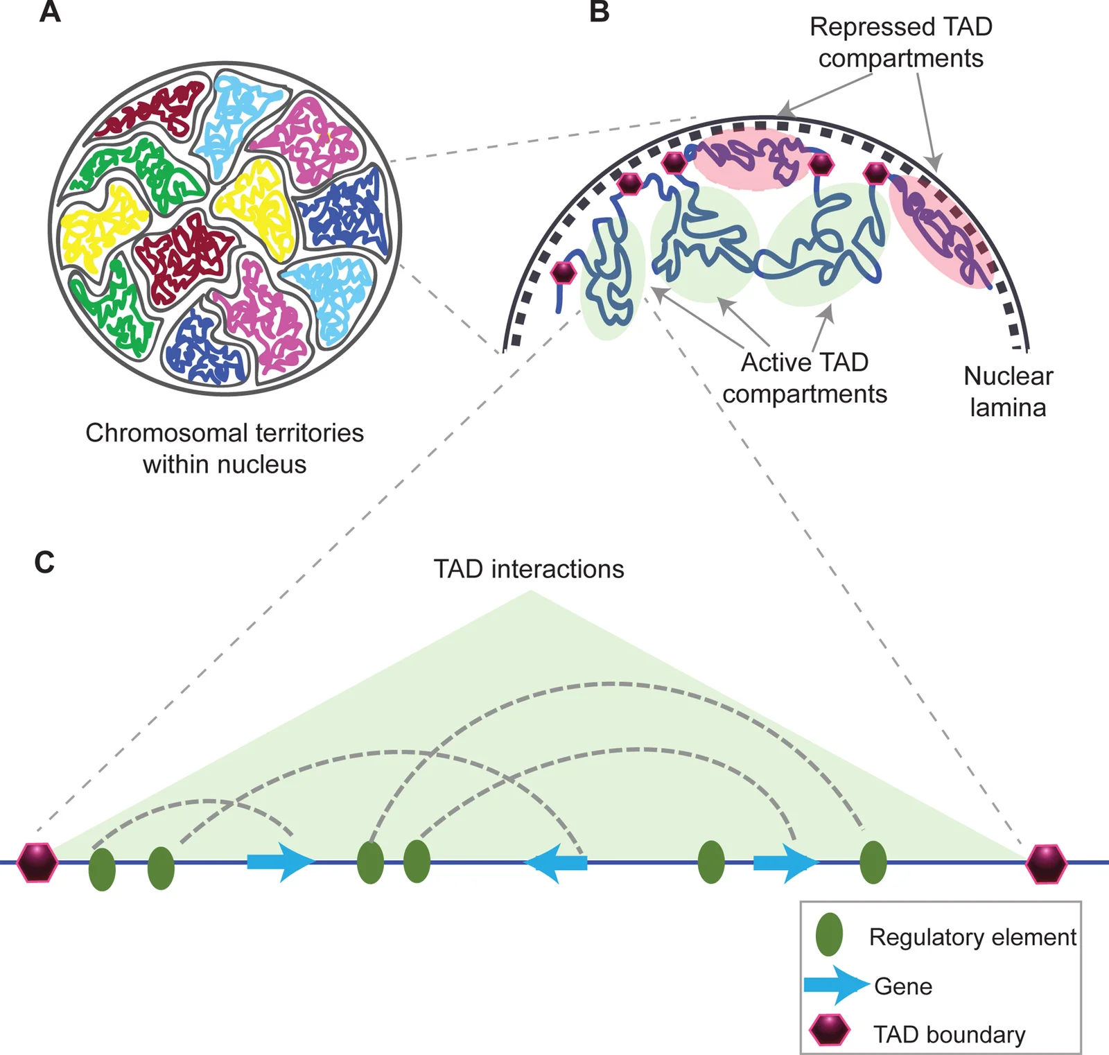
Long description
Author description: "Fig 1. Structural organization of chromatin (A) Chromosomes within an interphase diploid eukaryotic nucleus are found to occupy specific nuclear spaces, termed chromosomal territories. (B) Each chrom

DefinitionChromatin
Chromatin is the complex of DNA wrapped around histone proteins, forming the structural basis for genome organisation. [1]
The basic repeating unit is the nucleosome, which consists of DNA coiled around an octamer of the core histones H2A, H2B, H3 and H4. Each histone carries an amino‑terminal tail that protrudes from the nucleosome core and can be chemically modified (e.g., acetylation, methylation, phosphorylation, ubiquitination). These post‑translational modifications alter the physical properties of chromatin and influence fundamental DNA‑dependent processes such as transcription, replication and repair. [1]
Epigenetics is defined as the study of reversible, heritable changes in gene expression that do not involve alterations to the underlying DNA sequence. The principal epigenetic mechanisms are histone modifications, DNA methylation, chromatin remodelling and non‑coding RNAs. Together they dictate the accessibility of genomic regions to the transcriptional machinery.
Euchromatin and heterochromatin represent two functional states of chromatin distinguished by their compaction level, histone‑modification patterns and transcriptional activity.
- Euchromatin – loosely packed, enriched in histone marks associated with active transcription such as H3K4me3 and the histone variant H3.3. Genes located within euchromatic domains are generally expressed, and the DNA is more accessible to transcription factors.
- Heterochromatin – densely packed, containing repressive marks like H3K9me3 and histone variants H3.1/H3.2 that promote condensation. It is transcriptionally silent or low‑activity and often found at centromeres, telomeres and repeat‑rich regions.
The transition between these states is dynamic. During early embryogenesis, the one‑cell pronucleus is enriched in activating factors (H3K4me3, H3.3, H2A.X, H1foo) and lacks repressive variants, resulting in a globally relaxed chromatin landscape. By the two‑cell stage, the increase of H3 variants coincides with chromatin condensation, illustrating how histone composition drives structural change. [2]
- Transcription – Euchromatin permits RNA polymerase II recruitment; heterochromatin blocks access.
- DNA replication timing – Euchromatic regions replicate early in S‑phase; heterochromatic domains replicate late.
- Genome stability – Heterochromatin safeguards repetitive elements, preventing aberrant recombination.
DNA methylation is a covalent addition of a methyl group to the C5 position of cytosine, most frequently occurring at CpG dinucleotides. It is the most stable epigenetic mark and is generally linked to transcriptional repression. Two principal mechanisms underlie this repression: [3]
- Direct interference – Methylated cytosines hinder the binding of transcription factors to their cognate DNA motifs[1].
- Recruitment of methyl‑binding proteins – Proteins containing methyl‑CpG‑binding domains (MBDs) recognise methylated CpGs and attract chromatin‑remodelling complexes, including histone deacetylases (HDACs), leading to a closed chromatin conformation.
The DNA methyltransferase (DNMT) family catalyses the transfer of methyl groups from S‑adenosyl‑methionine to cytosine. In vertebrates, five members are described: DNMT1, DNMT2, DNMT3a, DNMT3b and DNMT3L. DNMT1 maintains existing methylation patterns during DNA replication, ensuring faithful inheritance of epigenetic information. DNMT3a and DNMT3b execute de novo methylation in response to developmental cues or cellular stress. DNMT3L lacks catalytic activity but assists DNMT3a/3b in establishing new methyl marks. [4] [5]
Methylated DNA and repressive histone marks often co‑occur. For example, promoter hypermethylation is frequently accompanied by histone deacetylation, resulting in synergistic silencing of tumour‑suppressor genes. MBD proteins can bind methylated CpGs and form complexes with HDACs (MeCP1, MeCP2), directly linking DNA methylation to histone deacetylation and chromatin compaction. [5] [4]
- Development and differentiation – DNA methylation patterns guide lineage commitment by silencing pluripotency genes and activating tissue‑specific programmes[1].
- Cancer – Aberrant hyper‑ or hypomethylation of CpG islands contributes to oncogene activation or tumour‑suppressor silencing, and methylation signatures can stratify leukaemia subtypes[1].
Illustrative: In a hypothetical tumour cell, the promoter of the CDKN2A tumour‑suppressor gene becomes hypermethylated. This methylation blocks transcription‑factor binding and recruits MeCP2‑HDAC complexes, leading to histone deacetylation, chromatin condensation and loss of CDKN2A expression, thereby removing a cell‑cycle checkpoint.
| Feature | Euchromatin | Heterochromatin |
|---|---|---|
| Compaction | Loosely packed | Densely packed |
| Dominant histone marks | H3K4me3, H3.3 | H3K9me3, H3.1/H3.2 |
| Transcriptional activity | High | Low/none |
| Replication timing | Early S‑phase | Late S‑phase |
| Typical locations | Gene‑rich regions | Centromeres, telomeres, repeats |
Epigenetic regulation of a gene promoter
- Signal (developmental cue)Activates DNMT3a/3b
- De novo DNA methylationMethyl groups added to CpG islands
- Question: Is promoter methylated?
Yes
- MBD protein bindsRecruits HDAC complex
- Histone deacetylationChromatin condenses
- Transcription silenced
No
- Transcription factors bindOpen chromatin
- Gene expressed
DefinitionHistone methylation
The covalent addition of one, two or three methyl groups to lysine or arginine residues on histone tails, catalysed by histone methyltransferases (HMTs).
Histone methylation is a versatile post‑translational modification that can either promote or repress transcription, depending on the residue that is modified and the degree of methylation. Unlike acetylation, which uniformly loosens chromatin, methyl marks act as docking sites for specific “reader” proteins that interpret the signal and recruit additional complexes. [1]
The enzymes that install methyl groups are called histone methyltransferases (HMTs); they transfer a methyl group from the co‑factor S‑adenosyl‑methionine to the ε‑amino group of lysine or the guanidino group of arginine. Conversely, histone demethylases remove methyl groups, restoring the unmodified state. Together, HMTs and demethylases constitute the “writers” and “erasers” of the methylation code, while proteins that recognise methylated residues are the “readers”. [4]
- Activating marks – Methylation of histone H3 at lysine 4 (H3K4me1/2/3) and lysine 36 (H3K36me2/3) is generally associated with open chromatin and active transcription. These marks recruit complexes such as the COMPASS family that facilitate RNA polymerase II recruitment.
- Repressive marks – Methylation of H3 at lysine 9 (H3K9me2/3) and lysine 27 (H3K27me3) creates a binding platform for heterochromatin protein 1 (HP1) and Polycomb repressive complexes, respectively, leading to chromatin compaction and transcriptional silencing.
The dual nature of methylation means that the same chemical group can have opposite functional outcomes, a principle that underlies the dynamic regulation of gene expression during development and disease.
Methylated lysines do not change the overall charge of the histone tail as dramatically as acetylation, but they provide a molecular address for reader domains such as Tudor, chromodomain, and PHD finger motifs. When a reader binds, it can:
- Recruit ATP‑dependent chromatin remodelers that slide or evict nucleosomes.
- Stabilise higher‑order folding of nucleosome arrays into heterochromatin.
- Facilitate the assembly of transcriptional co‑activator complexes.
Thus, the presence or absence of a specific methyl mark can tip the balance between an open, transcription‑permissive configuration and a closed, repressive state.
Methylation rarely acts in isolation. It frequently interacts with DNA methylation, histone acetylation and other modifications to fine‑tune gene expression. For example, H3K9 methylation often coincides with DNA methylation at promoters, reinforcing silencing, whereas H3K4 methylation can antagonise DNA methyltransferase recruitment, preserving an active state. This integrative network is essential for processes such as X‑chromosome inactivation, imprinting and lineage commitment.
DefinitionHistone acetylation
The addition of an acetyl group to the ε‑amino group of lysine residues on histone tails, neutralising positive charge and loosening DNA‑histone interactions.
Acetylation of lysine residues, particularly on the N‑terminal tails of histone H3, is a key determinant of chromatin openness. Acetylated lysines provide an open chromatin conformation, whereas deacetylated lysines provide a closed chromatin conformation and inhibit transcription. The reversible nature of this modification is controlled by two opposing enzyme families: [1]
| Enzyme type | Function | Representative members |
|---|---|---|
| Histone acetyltransferases (HATs) | Add acetyl groups to lysine residues (writers) | HAT1, p300/CBP [4] |
| Histone deacetylases (HDACs) | Remove acetyl groups (erasers) | HDAC1‑HDAC11 (18 members in total) |
Acetylation neutralises the positive charge of lysine, weakening the electrostatic attraction between the histone tail and the negatively charged DNA backbone. This relaxes nucleosome packing, allowing transcription factors and the basal transcriptional machinery to access promoter DNA. Conversely, deacetylation restores the positive charge, tightening DNA‑histone contacts and hindering transcriptional initiation.
- Developmental regulation – Dynamic acetylation patterns guide the activation of lineage‑specific genes during embryogenesis.
- Cancer – Aberrant acetylation is a hallmark of many tumours; high expression of HDACs correlates with poor prognosis in ovarian, endometrial, gastric, neuroblastoma and multiple myeloma cancers. [4]
In a cultured fibroblast line, treatment with the HDAC inhibitor trichostatin A leads to a rapid increase in H3K9ac levels within 30 minutes, accompanied by up‑regulation of the cyclin‑dependent kinase inhibitor p21, demonstrating how acetylation directly promotes transcription of growth‑suppressive genes.
Different HAT complexes target distinct lysine residues, conferring specificity:
- NuA4 complex acetylates H4 at lysines 5, 8, 12 and 16, facilitating DNA repair at stalled replication forks. [6]
- Gcn5p, a catalytic subunit of another HAT, acetylates H3 and H4 at defined N‑ε positions, promoting cell‑cycle progression. [6]
These examples illustrate that acetylation of particular lysines is essential for discrete cellular processes, such as DNA repair and proliferation.
While HDACs were first identified for histone deacetylation, many also act on non‑histone proteins. For instance, SIRT1 deacetylates p53, attenuating p53‑mediated apoptosis after stress exposure. This broad substrate range expands the impact of acetylation/deacetylation beyond chromatin to include signalling pathways and stress responses. [6]
Given the link between HDAC over‑activity and malignancy, HDAC inhibitors (HDACi) have entered clinical practice for certain haematological cancers. By restoring acetylation, HDACi reactivate tumour‑suppressor genes and induce cell‑cycle arrest or apoptosis. Ongoing research aims to develop isoform‑selective HDACi to minimise off‑target effects.
Chromatin modification decision tree
- Signal received (developmental cue, stress, etc.)
- Question: Is lysine to be acetylated?
Yes
- HAT recruitedAdds acetyl group → neutralises charge
- Chromatin relaxesTranscriptional activation
No
- HDAC recruitedRemoves acetyl group → restores charge
- Chromatin compactsTranscriptional repression
- Question: Is lysine to be methylated?
Activating methyl mark
- HMT adds methyl (e.g., H3K4me3)Reader recruits activator complex
- Gene expressed
Repressive methyl mark
- HMT adds methyl (e.g., H3K27me3)Reader recruits Polycomb
- Gene silenced
DefinitionEpigenetic drug
A small‑molecule that modifies the activity of enzymes or proteins that control DNA methylation, histone modification or chromatin structure, thereby altering gene expression without changing the DNA sequence.
Epigenetic drugs target the enzymes that write, erase or read chromatin marks. The most studied classes are DNA‑methyltransferase inhibitors (DNMTi) and histone deacetylase inhibitors (HDACi). DNMTi incorporate into DNA and trap DNMTs, leading to passive demethylation during replication. HDACi inhibit histone deacetylases, increasing histone acetylation and opening chromatin. Bromodomain inhibitors (BET inhibitors) block the recognition of acetyl‑lysine by bromodomains, preventing recruitment of transcriptional el [1]
The therapeutic potential of epigenetic drugs is expanding as specificity improves. When the correct combinations of agents are identified, it may be possible to reverse disease phenotypes, particularly if treatment is started early in the disease course. For example, epigenetic drugs have been proposed to prevent the formation of cancer progenitor cells while also killing drug‑resistant cancer cells. Future studies will demonstrate how much exploitation of epigenetic events could be useful for preventing and treating different diseases.
Clinical pearlLeukemia
In acute myeloid leukaemia, 5‑azacytidine and decitabine are approved for patients who are unfit for intensive chemotherapy, exploiting their DNMT inhibition to reactivate tumour‑suppressor genes.
Clinical pearlCutaneous T‑cell lymphoma
Vorinostat, an HDAC inhibitor, is licensed for cutaneous T‑cell lymphoma, where increased histone acetylation restores expression of genes that limit malignant T‑cell proliferation.
Neurodegenerative disorders also show promise for epigenetic therapy. Huntington’s disease is caused by an expanded polyglutamine repeat in huntingtin. The mutant protein enters the nucleus, binds and inhibits histone acetyltransferases (HATs), leading to a decrease in H3 and H4 acetylation and global silencing of neuronal genes. Drugs that correct these epigenetic imbalances could be a very promising treatment for such diseases in the future.
In cancer, BET inhibitors target BRD4, a protein that mediates transcriptional elongation of acetylated chromatin. NUT‑midline carcinoma, characterised by a BRD4 fusion protein, shows global hypoacetylation and transcriptional repression; BET inhibition restores acetylation and re‑activates silenced genes.
- Non‑specificity: Early epigenetic drugs often affect multiple targets, leading to off‑target effects and toxicity.
- Transient effects: Epigenetic marks can be rapidly re‑established after drug withdrawal, limiting long‑term benefit.
- Tumour heterogeneity: Different subclones may respond variably to the same epigenetic agent, necessitating combination therapy.
- Timing of intervention: Evidence suggests that early administration during disease progression yields the greatest reversal of the epigenetic switch, whereas late treatment may be less effective.
Clinical decision pathway for epigenetic therapy
- Patient presents with diseaseAssess eligibility for epigenetic therapy
- Question: Is the disease epigenetically driven?
Yes
- Select appropriate drug class
- Determine combination strategy
- Initiate therapy
No
- Consider alternative treatments
In haematological malignancies, epigenetic drugs are routinely used as first‑line or second‑line therapy, often in combination with conventional chemotherapy or targeted agents. In solid tumours, they are typically reserved for refractory disease or as part of clinical trials. In neurodegenerative disease research, epigenetic drugs remain experimental but are actively investigated in early‑phase studies.
Epigenetic changes involve histone and DNA modifications, which can result in drastic phenotypic changes—phenomena that are particularly interesting because these epigenetic events are inherently reversible.
- DNMT inhibitors and HDAC inhibitors are the most clinically established agents. [1]
- BET inhibitors target bromodomain proteins such as BRD4, important in transcriptional elongation. [1]
- Early intervention may maximise reversal of disease‑associated epigenetic changes. [1]
- Non‑specificity and transient effects are major challenges. [1]
- Epigenetic therapy is standard in some haematological cancers and experimental in neurodegeneration. [1]
Answer each one in your head first, then open the answer.
What is the primary mechanism of action of 5‑azacytidine?
Show answerHide answer
It incorporates into DNA and traps DNMT1, causing passive demethylation.Which epigenetic drug is approved for cutaneous T‑cell lymphoma?
Show answerHide answer
Vorinostat, an HDAC inhibitor.Why are BET inhibitors relevant to NUT‑midline carcinoma?
Show answerHide answer
Because the disease is driven by a BRD4 fusion protein that causes global hypoacetylation and transcriptional repression.What is a major pitfall of early epigenetic drugs?
Show answerHide answer
Non‑specificity leading to off‑target effects.When is epigenetic therapy most likely to reverse disease phenotypes?
Show answerHide answer
When administered early in disease progression.
- Heerboth S, Lapinska K, Snyder N, et al. Use of epigenetic drugs in disease: an overview. Genetics & epigenetics 2014;6:9-19. doi:10.4137/geg.s12270. Accessed .
- Funaya S, Aoki F. Regulation of zygotic gene activation by chromatin structure and epigenetic factors. The Journal of reproduction and development 2017;63(4):359-363. doi:10.1262/jrd.2017-058. Accessed .
- Akalin A, Kormaksson M, Li S, et al. methylKit: a comprehensive R package for the analysis of genome-wide DNA methylation profiles. Genome biology 2012;13(10):R87. doi:10.1186/gb-2012-13-10-r87. Accessed .
- Suraweera A, O'Byrne KJ, Richard DJ. Epigenetic drugs in cancer therapy. Cancer metastasis reviews 2025;44(1):37. doi:10.1007/s10555-025-10253-7. Accessed .
- Sinclair SH, Rennoll-Bankert KE, Dumler JS. Effector bottleneck: microbial reprogramming of parasitized host cell transcription by epigenetic remodeling of chromatin structure. Frontiers in genetics 2014;5:274. doi:10.3389/fgene.2014.00274. Accessed .
- Xia C, Tao Y, Li M, et al. Protein acetylation and deacetylation: An important regulatory modification in gene transcription (Review). Experimental and therapeutic medicine 2020;20(4):2923-2940. doi:10.3892/etm.2020.9073. Accessed .
Figure credits
Educational content for medical students and doctors. It is not a substitute for clinical judgement or local guidelines.