Chromosomal disorders and genetic syndromes
Chromosomal disorders arise from numerical or structural chromosome abnormalities affecting development.
Figure 11536px Քրոմոսոմներ և նրանց հետ կապակցված հիվանդություններ
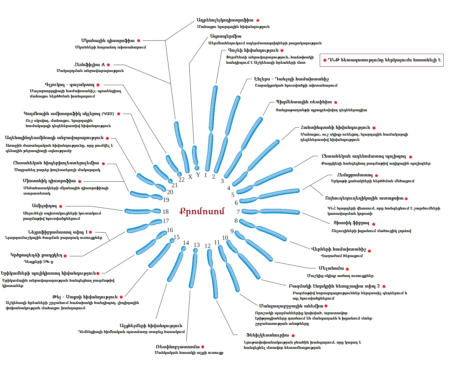
Long description
Մարդու քրոմոսոմները և նրանց հետ կապված գենետիկ հիվանդությունները

DefinitionChromosomal disorder
Chromosomal disorders encompass a spectrum from whole‑chromosome gains or losses (numerical defects) to deletions, duplications, translocations, and inversions (structural defects). Although each individual disorder is rare, together they represent a substantial proportion of genetic disease, rivaling multifactorial conditions in clinical importance[1].
Key points for clinicians:
- Detection – Conventional karyotyping remains the gold standard for identifying numerical abnormalities; newer techniques such as cell‑free fetal DNA (cfDNA) testing improve prenatal detection of common aneuploidies.
- Epidemiology – Chromosomal anomalies are implicated in about 50 % of spontaneous abortions, 6 % of stillbirths, roughly 5 % of couples with recurrent miscarriage, and approximately 0.5 % of live births[1]. [3]
- Clinical relevance – Manifestations may include growth restriction, congenital heart disease, distinctive facial features, neurodevelopmental delay, and oral findings that are important for dental practitioners.
Down syndrome (DS) is the archetypal numerical chromosomal disorder, resulting from an extra copy of chromosome 21.
Incidence
- Occurs in roughly 1 in 700 to 1 in 800 live births, making it the most common trisomy[1].
Genetic mechanisms
| Mechanism | Approximate contribution |
|---|---|
| Meiotic nondisjunction | 94 % of cases |
| Unbalanced Robertsonian translocation | 4 % |
| Mosaicism | 2 % |
These figures are derived from large population studies and underline that the majority of DS cases arise from errors in meiotic segregation[1].
Pathophysiology
The presence of a third chromosome 21 leads to dosage imbalance of over 200 genes, disrupting neurodevelopment, cardiac morphogenesis, and hematopoiesis.
Clinical features
- Characteristic facial profile (flat nasal bridge, epicanthal folds)
- Hypotonia and developmental delay
- Congenital heart defects (e.g., atrioventricular septal defect)
- Increased risk of early‑onset Alzheimer disease
Prenatal screening
- Traditional combined first‑trimester screening (ultrasound nuchal translucency + serum markers) has been standard since the 1990s. [4]
- cfDNA testing, introduced in the 2010s, offers higher sensitivity and specificity for T21, influencing diagnostic pathways and reducing invasive procedures.
Management principles
- Early multidisciplinary assessment (paediatrics, cardiology, developmental services).
- Routine monitoring for thyroid dysfunction, hearing loss, and vision problems.
- Family counselling emphasises recurrence risk (≈1 % for a subsequent pregnancy) and options for prenatal diagnosis.
Trisomy 18 (Edwards syndrome) and trisomy 13 (Patau syndrome) are the two other viable full‑trisomies seen at birth. Both are less common than DS and carry a markedly poorer prognosis.
Epidemiology
- While exact live‑birth rates are not provided in the source material, these trisomies together account for a minority of aneuploid births, with most affected pregnancies ending in miscarriage or stillbirth.
Genetic basis
- Like DS, the extra chromosome originates primarily from meiotic nondisjunction.
Phenotypic spectrum
- Trisomy 18: severe growth restriction, rocker‑bottom feet, clenched fists with overlapping fingers, congenital heart defects, and renal anomalies.
- Trisomy 13: midline facial clefts, polydactyly, holoprosencephaly, and severe cardiac malformations.
Both conditions often present with multiple organ system involvement, reflecting the global gene‑dosage effect of an extra chromosome.
Prenatal detection
- cfDNA testing includes T18 as a primary target, offering earlier and more accurate risk stratification compared with serum‑based screening alone. [4]
- Positive cfDNA results are typically confirmed by invasive diagnostic testing (chorionic villus sampling or amniocentesis) before definitive counselling.
Prognosis and care
- Median survival is measured in weeks to months; however, a small subset of infants survive beyond the first year with intensive supportive care.
- Management focuses on comfort, addressing feeding difficulties, cardiac support, and family psychosocial support.
Counselling considerations
- Recurrence risk for a subsequent pregnancy is low (<1 %) but higher for parents who are carriers of balanced translocations involving chromosomes 18 or 13.
- Preimplantation genetic diagnosis (PGD) can be offered to couples with known translocation carriers to select embryos without the trisomy, reducing the risk of affected pregnancy. [5]
| Trisomy | Live‑birth incidence (approx.) | Main genetic mechanism |
|---|---|---|
| 21 (Down syndrome) | 1 in 700–800 | Meiotic nondisjunction (94 %), Robertsonian translocation (4 %), mosaicism (2 %) [3] |
| 18 (Edwards) | < 1 in 5,000 (estimated) | Meiotic nondisjunction |
| 13 (Patau) | < 1 in 10,000 (estimated) | Meiotic nondisjunction |
Prenatal pathway for common trisomies
- First‑trimester visitOffer combined screening (ultrasound + serum)
- Question: High risk on combined screen?
Yes
- Discuss cfDNA testingNon‑invasive, higher sensitivity for T21, T18, T13
- Question: cfDNA positive?
Yes
- Offer invasive diagnostic testCVS or amniocentesis for definitive karyotype
No
- Reassure, continue routine care
No
- Routine prenatal careStandard monitoring
DefinitionRobertsonian translocation
Robertsonian translocations occur in approximately 0.19 % of the general population and 2.2 % of patients undergoing IVF with recurrent miscarriage or repeated IVF failure. The most common carriers are those with a 14;21 translocation, which accounts for 24.3 % of all translocation carriers in a tertiary centre. The incidence of carriers in the general population is estimated at 0.1 %. [6]
During meiosis I, a quadrivalent structure forms when the translocated chromosome pairs with its normal homologues. The quadrivalent can segregate in three main ways:
- 2:2 (balanced) – each gamete receives one normal and one translocated chromosome, resulting in a balanced karyotype.
- 3:1 (unbalanced) – one gamete receives three chromosomes (two normal and one translocated) and the other receives only one, leading to partial trisomy or monosomy.
- 4:0 (unbalanced) – all four chromosomes go to one gamete, producing a severely unbalanced product.
The aneuploidy rate in embryos of Robertsonian translocation carriers is 69.81 % compared with 1.096 % relative risk of an unrelated error producing an abnormal cleavage‑stage embryo[1]. Thus, out of every 11 euploid zygotes produced by a carrier, one is expected to become abnormal by the cleavage stage due to an independent chromosomal error. [5]
Carriers are usually phenotypically normal, but they have a high risk of producing unbalanced gametes. The most common outcomes are:
- Down syndrome (trisomy 21) when the translocation involves chromosome 21.
- Partial trisomy or monosomy of the involved chromosomes, leading to variable phenotypes.
Because of the high risk of miscarriage and congenital abnormalities, couples with a known Robertsonian translocation are often offered preimplantation genetic testing (PGT) to select embryos that are either normal or balanced before transfer.
Preimplantation genetic diagnosis (PGD) has evolved from fluorescent in‑situ hybridisation (FISH) to array‑based and next‑generation sequencing (NGS) platforms. Array‑based PGD can improve pregnancy rates in translocation carrier couples to between 45 % and 70 % per transfer. However, these methods cannot reliably distinguish balanced embryos from structurally normal embryos in carriers. [6]
Preimplantation genetic haplotyping (PGH) has been introduced as a more accurate approach. PGH uses short tandem repeat (STR) or single‑nucleotide polymorphism (SNP) markers to establish haplotypes that span the breakpoint regions and the entire chromosomes involved. By comparing the haplotype of each embryo with that of the carrier and a reference (family member or unbalanced embryo), PGH can differentiate:
- Normal embryos (no translocation).
- Balanced embryos (carry the translocation but are phenotypically normal).
- Unbalanced embryos (carry extra or missing chromosomal material).
PGH can be completed within two days and is applicable to both reciprocal and Robertsonian translocations. Its sensitivity and specificity require further validation in larger cohorts, but it offers a promising tool for couples seeking to avoid transmission of chromosomal rearrangements.
Deletion syndromes arise when a segment of a chromosome is lost, leading to haploinsufficiency of the genes within that region. The clinical picture depends on the size of the deletion, the genes involved, and the chromosomal location. Three well‑characterised deletion syndromes are discussed below.
Cri‑du‑chat (also known as 5p‑ syndrome) is caused by a deletion of the short arm of chromosome 5. The hallmark features include:
- Characteristic cry – a high‑pitched, cat‑like cry in infancy.
- Microcephaly – small head circumference.
- Hypotonia – reduced muscle tone.
- Developmental delay – both motor and cognitive.
- Distinctive facial features – large ears, micrognathia, and a broad nasal bridge.
The size of the deletion can vary, but the minimal critical region is located at 5p15.3. The syndrome is usually sporadic, with de novo deletions occurring in approximately 90 % of cases. Prenatal diagnosis can be achieved by chorionic villus sampling or amniocentesis followed by fluorescence in‑situ hybridisation (FISH) or array‑comparative genomic hybridisation (aCGH).
Williams‑Beuren syndrome (WBS) results from a hemizygous deletion of approximately 1.5 Mb at 7q11.23. The deletion removes the elastin gene (ELN) and several other genes that contribute to the phenotype. Key clinical features include:
- Cardiovascular disease – supravalvular aortic stenosis is the most common cardiac abnormality.
- Distinctive facial appearance – “elfin” facies with a broad nasal bridge, full cheeks, and a small mouth.
- Intellectual disability – mild to moderate, with relative strengths in verbal ability.
- Socially outgoing personality – often described as “cocktail‑party” syndrome.
- Hypercalcaemia – elevated calcium levels in infancy.
The prevalence of WBS is estimated at 1 in 7,500 live births. Early intervention with speech, occupational, and physical therapy can improve developmental outcomes. [6]
DiGeorge syndrome (also known as 22q11.2 deletion syndrome) is caused by a microdeletion of 3 Mb at 22q11.2. The deletion encompasses the TBX1 gene, which is critical for pharyngeal arch development. The syndrome is highly variable, but common manifestations include:
- Cleft palate or bifid uvula – affecting feeding and speech.
- Congenital heart defects – the most frequent cardiac abnormality is truncus arteriosus or interrupted aortic arch.
- Hypocalcaemia – due to hypoparathyroidism.
- Craniofacial anomalies – low‑set ears, micrognathia, and a small, broad nasal bridge.
- Immunodeficiency – T‑cell deficiency leading to recurrent infections.
- Developmental delay – ranging from mild learning difficulties to severe intellectual disability.
The incidence of DiGeorge syndrome is approximately 1 in 4,000 live births. Prenatal diagnosis is possible via amniocentesis or chorionic villus sampling with FISH or aCGH. Post‑natal management focuses on multidisciplinary care, including cardiology, endocrinology, immunology, and developmental services. [6] [3]
Robertsonian translocations are balanced chromosomal rearrangements that carry a high risk of producing unbalanced gametes, leading to miscarriages or congenital abnormalities. Modern preimplantation genetic testing, particularly PGH, offers a promising strategy to select embryos that are either normal or balanced. Deletion syndromes such as cri‑du‑chat, Williams‑Beuren, and DiGeorge arise from loss of chromosomal material and present with distinct clinical features that guide diagnosis and management. Understanding the mechanisms, epidemiology, and diagnostic approaches to these chromosomal disorders is essential for effective counselling and care of affected individuals and their families.
Sex chromosome disorders arise from numerical or structural abnormalities of the X and Y chromosomes. The most common are Turner syndrome (TS) and Klinefelter syndrome (KS), but other rarer conditions such as 47,XXY mosaicism, 45,XO mosaicism, and structural rearrangements (e.g., isochromosomes, ring chromosomes) also occur.
DefinitionTurner syndrome
DefinitionKlinefelter syndrome
Turner syndrome is present in about 1 in 8 000 female births and is not related to maternal age. Approximately 50 % of TS patients have a 45,X0 karyotype, 30 % are mosaics and the remainder have other X‑chromosome abnormalities such as isochromosomes or ring chromosomes. The loss of the paternal X chromosome occurs in roughly 75 % of cases. [3]
Klinefelter syndrome occurs in about 1 in 500 to 1 000 live male births, though the exact prevalence varies with detection methods. It is usually diagnosed after puberty when hypogonadism and infertility become apparent.
Other sex‑chromosome anomalies include 47,XXX (triploidy of the X chromosome), 47,XYY and mosaic forms such as 45,X/46,XX or 45,X/47,XXY. These are generally less common and may present with variable phenotypes, often milder than classic TS or KS.
Prenatal screening aims to identify pregnancies at increased risk of chromosomal abnormalities so that invasive diagnostic testing can be offered. In Ontario, the government‑funded Prenatal Screening Ontario programme coordinates multiple‑marker screening, cell‑free DNA (cfDNA) screening and invasive testing, and uses linked registry data for quality assurance. [4]
First‑trimester screening combines maternal age, nuchal translucency and serum markers (free β‑hCG, PAPP‑A). Second‑trimester screening uses AFP, hCG, estriol and inhibin‑A. The Ontario programme uses stringent cut‑offs to balance sensitivity and specificity; a lower cut‑off improves sensitivity but reduces specificity, increasing false‑positives and unnecessary invasive procedures.
cfDNA screening detects fetal aneuploidies by analysing cell‑free DNA fragments in maternal plasma. In Ontario, cfDNA is offered as a first‑tier test for high‑risk pregnancies (e.g., maternal age > 40 y) or after a positive multiple‑marker result. Uptake of cfDNA screening has increased markedly, with 86.5 % of screen‑positive pregnancies undergoing cfDNA and 13.5 % proceeding directly to invasive testing. [4]
Amniocentesis and chorionic villus sampling (CVS) provide definitive karyotype or microarray results. In Ontario, 65.2 % of pregnancies with a positive cfDNA result underwent invasive testing, reflecting a high uptake of confirmatory diagnostics. [4]
Ontario’s real‑world data show higher uptake and better detection rates than earlier modelled estimates, and a twofold reduction in invasive testing since cfDNA integration. Continuous monitoring of cut‑offs and test pathways allows the programme to optimise sensitivity, specificity and cost‑effectiveness. [4]
- Limited accuracy in multifetal pregnancies – First‑trimester screening can be used in multifetal pregnancies, but clinicians must counsel patients on the limitations in this setting. [2]
- False‑positive results – Lower cut‑offs increase sensitivity but also false‑positive rates, leading to unnecessary anxiety and invasive procedures.
- Incomplete follow‑up – In Ontario, 14.6 % of screen‑positive pregnancies did not receive further testing, potentially missing affected cases.
- Misinterpretation of cfDNA results – cfDNA can miss rare structural abnormalities and mosaicism; a negative result does not guarantee a normal karyotype.
- Over‑reliance on maternal age – While maternal age > 40 y is a strong risk factor, it is not the sole determinant; other markers and ultrasound findings are essential.
Prenatal screening programmes are now standard in most high‑income countries. In Ontario, the coordinated system links screening, cfDNA, and diagnostic testing with the Better Outcomes Registry & Network, enabling real‑time quality improvement. Clinicians use the data to counsel patients, adjust cut‑offs, and decide when to offer cfDNA or invasive testing. The flowchart below illustrates a typical decision pathway for a screen‑positive pregnancy.
Prenatal Screening Decision Pathway
- First‑trimester screeningMaternal age, NT, serum markers
- Question: Screen‑positive?
Yes
- Offer cfDNA screening
- Question: cfDNA positive?
Yes
- Offer invasive diagnostic testing
No
- Counsel on residual risk
No
- Consider cfDNA if high risk (age > 40 y or abnormal US)
- Question: cfDNA positive?
Yes
- Offer invasive diagnostic testing
No
- Routine antenatal care
- Sex chromosome disorders such as Turner and Klinefelter are common and often diagnosed postnatally.
- Prenatal screening programmes combine multiple‑marker tests, cfDNA and invasive diagnostics.
- Ontario’s data show high uptake of cfDNA and a reduction in invasive testing.
- Multifetal pregnancies reduce screening accuracy; counselling is essential.
- Continuous quality monitoring allows optimisation of cut‑offs and pathways.
Answer each one in your head first, then open the answer.
What is the approximate prevalence of Turner syndrome in female births?
Show answerHide answer
1 in 8 000Which test is used to detect fetal aneuploidy by analysing maternal plasma?
Show answerHide answer
cfDNA screeningWhat proportion of screen‑positive pregnancies in Ontario underwent invasive testing after a positive cfDNA result?
Show answerHide answer
65.2 %Name one common pitfall of first‑trimester screening in multifetal pregnancies.
Show answerHide answer
Limited accuracyWhich registry provides real‑world data for Ontario’s prenatal screening programme?
Show answerHide answer
Better Outcomes Registry & Network (BORN) Ontario
- Lu L, Luo F, Wang X. Gonadal tumor risk in pediatric and adolescent phenotypic females with disorders of sex development and Y chromosomal constitution with different genetic etiologies. Frontiers in pediatrics 2022;10:856128. doi:10.3389/fped.2022.856128. Accessed .
- Driscoll DA, Gross SJ, Professional Practice and Guidelines Committee. First trimester diagnosis and screening for fetal aneuploidy. Genetics in medicine : official journal of the American College of Medical Genetics 2008;10(1):73-75. doi:10.1097/gim.0b013e31815efde8. Accessed .
- Patil S, Rao RS, Majumdar B. Chromosomal and multifactorial genetic disorders with oral manifestations. Journal of international oral health : JIOH 2014;6(5):118-125. Accessed .
- Dougan SD, Okun N, Bellai-Dussault K, et al. Performance of a universal prenatal screening program incorporating cell-free fetal DNA analysis in Ontario, Canada. CMAJ : Canadian Medical Association journal = journal de l'Association medicale canadienne 2021;193(30):E1156-E1163. doi:10.1503/cmaj.202456. Accessed .
- Alfarawati S, Fragouli E, Colls P, Wells D. Embryos of robertsonian translocation carriers exhibit a mitotic interchromosomal effect that enhances genetic instability during early development. PLoS genetics 2012;8(10):e1003025. doi:10.1371/journal.pgen.1003025. Accessed .
- Zhang S, Lei C, Wu J, et al. The establishment and application of preimplantation genetic haplotyping in embryo diagnosis for reciprocal and Robertsonian translocation carriers. BMC medical genomics 2017;10(1):60. doi:10.1186/s12920-017-0294-x. Accessed .
Figure credits
Educational content for medical students and doctors. It is not a substitute for clinical judgement or local guidelines.