Collagen, elastin and inherited connective tissue disease
The extracellular matrix’s fibre proteins collagen and elastin give strength and elasticity to connective tissues.
Figure 1Collagen-synthesis
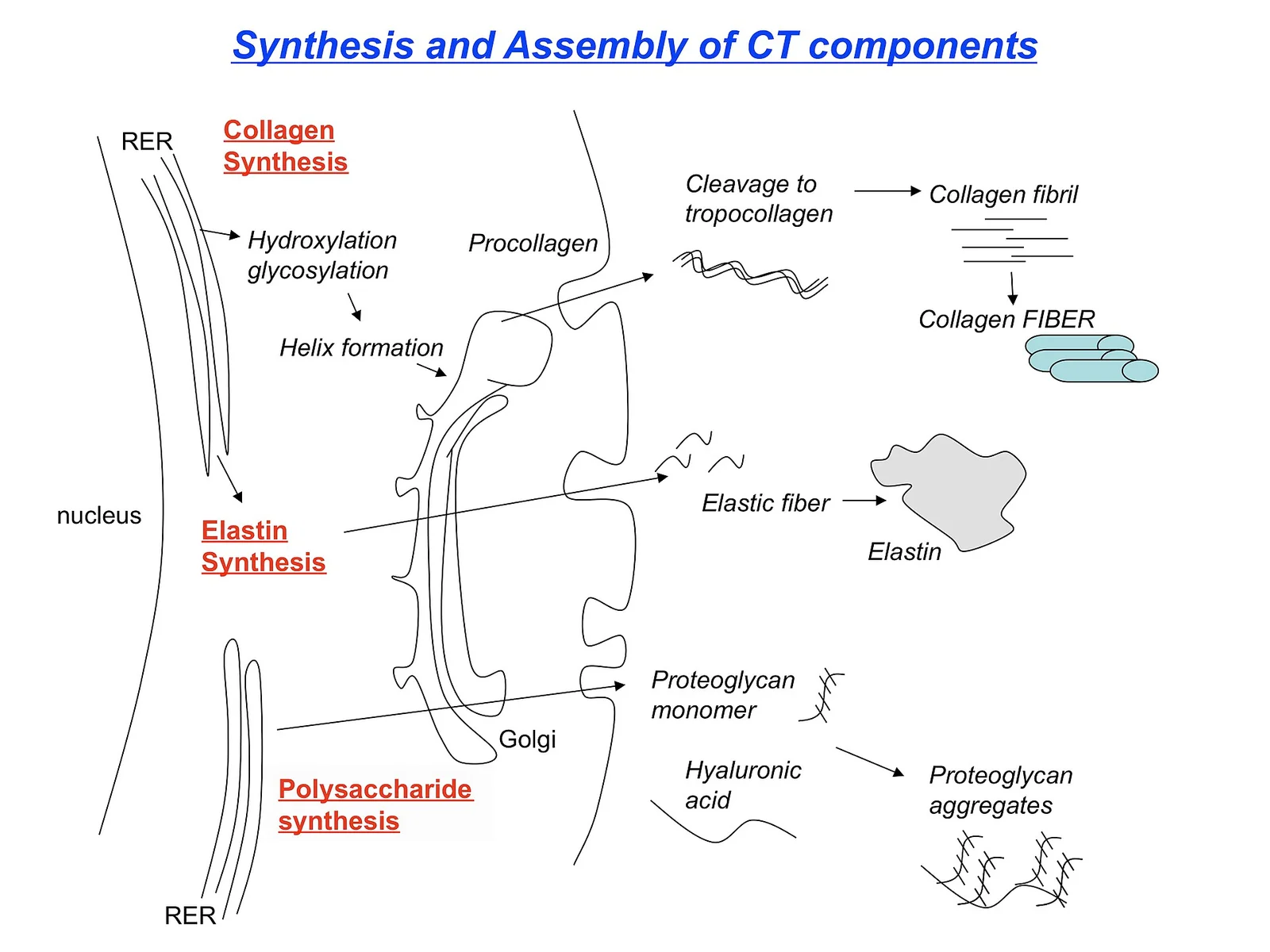
Long description
Illustration for biochemistry/collagen elastin and connective tissue disease from Wikimedia Commons, titled 'Collagen-synthesis': Synthesis of collagen, schematic. The author and licence are given in the credit line.

DefinitionCollagen
DefinitionElastin
Collagens and elastin together create a resilient network that links bone, tendon, ligament, blood‑vessel wall, heart, eye and many other structures. The most abundant collagen in the body is type I, which accounts for the bulk of bone and skin matrix. Type III is a key component of the pulmonary interstitium and vascular wall, where it works alongside type I to maintain tissue integrity. [2] [4]
Collagen biosynthesis proceeds through a series of tightly regulated intracellular events before the mature fibre is deposited in the extracellular matrix (ECM).
| Step | Key Molecular Events |
|---|---|
| 1. Gene transcription | COL1A1, COL1A2, COL3A1 etc. are transcribed in the nucleus; transcription is influenced by upstream regulators such as FMRP and MECP2, whose loss can reduce collagen I synthesis. [1] |
| 2. Translation & pre‑pro‑peptide formation | Nascent polypeptides acquire N‑terminal signal peptides and are translocated into the rough endoplasmic reticulum (RER). |
| 3. Post‑translational modifications | Hydroxylation of proline and lysine residues (vitamin C‑dependent) and glycosylation of hydroxy‑lysine occur in the RER. |
| 4. Triple‑helix assembly | Three modified α‑chains align and fold into a procollagen triple helix; this step is assisted by molecular chaperones. |
| 5. Secretion | Procollagen is packaged into vesicles and exported to the Golgi, then released into the extracellular space. |
| 6. Extracellular processing | N‑ and C‑propeptidases cleave the terminal extensions, yielding tropocollagen. |
| 7. Fibril formation & cross‑linking | Tropocollagen self‑assembles into fibrils; lysyl oxidase, a copper‑dependent enzyme, catalyses oxidative deamination of lysine residues to form covalent cross‑links that stabilise the fibrils. Reduced lysyl‑oxidase activity in Menkes disease leads to defective cross‑linking and connective‑tissue signs that can include pectus deformities and spontaneous fractures. [3] |
Copper delivery to lysyl oxidase is essential. Cellular copper is imported via the CTR1 transporter, bound to chaperones (ATOX1, COX17, CCS) and shuttled to the trans‑Golgi network where ATP7A incorporates it into copper‑dependent enzymes including lysyl oxidase. Mutations in ATP7A cause Menkes disease, a systemic disorder with connective‑tissue manifestations that stem from impaired collagen and elastin cross‑linking. [3]
Osteogenesis imperfecta (OI) is a hereditary disorder characterised by bone fragility, short stature and dentin abnormalities. The disease is most often caused by dominant mutations in the genes encoding the α‑chains of type I collagen (COL1A1, COL1A2), leading to quantitative or qualitative defects in the collagen triple helix. The resulting fibrils are fewer, thinner and less cross‑linked, compromising tensile strength of bone and other type I‑rich tissues.
Pathophysiology
- Quantitative defects: Null alleles reduce the amount of normal type I collagen produced, yielding a milder “type I” OI phenotype.
- Qualitative defects: Glycine substitutions disrupt the tight packing of the triple helix, producing over‑modified collagen that is prone to intracellular retention and extracellular instability.
- Secondary effects: Impaired collagen scaffolding alters osteoblast differentiation and mineralisation, and may provoke an abnormal wound‑repair response similar to that observed in some EDS subtypes.
Clinical spectrum
- Recurrent low‑impact fractures from birth or early childhood.
- Blue sclerae, dentinogenesis imperfecta, hearing loss.
- Variable degrees of short stature and scoliosis.
- In severe forms, respiratory compromise due to rib cage fragility.
Diagnostic clues
- Radiographs show osteopenia, wormian bones and cortical thinning.
- Biochemical analysis of skin fibroblasts demonstrates reduced type I collagen secretion or abnormal electrophoretic migration.
- Genetic testing confirms pathogenic COL1A1/2 variants.
Management principles
- First‑line therapy: Bisphosphonate infusion to increase bone mineral density and reduce fracture rate (standard of care in paediatric OI).
- Adjuncts: Physiotherapy, orthopaedic surgery for deformity correction, and careful monitoring for hearing loss.
- Emerging approaches: Gene‑editing tools and allele‑specific silencing are under investigation, with the Aga2 cell line providing a platform to test novel therapeutics for OI. [4]
Relation to other connective‑tissue disorders
The molecular defect in OI (type I collagen) contrasts with the collagen‑III deficiency that predisposes to pulmonary fragility in vascular EDS, yet both illustrate how specific collagen isoform loss translates into organ‑specific vulnerability. Moreover, upstream regulators of collagen synthesis such as FMRP and MECP2 can modulate collagen I output, linking neurodevelopmental syndromes to hypermobile phenotypes and highlighting the broader network that governs connective‑tissue homeostasis.
Collagen‑related disease pathway
- Genetic mutation or regulatory defecte.g., COL1A1/2, ATP7A, FMR1, MECP2
- Question: Is collagen synthesis affected?
Yes
- Altered procollagen formationhydroxylation, helix assembly
- Impaired secretion or cross‑linkinglysyl oxidase deficiency, MMP9 dysregulation
- Clinical phenotypeOI, EDS, Menkes, hypermobility
No
- Investigate alternative pathwaysimmune dysregulation, elastin defects
DefinitionHereditary connective tissue disorder
The International EDS Consortium recognises fourteen distinct subtypes of the condition, thirteen of which are classified as rare, each occurring in no more than one in two thousand individuals and usually far rarer. The most common clinical presentation is hypermobile EDS (hEDS), which accounts for 80–90 % of all EDS diagnoses. Because hEDS lacks a single pathogenic gene, it is thought to arise from polygenic risk factors of small effect together with environmental influences. [1]
- Joint hypermobility affecting large and small joints, often with chronic pain and recurrent dislocations.
- Skin manifestations ranging from hyperextensibility to easy bruising and poor wound healing.
- Vascular and organ fragility in the vascular, arterial, and some other subtypes, predisposing to aneurysms and organ rupture.
Patients with EDS/HSD frequently display a neurodevelopmental profile that includes coordination difficulties resembling developmental coordination disorder, as well as learning and other disabilities. These features add to the physical burden of the connective tissue disorder and can affect the well‑being of children and families.
A growing body of evidence highlights a phenotypic overlap between EDS/HSD and autism spectrum disorder (ASD). Both conditions are complex, multisystemic spectrums, and genetic studies have identified shared molecular pathways, particularly those involving collagen‑related genes. Epidemiological observations suggest that hereditary connective tissue disorders may represent a subtype of autism, although the exact prevalence remains unknown. The proposed mechanisms include direct effects of altered extracellular matrix on brain development and indirect influences via maternal immune dysregulation.
- When evaluating a patient with joint hypermobility, screen for associated neurodevelopmental or psychiatric features, especially if there is a family history of ASD.
- Recognise that vascular complications may be silent; a low threshold for imaging is warranted in subtypes with known arterial involvement.
- Management is multidisciplinary: physiotherapy for joint stability, dermatology for skin care, genetics for counselling, and psychology/psychiatry for neurodevelopmental comorbidities.
DefinitionCopper‑transport disorder
The ATP7A protein normally transports copper from the cytosol into the secretory pathway, enabling the metalation of copper‑dependent enzymes. In Menkes disease, ATP7A dysfunction results in deficient activity of enzymes such as lysyl oxidase (LOX), which is essential for cross‑linking collagen and elastin fibres[1]. Consequently, connective tissue abnormalities arise, mirroring features seen in other collagenopathies.
Recent cellular studies have expanded the spectrum of affected pathways:
| Affected process | Consequence in Menkes disease |
|---|---|
| LOX dysfunction | Impaired collagen/elastin cross‑linking, leading to fragile vessels and skin |
| LMAN1 deficiency (liver) | Reduced coagulation factors V + VIII and α‑1‑antitrypsin |
| LMAN1 deficiency (brain) | Poor trafficking of neuroreceptors, contributing to neurological signs |
| Collagen‑like receptors (COLQ, CCBE1) | Muscle weakness and lymphedema |
| Cq1 deficiency | Defects in innate immunity and lung surfactant |
| Peroxisomal SOD1 pool | Requires ER for metalation; its loss contributes to Zellweger‑like features |
| SMAX3 (mildest form) | Motor neuron disease is characteristic [2] |
These findings illustrate that copper‑dependent processes extend far beyond classic neuro‑developmental impairment, encompassing coagulation, immunity, lung function and connective tissue integrity[1].
- Neurological: Developmental delay, seizures, progressive neurodegeneration; motor neuron disease may dominate in milder forms (SMAX3)[1].
- Dermatological: Sparse, kinky hair (pili torti), hypopigmentation, and skin laxity due to defective collagen cross‑linking.
- Vascular: Fragile arteries predispose to aneurysms and ruptures, echoing findings in animal models with “viable brindled” and “blotchy” mutants that develop aortic and abdominal aneurysms. [3]
- Skeletal and muscular: Osteoarthrosis, muscle weakness, and lymphedema linked to COLQ and CCBE1 abnormalities[1].
- Immune and pulmonary: Recurrent infections, impaired innate immunity, and surfactant defects[1].
- Hair microscopy showing pili torti and trichorrhexis nodosa.
- Serum copper and ceruloplasmin markedly low.
- Radiographic signs of osteopenia and wormian bones.
- Genetic testing confirming pathogenic ATP7A variants.
- Early parenteral copper histidine can improve neurodevelopment if started before irreversible damage occurs.
- Multidisciplinary care addressing neurologic, dermatologic, vascular and immunologic complications.
- Genetic counselling for families, given the X‑linked inheritance pattern.
- Menkes disease exemplifies how a single metal‑transport defect can disrupt multiple copper‑dependent enzymes, leading to a broad connective tissue phenotype.
- Recognise the overlap with other collagen‑related disorders (e.g., EDS) through shared features such as vascular fragility and skin laxity, but note the distinct neuro‑developmental trajectory driven by copper deficiency.
- Early diagnosis and copper replacement are time‑critical; delayed treatment results in irreversible neurodegeneration.
Elastin is the principal protein that confers reversible stretch to arteries, lungs, skin and ligaments. It is produced as tropoelastin, secreted into the extracellular matrix and cross‑linked by lysyl oxidase (LOX) to form insoluble elastic fibres. These fibres are organised into a core of elastin surrounded by a sheath of microfibrils that contain fibrillin‑1. The microfibrillar scaffold not only provides structural support but also regulates the bioavailability of transforming growth factor‑β (TGF‑β) by sequestering latent complexes.
Marfan syndrome (MFS) is an autosomal‑dominant connective‑tissue disorder caused by pathogenic variants in the FBN1 gene, which encodes fibrillin‑1. Loss of functional fibrillin‑1 weakens the microfibrillar scaffold, leading to fragmented elastic fibres and dysregulated TGF‑β signalling. The clinical phenotype reflects the tissues that rely heavily on elastin‑rich structures:
- Cardiovascular – progressive aortic root dilatation, annulo‑aortic ectasia and risk of dissection; mitral valve prolapse.
- Ocular – ectopia lentis, myopia, increased risk of retinal detachment.
- Skeletal – tall stature, arachnodactyly, pectus excavatum or carinatum, scoliosis, joint hypermobility.
From a mechanistic perspective, the deficiency of fibrillin‑1 reduces the tensile strength of the microfibrillar sheath, making elastic lamellae more susceptible to mechanical stress. Simultaneously, excess TGF‑β activity promotes matrix remodelling and inflammation, further compromising vessel wall integrity. The combination of structural weakness and maladaptive signalling underlies the propensity for aortic aneurysm formation.
Therapeutic strategies therefore target two complementary aspects:
- Reduction of haemodynamic stress – strict blood‑pressure control (β‑blockers or angiotensin‑II receptor blockers) to lessen wall tension.
- Modulation of TGF‑β signalling – angiotensin‑II receptor blockers (e.g., losartan) have been shown to attenuate TGF‑β‑mediated matrix degradation in animal models and are now considered first‑line disease‑modifying therapy.
Homocystinuria is an inherited metabolic disorder most commonly caused by deficiency of cystathionine β‑synthase (CBS), an enzyme that catalyses the conversion of homocysteine to cystathionine in the transsulfuration pathway. Accumulation of homocysteine and its metabolites leads to widespread connective‑tissue abnormalities that mimic Marfan syndrome, but with distinct features.
- Enzyme deficiency – loss of CBS activity prevents the normal clearance of homocysteine, resulting in markedly elevated plasma homocysteine levels.
- Disruption of collagen cross‑linking – homocysteine interferes with lysyl oxidase, the copper‑dependent enzyme that cross‑links collagen and elastin. Impaired cross‑linking weakens the extracellular matrix, particularly in vascular and skeletal tissues.
- Oxidative stress – excess homocysteine generates reactive oxygen species, further damaging endothelial cells and extracellular matrix components.
- Ocular – ectopia lentis (often downward), severe myopia.
- Skeletal – tall stature, arachnodactyly, pectus deformities, osteoporosis.
- Vascular – premature atherosclerosis, thromboembolic events, risk of aortic dilatation.
- Neurological – developmental delay, seizures in severe cases.
- Pyridoxine (vitamin B6) responsiveness – a proportion of patients respond to high‑dose pyridoxine, which acts as a co‑factor for residual CBS activity.
- Dietary restriction – low‑methionine diet supplemented with cysteine reduces homocysteine production.
- Betaine – serves as a methyl donor to remethylate homocysteine to methionine, lowering plasma homocysteine.
- Antithrombotic prophylaxis – indicated in patients with a history of thrombosis or markedly elevated homocysteine.
Early diagnosis through newborn screening and prompt metabolic control can markedly reduce the risk of vascular complications and improve long‑term outcomes.
Exam trapAssuming identical management for Marfan syndrome and homocystinuria
- Over‑reliance on phenotypic similarity – Ectopia lentis and aortic dilatation occur in both disorders, yet only homocystinuria benefits from vitamin B6, dietary methionine restriction and betaine.
- Neglecting TGF‑β modulation in Marfan syndrome – β‑blockers alone do not address the pathogenic signalling cascade; omission of an angiotensin‑II receptor blocker may leave patients at higher risk of aortic events.
- Ignoring copper‑dependent enzymes – Lysyl oxidase requires copper; in disorders affecting copper metabolism (e.g., Menkes disease) elastic‑fibre formation is impaired, compounding connective‑tissue fragility.
- Failure to screen for thrombosis in homocystinuria – Elevated homocysteine is a potent pro‑thrombotic factor; lack of anticoagulation prophylaxis can result in preventable strokes or pulmonary emboli.
- Misinterpretation of genetic testing – Pathogenic variants in FBN1 are not synonymous with CBS mutations; comprehensive gene panels are essential when the phenotype is ambiguous.
Clinicians encounter elastin‑related pathology across several specialties:
- Cardiology – Surveillance of aortic root dimensions in Marfan patients using echocardiography or MRI; initiation of β‑blocker or angiotensin‑II receptor blocker therapy based on guideline thresholds.
- Genetics – Cascade testing of first‑degree relatives for FBN1 or CBS mutations; counselling regarding reproductive options and prenatal diagnosis.
- Metabolic medicine – Implementation of newborn screening programs for homocystinuria; monitoring plasma homocysteine and adjusting pyridoxine, diet, and betaine regimens.
- Ophthalmology – Early detection of ectopia lentis; referral for lens‑sparing surgery when visual acuity is threatened.
- Orthopaedics – Management of scoliosis and pectus deformities; consideration of prophylactic bracing versus surgical correction.
The following flowchart summarises the diagnostic pathway when a patient presents with Marfanoid habitus and cardiovascular findings.
Diagnostic pathway for Marfanoid phenotype
- Patient presents with tall stature, arachnodactyly, and aortic root dilatation
- Question: Is ectopia lentis present?
Yes
- Obtain genetic testing for FBN1 and CBS
No
- Measure plasma homocysteine
- Question: Homocysteine elevated?
Yes
- Start pyridoxine trial and dietary methionine restriction
No
- Proceed with FBN1 sequencing
- Elastin fibres consist of cross‑linked tropoelastin surrounded by fibrillin‑rich microfibrils.
- Fibrillin‑1 deficiency in Marfan syndrome weakens elastic lamellae and dysregulates TGF‑β signalling.
- First‑line disease‑modifying therapy for Marfan syndrome includes angiotensin‑II receptor blockers to curb TGF‑β activity.
- Homocystinuria results from cystathionine β‑synthase deficiency, leading to homocysteine accumulation and impaired collagen/elastin cross‑linking.
- Vitamin B6 responsiveness, low‑methionine diet and betaine are cornerstone treatments for homocystinuria.
- Thrombosis risk is markedly increased in homocystinuria; prophylactic anticoagulation may be required.
- Phenotypic overlap can mislead clinicians; genetic confirmation is essential for targeted therapy.
- Multidisciplinary follow‑up (cardiology, genetics, ophthalmology, metabolic medicine) optimises outcomes.
Answer each one in your head first, then open the answer.
Which enzyme cross‑links elastin and requires copper as a co‑factor?
Show answerHide answer
Lysyl oxidaseWhat signalling pathway is over‑active in Marfan syndrome due to fibrillin‑1 deficiency?
Show answerHide answer
Transforming growth factor‑β (TGF‑β) pathwayName a first‑line metabolic therapy for pyridoxine‑responsive homocystinuria.
Show answerHide answer
High‑dose vitamin B6 (pyridoxine)Which ocular feature helps differentiate homocystinuria from Marfan syndrome?
Show answerHide answer
Downward ectopia lentisWhat class of drug is recommended alongside β‑blockers to modify disease progression in Marfan syndrome?
Show answerHide answer
Angiotensin‑II receptor blockers
- Casanova EL, Baeza-Velasco C, Buchanan CB, Casanova MF. The Relationship between Autism and Ehlers-Danlos Syndromes/Hypermobility Spectrum Disorders. Journal of personalized medicine 2020;10(4):E260. doi:10.3390/jpm10040260. Accessed .
- Horn N, Wittung-Stafshede P. ATP7A-Regulated Enzyme Metalation and Trafficking in the Menkes Disease Puzzle. Biomedicines 2021;9(4):391. doi:10.3390/biomedicines9040391. Accessed .
- Ojha R, Prasad AN. Menkes disease: what a multidisciplinary approach can do. Journal of multidisciplinary healthcare 2016;9:371-385. doi:10.2147/jmdh.s93454. Accessed .
- Lisse TS, Thiele F, Fuchs H, et al. ER stress-mediated apoptosis in a new mouse model of osteogenesis imperfecta. PLoS genetics 2008;4(2):e7. doi:10.1371/journal.pgen.0040007. Accessed .
Figure credits
Educational content for medical students and doctors. It is not a substitute for clinical judgement or local guidelines.