DNA replication, repair and mutations
The coordinated enzymatic steps that copy DNA, fix errors and maintain chromosome ends.
Figure 1DNA replication
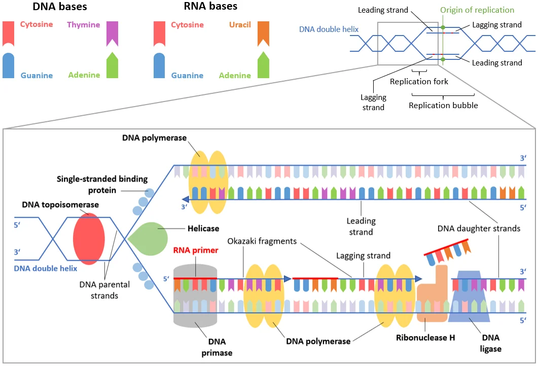
Long description
This image features the most important intracellular structures (such as proteins/enzymes, the DNA, etc.) participating in DNA replication.

DefinitionDNA replication
The process by which a cell copies its entire genome before cell division, producing two identical daughter DNA molecules.
DefinitionDNA repair
A collection of mechanisms that detect and correct DNA lesions, preserving genomic integrity.
DefinitionMutation
A permanent alteration in the DNA sequence that can arise spontaneously or after exposure to damaging agents.
Replication initiates at specific origins of replication, where the double helix is locally unwound to form a replication fork. The fork is the moving junction between parental double‑stranded DNA and newly synthesised strands. Accurate duplication requires a suite of enzymes that unwind DNA, synthesise new strands, join fragments and resolve the special structures at chromosome ends. When replication stalls or DNA is damaged, specialised repair systems intervene to restore continuity and prevent mutagenesis.
Origin, fork, helicase, primase, polymerases, ligase, telomerase, repair systems (NHEJ, homologous recombination, nucleotide and base excision, mismatch) and their diseases
At each origin, the origin recognition complex (ORC) recruits additional factors that load the MCM helicase onto DNA. The helicase then separates the strands, generating the replication fork. The leading strand is synthesised continuously, while the lagging strand is produced as short Okazaki fragments.
DNA helicases unwind duplex DNA using ATP hydrolysis. Several helicases contain an iron‑sulphur (Fe‑S) cluster essential for activity. Mutations that disrupt this cluster abolish helicase function and can lead to severe genetic disorders. Other Fe‑S helicases (Rad3, ChlR1, RTEL1, Dna2) similarly preserve genome stability, and their dysfunction is associated with repair‑defect diseases.
Primase synthesises short RNA primers that provide a 3′‑OH group for DNA polymerases to extend. In eukaryotes, a heterodimeric primase is part of the DNA polymerase α‑primase complex, which initiates synthesis on both strands.
Multiple polymerases act at the fork. Polymerase α extends the RNA primer; polymerase δ carries out bulk lagging‑strand synthesis; polymerase ε primarily synthesises the leading strand. All possess proofreading exonuclease activity that removes misincorporated nucleotides, reducing the mutation rate.
After Okazaki fragments are synthesised, DNA ligase I seals the nicks between adjacent fragments, creating a continuous phosphodiester backbone.
Chromosome ends cannot be fully replicated by conventional polymerases, leading to progressive shortening. Telomerase, a ribonucleoprotein reverse transcriptase, adds telomeric repeats to the 3′ end, counteracting attrition and maintaining cellular proliferative capacity.
NHEJ ligates DNA double‑strand breaks (DSBs) without a homologous template, operating throughout interphase. It is rapid but can be error‑prone, occasionally joining ends with small insertions or deletions. [1]
HR uses a sister chromatid as a template for error‑free repair, predominating in S and G2 phases. Initiation requires DNA end resection, where the MRN‑CtIP complex creates initial 3′‑ssDNA overhangs. This step is essential for recruiting HR proteins and preventing NHEJ. Extension of the resected strand is performed by nucleases and helicases such as EXO1, DNA2 and BLM, generating longer ssDNA tracts that are coated by RPA and later replaced by RAD51 to mediate strand invasion. [1]
NER removes bulky lesions (e.g., UV‑induced pyrimidine dimers). The XPD helicase, a member of the SF2 family, unwinds DNA around the lesion to allow excision and resynthesis.
BER corrects small, non‑bulky base modifications (e.g., deamination, oxidation). A DNA glycosylase excises the damaged base, creating an abasic site that is processed by AP endonuclease, DNA polymerase β and DNA ligase III.
MMR rectifies base‑base mismatches and insertion‑deletion loops that escape polymerase proofreading. Core proteins (MSH2‑MSH6, MLH1‑PMS2) recognise the error, recruit exonucleases and DNA polymerase δ for resynthesis, and seal the nick with ligase I.
- Fanconi anaemia results from defective FANCJ helicase activity, leading to impaired DNA repair and chromosomal breakage.
- Trichothiodystrophy stems from XPD mutations that abolish NER helicase function, causing photosensitivity and developmental abnormalities.
- Defects in DNA end resection (e.g., mutations in MRN components, CtIP, EXO1, DNA2, BLM) compromise HR, increase reliance on error‑prone NHEJ and predispose to genomic instability, embryonic lethality, ageing, immunodeficiency, neurological disease and cancer.
- Dysregulation of RIF1 or the CST complex interferes with NHEJ suppression and fork protection, leading to excessive fork degradation and replication stress‑induced genome instability.
Stalled forks generate ssDNA that is rapidly coated by the RPA complex. This triggers the ATR‑CHK1 checkpoint, pausing the cell cycle to allow fork remodeling. Fork reversal converts a three‑way junction into a four‑way structure, facilitating repair. However, uncontrolled end resection by MRE11 or EXO1 can degrade reversed forks, causing collapse. Protective factors such as BRCA1/2 and RAD51 limit resection and preserve fork integrity. [1]
Point mutations involve a single nucleotide change and can be classified by their effect on the coding sequence:
| Type | Description | Typical consequence |
|---|---|---|
| Transition | Purine ↔ purine (A↔G) or pyrimidine ↔ pyrimidine (C↔T) substitution | Often silent or cause conservative amino‑acid change |
| Transversion | Purine ↔ pyrimidine substitution (A↔C, A↔T, G↔C, G↔T) | More likely to alter protein structure |
| Missense | Single base change that substitutes one amino acid for another | Can be benign, deleterious or gain‑of‑function |
| Nonsense | Substitution creates a premature stop codon | Truncates protein, usually loss‑of‑function |
| Silent (synonymous) | Change does not alter the encoded amino acid | Generally neutral, but may affect splicing or translation efficiency |
| Splice‑site | Mutation at intron‑exon boundaries affecting mRNA processing | Can lead to exon skipping or intron retention, often pathogenic |
Polymerase proofreading and MMR reduce the frequency of these events. When repair fails, the resulting mutations can accumulate, driving oncogenesis or hereditary disease.
| Enzyme / Complex | Primary Function | Representative Disease |
|---|---|---|
| XPD (SF2 helicase) | Nucleotide excision repair helicase | Trichothiodystrophy |
| FANCJ (Fe‑S helicase) | Unwinds duplex and G‑quadruplex DNA | Fanconi anaemia |
| MRN‑CtIP | Initiates DNA end resection for HR | Genomic instability syndromes |
| RIF1‑CST | Suppresses resection, promotes NHEJ | Replication‑stress disorders |
| BRCA1/2 | Protects stalled forks, facilitates HR | Breast/ovarian cancer predisposition |
DefinitionFrameshift mutation
A frameshift mutation is an insertion or deletion of nucleotides whose length is not a multiple of three, thereby shifting the reading frame of the coding sequence downstream of the lesion.
DefinitionSplice‑site mutation
A splice‑site mutation alters the conserved sequences at intron‑exon boundaries (typically the GU‑AG dinucleotides) and can disrupt normal removal of introns during pre‑mRNA processing.
Frameshift mutations arise most often when the DNA replication machinery slips on repetitive sequences or when a DNA lesion is bypassed by an error‑prone polymerase. Because the genetic code is read in triplets, adding or losing nucleotides that are not in multiples of three changes every codon after the lesion. The new reading frame usually encounters a premature stop codon within a few dozen codons, producing a truncated protein that is often non‑functional or deleterious. In the clinical context, frameshifts are a common cause of loss‑of‑function alleles in tumour suppressor genes such as TP53 or APC.
Splice‑site mutations affect the consensus sequences at the 5′ donor site (usually GU) and the 3′ acceptor site (usually AG). Disruption of these motifs can lead to several abnormal splicing outcomes:
- Exon skipping – the spliceosome bypasses the affected exon, joining the flanking exons directly.
- Intron retention – the intron is retained in the mature mRNA, often introducing premature stop codons.
- Activation of cryptic splice sites – nearby sequences that resemble the consensus are used, producing aberrant transcripts.
Both classes of mutation can generate premature termination codons (PTCs). Cellular quality‑control mechanisms, notably the nonsense‑mediated decay (NMD) pathway, recognise PTC‑containing transcripts and target them for rapid degradation, reducing the amount of mutant protein that reaches the cytoplasm.
| Consequence | Frameshift | Splice‑site |
|---|---|---|
| Change in reading frame | Yes (if indel not multiple of 3) | No (coding frame unchanged) |
| Production of premature stop codon | Common | Common (via exon skipping or intron retention) |
| Potential activation of NMD | Frequent | Frequent |
| Effect on protein domains | Truncation, loss of C‑terminal domains | May delete whole exons, alter domain composition |
The pathogenic impact depends on where the mutation occurs. Early‑coding region frameshifts usually abolish most of the protein, whereas those near the 3′ end may retain partial function. Similarly, splice‑site mutations that cause skipping of a critical exon can be more damaging than those affecting a non‑essential region.
Exam trapAssuming all indels are pathogenic
Not every insertion or deletion causes a frameshift; indels that are multiples of three preserve the reading frame and may be benign if they do not disrupt functional motifs.
Exam trapOver‑reliance on canonical splice motifs
Splice‑site mutations can occur outside the strict GU‑AG dinucleotides, affecting branch points or auxiliary splicing enhancers; ignoring these can miss pathogenic variants.
- Mis‑annotation of variant effect – Bioinformatic tools may predict a frameshift, but experimental validation (e.g., cDNA sequencing) is essential because alternative splicing can rescue the reading frame.
- Neglecting NMD – A transcript harbouring a PTC may be degraded, leading to haploinsufficiency rather than a dominant‑negative protein; clinical interpretation must consider this.
- Ignoring tissue‑specific splicing – Some splice‑site mutations have differential effects in distinct cell types, complicating genotype‑phenotype correlation.
- Confusing somatic and germline events – In cancer genomics, frameshifts may be acquired somatically; distinguishing them from inherited variants is crucial for counselling.
Frameshift and splice‑site mutations are central to several diagnostic and therapeutic arenas:
- Molecular diagnostics – Targeted next‑generation sequencing panels for hereditary cancer routinely report frameshift and splice‑site variants, guiding risk assessment and management.
- Pharmacogenomics – Certain drug‑metabolising enzymes (e.g., DPYD) harbour clinically relevant frameshifts that predict severe toxicity to fluoropyrimidines; testing prevents adverse reactions.
- Gene therapy – Strategies such as exon skipping (e.g., in Duchenne muscular dystrophy) aim to bypass pathogenic splice‑site mutations, restoring an in‑frame transcript.
- CRISPR‑based editing – Introducing frameshift indels via non‑homologous end joining can knock out disease‑causing genes in experimental models or, potentially, in vivo therapies.
- Cancer immunotherapy – Frameshift‑derived neoantigens are highly immunogenic; they are being explored as personalised vaccine targets.
Assessing a suspected frameshift or splice‑site mutation
- Identify variant from sequencingIndel or splice‑site change?
- Question: Is the indel length % 3 ≠ 0?
Yes
- Predict frameshiftCheck for premature stop codon
- Assess NMD likelihoodPosition of PTC relative to exon‑junction complex
No
- In‑frame indelEvaluate impact on functional domains
- Question: Splice‑site variant?
Yes
- Run splice prediction toolsAssess donor/acceptor loss
- Validate by RT‑PCRDetect exon skipping or intron retention
No
- Proceed with other variant classification steps
- Frameshift mutations shift the codon reading frame, usually creating premature stop codons.
- Splice‑site mutations disrupt canonical GU‑AG motifs, leading to exon skipping, intron retention or cryptic splice‑site use.
- Both mutation types often trigger nonsense‑mediated decay, reducing mutant protein levels.
- Not all indels are pathogenic; only those not divisible by three cause frameshifts.
- Splice‑site effects can extend beyond the immediate GU‑AG dinucleotides.
- Clinical applications include diagnostic panels, pharmacogenomics, gene‑editing, and neoantigen‑based immunotherapy.
Answer each one in your head first, then open the answer.
What defines a frameshift mutation?
Show answerHide answer
An insertion or deletion whose length is not a multiple of three, shifting the reading frame.Which consensus nucleotides mark the canonical splice donor and acceptor sites?
Show answerHide answer
GU at the 5′ donor and AG at the 3′ acceptor.How does nonsense‑mediated decay influence the phenotype of a frameshift mutation?
Show answerHide answer
It degrades transcripts with premature stop codons, often causing haploinsufficiency.Name one therapeutic strategy that exploits a splice‑site mutation.
Show answerHide answer
Exon‑skipping therapy, as used in Duchenne muscular dystrophy.Why must a frameshift indel be experimentally validated despite in‑silico prediction?
Show answerHide answer
Because alternative splicing or rescue mechanisms may mitigate the predicted effect.
- Zhao F, Kim W, Kloeber JA, Lou Z. DNA end resection and its role in DNA replication and DSB repair choice in mammalian cells. Experimental & molecular medicine 2020;52(10):1705-1714. doi:10.1038/s12276-020-00519-1. Accessed .
Figure credits
Educational content for medical students and doctors. It is not a substitute for clinical judgement or local guidelines.