Energy balance, obesity and thermogenesis
Energy balance, obesity and thermogenesis: BMR, brown fat and UCP1, leptin and ghrelin appetite control, starvation adaptation and refeeding syndrome.
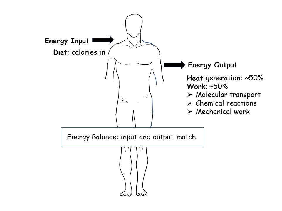
- Energy balance is the difference between the energy taken in as food and the energy the body spends; when intake exceeds expenditure over time, the surplus is stored, mostly as fat.
- Total daily energy expenditure has three parts: basal metabolism (about 60%), the thermic effect of food (about 10%) and physical activity, including non-exercise movement (about 30%).[2]
- Brown adipose tissue burns fuel to make heat rather than ATP, using uncoupling protein 1 (UCP1) to leak protons across the mitochondrial membrane; this is non-shivering thermogenesis.
- Leptin, made by fat cells, signals energy sufficiency to the hypothalamus and suppresses appetite; ghrelin, made by the stomach during fasting, signals hunger.
- Obesity is a chronic disease defined by excess adiposity; the WHO adult cut-offs are a body mass index (BMI) of 25 or more for overweight and 30 or more for obesity.[1]
- After prolonged starvation, rapid refeeding can cause refeeding syndrome: an insulin surge drives phosphate, potassium and magnesium into cells, producing dangerous hypophosphataemia, hypokalaemia and hypomagnesaemia.[7]
By the end of this page you should be able to:
- Define energy balance and name the three components of total energy expenditure with their approximate shares.
- Explain how brown adipose tissue generates heat through UCP1 and how it is regulated.
- Describe the hypothalamic circuits that control appetite, and the roles of leptin and ghrelin in them.
- Explain why obesity develops and persists, including leptin resistance.
- Outline the metabolic adaptations to starvation and the pathophysiology, diagnosis and prevention of refeeding syndrome.
- Apply BMI cut-offs and refeeding risk criteria in a clinical setting.
Figure 1Balance between energy into the body and energy usage
Long description
Balance between energy into the body and energy usage

Energy balance is the arithmetic of the body: energy in minus energy out. For a medical student this is not an abstract accounting exercise. It underlies obesity, the most common chronic disease in many countries, and its complications (type 2 diabetes, cardiovascular disease, several cancers). It also underlies the opposite problem: starvation, whether from famine, eating disorders, malabsorption or critical illness, and the dangerous transition back to feeding. Understanding the biochemistry turns both into mechanisms that can be reasoned about rather than lists to memorise.
This page is a biochemistry pathway topic. It covers the molecules and circuits, not the clinical management of obesity in full, which is a separate topic.
Energy balance is regulated in three places that talk to each other:
- Adipose tissue, which stores energy as triglyceride and secretes leptin and other signalling molecules.
- The gastrointestinal tract and pancreas, which release ghrelin, insulin, glucagon-like peptide-1 (GLP-1), peptide YY (PYY) and other gut hormones in response to food.
- The hypothalamus, which integrates these signals and controls both appetite and energy expenditure, including sympathetic output to brown adipose tissue.
The biochemical machinery of energy storage and release is in every cell, but the key regulated steps are in liver, skeletal muscle, adipose tissue and the mitochondrial inner membrane of brown adipocytes.
The basal metabolic rate (BMR) is the energy used by the body at complete rest, in a thermoneutral environment, after an overnight fast. In practice clinicians measure the resting metabolic rate (RMR), which is slightly higher. BMR is the largest single component of daily energy use.
BMR depends on lean body mass (the metabolically active tissue), age, sex, body size and hormonal state. Thyroid hormone raises it; starvation lowers it. The classic way to estimate it is with predictive equations such as Harris-Benedict or Mifflin-St Jeor, but these are approximations and can be off by 10% or more in an individual. The gold standard is indirect calorimetry, which measures oxygen consumption and carbon dioxide production.
Digesting, absorbing and storing food costs energy. This is the thermic effect of food (TEF), also called diet-induced thermogenesis. It contributes about 10% of total daily energy expenditure.[2] Protein has the highest thermic effect (20 to 30% of its energy), carbohydrate an intermediate one (5 to 10%) and fat the lowest (0 to 3%).[2] Meal size, composition and palatability all influence it.
Intentional exercise accounts for 0 to 10% of total energy expenditure in most people. Non-exercise activity thermogenesis (NEAT) - fidgeting, posture, walking, standing - accounts for roughly the remaining 20%.[2] NEAT varies enormously between individuals and is a major, under-recognised contributor to whether someone gains or loses weight on a given intake.
Putting it together: basal metabolism about 60%, thermic effect of food about 10%, and physical activity plus NEAT about 30% of total daily energy expenditure.[2] A sustained mismatch of even 100 kcal per day (about half a chocolate bar) accumulates to roughly 5 kg of fat over five years.
Energy balance is regulated by a hormonal conversation between the periphery and the brain.
The arcuate nucleus (ARC) of the hypothalamus contains two opposing neuronal populations:
- NPY/AgRP neurons are orexigenic: they drive hunger, reduce energy expenditure and promote weight gain. They are most active during energy deficit.
- POMC/CART neurons are anorexigenic: they promote satiety and increase energy expenditure, partly through alpha-melanocyte-stimulating hormone acting on melanocortin-4 receptors (MC4R).
The paraventricular nucleus (PVN) is the main output centre, linking ARC signals to feeding behaviour and to sympathetic output. The lateral hypothalamus drives hunger and reward; the ventromedial hypothalamus integrates satiety and glucose signals.[3]
Leptin is a hormone secreted by adipocytes in proportion to fat mass. It signals energy sufficiency. It acts through the long form of its receptor (LepRb) in ARC neurons, activating Janus kinase 2 (JAK2) and signal transducer and activator of transcription 3 (STAT3). The net effect is to suppress NPY/AgRP neurons and activate POMC/CART neurons, reducing food intake and raising energy expenditure.
Leptin's dose-response curve is steep between starvation and sufficiency, then flat: further increases in an already well-nourished person add little. This is why leptin deficiency causes severe obesity and hyperphagia, while common obesity is a state of high leptin with blunted response (leptin resistance). Mechanisms of resistance include impaired transport across the blood-brain barrier and negative feedback via suppressor of cytokine signalling 3 (SOCS3).[5]
Leptin also lowers during fasting within hours, before fat mass changes, which is why it acts as a signal of acute energy status as well as of fat stores. Low leptin suppresses the reproductive and thyroid axes and sympathetic tone - an adaptive response to starvation.[5]
Ghrelin is a 28-amino-acid peptide secreted mainly by the stomach during fasting.[4] It is the principal circulating orexigenic hormone. It is acylated (octanoylated) on serine 3, which is required for activity; the acyl group comes from medium-chain fatty acids.[4] Ghrelin acts on NPY/AgRP neurons to stimulate appetite and on the growth hormone secretagogue receptor (GHSR). Levels rise before meals and fall after eating. Unlike leptin, ghrelin has not yet yielded an effective anti-obesity drug.
Insulin (fed state), GLP-1, PYY, cholecystokinin (CCK) and amylin are anorexigenic; they are released after meals and promote satiety. The gut-brain axis uses vagal afferents and circulating hormones. Hedonic (reward) pathways involving dopamine can override homeostatic signals, which is why palatable food is eaten beyond need.
Fat is the most energy-dense macronutrient, which is why high-fat foods deliver a large energy surplus in a small volume and why excess fat intake is stored so efficiently.
Brown adipose tissue (BAT) is specialised for heat production. Its adipocytes contain many small lipid droplets and abundant mitochondria with cristae. The key protein is uncoupling protein 1 (UCP1, also called thermogenin), a proton channel in the inner mitochondrial membrane.
Normally, the electron transport chain pumps protons to create a gradient that drives ATP synthase. UCP1 provides an alternative route: protons leak back across the membrane, dissipating the gradient as heat instead of making ATP. This is "uncoupled" respiration. The fatty acid cycle hypothesis proposes that UCP1 transports fatty acid anions rather than protons directly, but the functional result is the same: heat.[6]
Regulation:
- Cold exposure activates sympathetic nerves, releasing noradrenaline, which acts mainly via beta-3 adrenergic receptors in rodents and beta-1/beta-2 in humans. This raises cyclic AMP and protein kinase A, leading to lipolysis and UCP1 activation.
- Thyroid hormone, via type 2 deiodinase in BAT, amplifies thermogenesis.
- Leptin and insulin, acting through hypothalamic melanocortin circuits, also modulate sympathetic output to BAT.
Humans have functional BAT, mainly in the supraclavicular and paraspinal regions, and its activity declines with age and obesity. Beige (brite) adipocytes can appear in white fat depots under cold or hormonal stimulation, adding thermogenic capacity.
There are also UCP1-independent routes: futile cycles such as creatine cycling, calcium cycling (SERCA/RyR) and triglyceride-fatty acid cycling consume ATP and release heat. These matter especially when UCP1 is limited or in beige adipocytes.
Energy balance sits at the centre of metabolism. It connects to:
- Glycolysis and the citric acid cycle, which provide the ATP and reducing equivalents whose demand drives respiration.
- Fatty acid synthesis and oxidation, which determine whether surplus energy is stored or burned.
- The leptin-insulin axis and glucose homeostasis (see endocrine topics on diabetes and insulin resistance).
- Mitochondrial oxidative phosphorylation and the chemiosmotic theory.
Obesity is a chronic disease of excess adiposity. The WHO adult cut-offs are a BMI of 25 or more for overweight and 30 or more for obesity; for children, BMI-for-age is used. In 2024 about 1 in 7 people worldwide (1.1 billion) lived with obesity, and 45% of adults were overweight.[1]
Biochemically, obesity is not simply "eating too much". It is a state of positive energy balance maintained by a defended, elevated body weight. Leptin resistance, reduced NEAT, and an environment of cheap, energy-dense food all contribute. The biochemistry of the adipocyte matters: enlarged adipocytes become inflamed and insulin resistant, and ectopic fat in liver and muscle drives metabolic disease.
Monogenic forms exist (leptin deficiency, leptin receptor deficiency, MC4R deficiency, POMC deficiency) and present with severe early-onset obesity and hyperphagia. These are rare but instructive, because they prove the circuitry.
During starvation, the body switches from glucose to fat and ketone bodies:
- Glycogen stores are depleted within about a day.
- Gluconeogenesis in the liver uses amino acids, glycerol and lactate.
- Lipolysis releases free fatty acids; the liver converts them to ketone bodies (beta-hydroxybutyrate and acetoacetate), which become the brain's main fuel after several days.
- Basal metabolic rate falls, partly through reduced thyroid hormone conversion and reduced sympathetic tone.
- Leptin falls sharply, suppressing reproduction, thyroid function and immunity to conserve energy.
- Protein breakdown is slowed to spare muscle once ketones are available.
This is why prolonged starvation is survivable but why refeeding is dangerous: the body has adapted to a low-insulin, ketone-burning state.
Refeeding syndrome is the metabolic disturbance caused by reintroducing nutrition after a period of significant undernutrition. The central event is an insulin surge in response to glucose, which drives phosphate, potassium and magnesium into cells and causes extracellular depletion.
Clinical features: hypophosphataemia (the hallmark), hypokalaemia, hypomagnesaemia, thiamine deficiency (Wernicke encephalopathy), fluid overload and oedema, and cardiac, respiratory and neurological dysfunction. It can be fatal.
Risk factors include low BMI, unintentional weight loss, little or no intake, pre-existing electrolyte abnormalities and high-risk comorbidities.[7]
Diagnosis: there is no single definition. Friedli criteria include changes in phosphate after starting nutrition, with or without symptoms.[7] ASPEN criteria grade severity based on changes in phosphate, potassium and magnesium.[7]
Prevention and management:
- Identify at-risk patients and check and correct electrolytes before feeding.
- Give thiamine before glucose. ASPEN, ESPEN and Australasian guidance recommend thiamine supplementation during refeeding; Australasian guidance also recommends a multivitamin.[9][8]
- Start feeding at 50% or more of estimated energy requirements with close monitoring; the old "start low, go slow" approach is no longer recommended, because underfeeding is also harmful.[8]
- Monitor phosphate, potassium, magnesium and fluid status daily until stable.
- Replace electrolytes aggressively and watch for cardiac and respiratory failure.
- Thyroid hormone raises BMR and is used in hypothyroidism.
- Beta-3 adrenergic agonists (such as mirabegron) and cold-mimetic drugs are research targets for activating BAT; none is standard obesity treatment.
- GLP-1 receptor agonists (e.g. semaglutide) and GIP/GLP-1 co-agonists (e.g. tirzepatide) reduce appetite and are now central to obesity pharmacotherapy.
- Orlistat reduces fat absorption.
- Metformin improves insulin sensitivity and modestly reduces weight.
- BMI and waist circumference for adiposity.
- Fasting glucose, HbA1c and lipid profile for metabolic complications.
- Leptin levels are not routine; they are high in common obesity and low in leptin deficiency.
- In refeeding: phosphate, potassium, magnesium, thiamine status, renal and liver function, full blood count.
- Indirect calorimetry for energy expenditure when precise measurement is needed.
- Energy balance is intake minus expenditure; sustained surplus is stored as fat.
- Total daily energy expenditure: basal metabolism about 60%, thermic effect of food about 10%, physical activity and NEAT about 30%.
- BMR is the largest component and falls during starvation.
- Brown adipose tissue generates heat by uncoupling oxidative phosphorylation through UCP1, regulated by cold, sympathetic tone and thyroid hormone.
- Leptin signals energy sufficiency and suppresses appetite; ghrelin signals hunger; both act on ARC NPY/AgRP and POMC/CART neurons.
- Common obesity is a state of leptin resistance with high leptin levels.
- Starvation switches the body to fat and ketones and lowers metabolic rate.
- Refeeding syndrome is driven by an insulin surge causing hypophosphataemia, hypokalaemia and hypomagnesaemia; thiamine is given before glucose.
- WHO adult BMI cut-offs: overweight 25 or more, obesity 30 or more.
Answer each one in your head first, then open the answer.
What are the three components of total daily energy expenditure and their approximate shares?
Show answerHide answer
Basal metabolism about 60%, the thermic effect of food about 10%, and physical activity plus non-exercise activity thermogenesis (NEAT) about 30%. NEAT varies greatly between individuals and is a major determinant of weight change on a given intake.How does brown adipose tissue generate heat, and what is the role of UCP1?
Show answerHide answer
BAT oxidises fuel but, instead of making ATP, UCP1 (thermogenin) in the inner mitochondrial membrane lets protons leak back across, dissipating the proton gradient as heat. This is non-shivering thermogenesis, triggered by cold via sympathetic noradrenaline.A patient has a BMI of 32 and a leptin level three times the upper limit of normal. Why is she still hungry, and what does this tell you about common obesity?
Show answerHide answer
She has leptin resistance: high leptin but a blunted hypothalamic response, so satiety signalling fails. This shows common obesity is not leptin deficiency; the circuit is intact but insensitive, unlike rare monogenic leptin deficiency.Why is rapid refeeding after starvation dangerous, and which electrolyte fall is the hallmark?
Show answerHide answer
Refeeding triggers an insulin surge that drives phosphate, potassium and magnesium into cells. The hallmark is hypophosphataemia, which can cause cardiac, respiratory and neurological failure. Thiamine must be given before glucose.According to current consensus, how should feeding be started in a patient at risk of refeeding syndrome?
Show answerHide answer
Start at 50% or more of estimated energy requirements with close electrolyte monitoring and thiamine supplementation. The old "start low, go slow" approach is no longer recommended, because underfeeding is also harmful.
- Biochemistry of carbohydrates and glucose homeostasis
- Fatty acid metabolism and ketone bodies
- Mitochondrial oxidative phosphorylation
- Endocrine control of appetite and body weight
- Clinical nutrition and malnutrition
- World Health Organization. Obesity and overweight. WHO fact sheet, 7 October 2026. Accessed .
- Tzeravini E, Tentolouris A, Kokkinos A, et al. Diet induced thermogenesis, older and newer data with emphasis on obesity and diabetes mellitus - A narrative review. Metabolism Open 2024;22:100291. doi:10.1016/j.metop.2024.100291. Accessed .
- Khatun MM, Sarker MNA, Biswas MS, et al. Neuroendocrine regulation of appetite and energy balance in metabolic disorders. IBRO Neuroscience Reports 2026. doi:10.1016/j.ibneur.2026.09.017. Accessed .
- Abdollahi S, Adam H, Al Musaimi O, et al. Peptide Hormones in Appetite Regulation: A Complex Network. Pharmaceuticals (Basel) 2026;19(7):989. doi:10.3390/ph19070989. Accessed .
- Flier JS. Starvation in the Midst of Plenty: Reflections on the History and Biology of Insulin and Leptin. Endocrine Reviews 2019;40(1):1-20. doi:10.1210/er.2018-00179. Accessed .
- Deng X, Shen Y, Sun Y, et al. Brown Adipose Tissue: Molecular Mechanisms of Regulation, Physiological Functions, and Therapeutic Targets. MedComm (2020) 2026. doi:10.1002/mco2.70976. Accessed .
- Borriello R, Esposto G, Ainora ME, et al. Understanding Refeeding Syndrome in Critically Ill Patients: A Narrative Review. Nutrients 2025;17(11):1866. doi:10.3390/nu17111866. Accessed .
- AuSPEN working group. The Australasian Society of Parenteral and Enteral Nutrition: Consensus statements on refeeding syndrome. Nutrition and Dietetics 2025;82(2):128-142. doi:10.1111/1747-0080.70003. Accessed .
- Rosewarne RE, Farina N. Thiamine Use in Hospitalized Patients: A Clinical Review. Journal of Nutrition and Metabolism 2026. doi:10.1155/jnme/7143046. Accessed .
Figure credits
Educational content for medical students and doctors. It is not a substitute for clinical judgement or local guidelines.