Fatty acid oxidation, ketone bodies and fasting
Physiological overview of fatty acid oxidation, ketone body formation, carnitine shuttle defects and MCAD deficiency.
Figure 1Beta oxidation of fatty acids
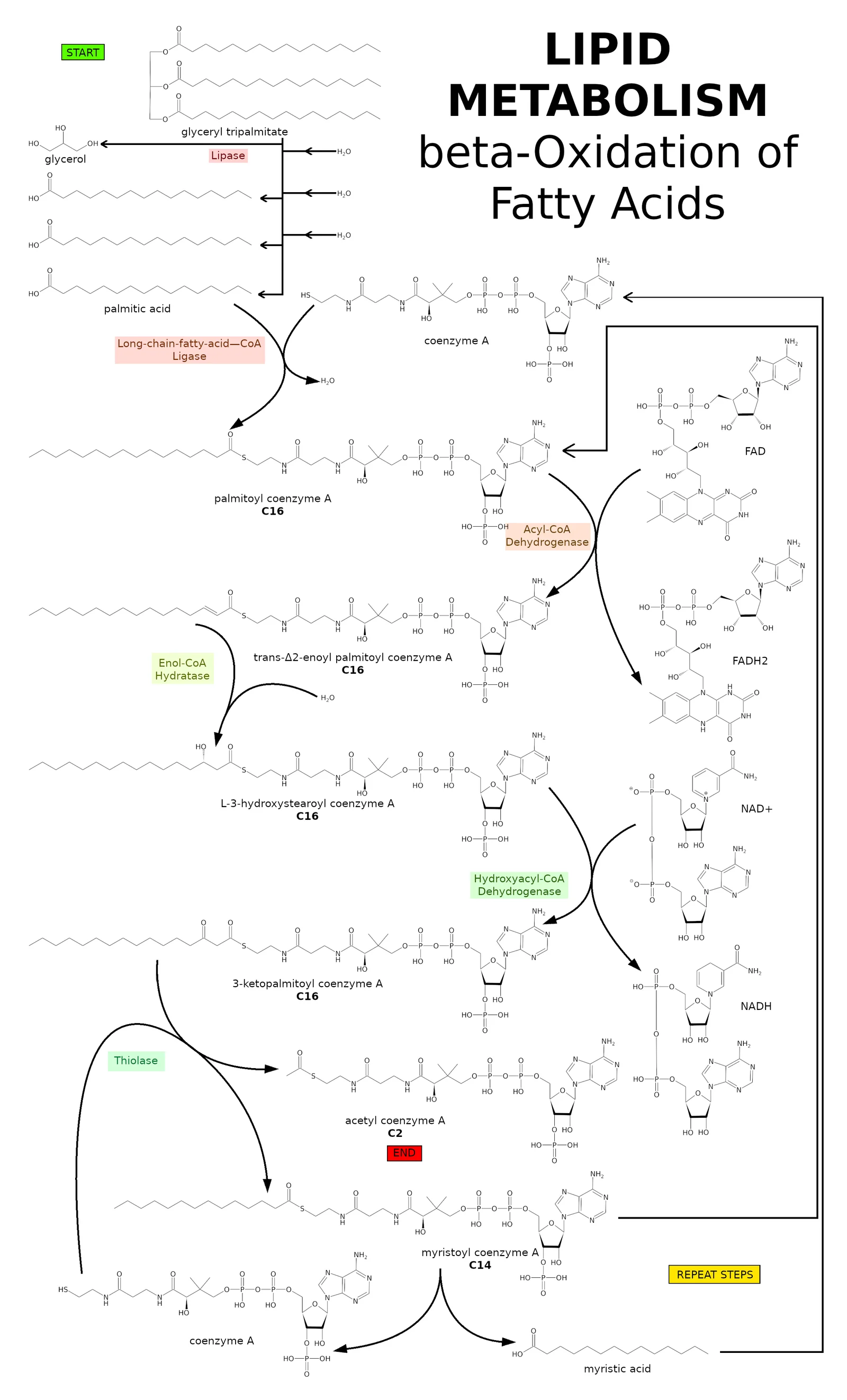
Long description
The beta-Oxidation cycle pathway diagram illustrates the metabolic reactions that allow for the breakdown of fatty acids into NADH and ATP, often taught in connection with the electron transport chain and ATP synthase. T

Glucose, fatty acids and amino acids are the three major substrates that a living organism can utilise to preserve metabolic homoeostasis. These fundamental substrates are required not only for the generation of cellular energy, but also to supply essential building blocks for the biosynthesis of diverse macromolecules. Among these metabolic pathways, the primary mechanism for the biological degradation of fatty acids is mitochondrial fatty acid beta-oxidation. This biochemical pathway functions as a vital pillar for energy homoeostasis across highly metabolically active organs, including the liver, heart and skeletal muscle. [3]
During periods of fasting or when exogenous carbohydrate supply becomes severely limited, mitochondrial fatty acid beta-oxidation assumes paramount importance. Under such catabolic conditions, most peripheral tissues throughout the body are capable of utilizing fatty acids directly to generate necessary ATP. Concurrently, the liver processes these lipid substrates to synthesize ketone bodies, a specialized metabolic pathway for which beta-oxidation is entirely indispensable. Ketone bodies then serve as a critical alternative energy source that can be readily consumed by all major tissues, including the central nervous system and the brain. [3]
DefinitionBeta-oxidation pathway
The classical biochemical degradation of fatty acids proceeds through the successive removal of two-carbon units via oxidation at the beta carbon atom.
The historical discovery of this pathway traces back to classical physiological experiments. Georg Franz Knoop first discovered fatty acid beta-oxidation in 1904 by administering odd- and even-chain omega-phenyl fatty acids, such as omega-phenylvaleric acid and omega-phenylbutyric acid, to dogs and analysing their resultant urinary metabolites. Animals fed odd-chain fatty acids excreted hippuric acid, whereas those fed even-chain fatty acids excreted phenaceturic acid. From these landmark observations, Knoop deduced that fatty acid metabolism proceeds via the successive removal of two-carbon fragments, postulating that oxidation occurred specifically at the beta carbon atom. This pioneering theory was subsequently confirmed and further developed by Henry Drysdale Dakin using analogous phenyl derivatives. [3]
| Metabolic State | Primary Substrate | Major Organ Pathways |
|---|---|---|
| Fed state | Glucose | Glycolysis and systemic carbohydrate utilisation |
| Early fasting | Glycogen stores | Hepatic glycogenolysis supporting euglycemia |
| Prolonged fasting | Fatty acids and ketones | Beta-oxidation and hepatic ketogenesis |
Inherited defects in this pathway encompass almost twenty distinct inborn enzymopathies collectively known as fatty acid oxidation disorders. Long-chain fatty acid oxidation disorders represent a specific group of inherited metabolic conditions characterised by impaired mitochondrial beta-oxidation of long-chain fatty acids, which are essential for maintaining energy production particularly during periods of fasting or increased physiological energy demand. These enzymatic defects arise from pathogenic genetic variants encoding proteins involved in the carnitine cycle and the beta-oxidation cascade. Specific named conditions within this category include carnitine palmitoyltransferase 1a deficiency, carnitine-acylcarnitine translocase deficiency, carnitine palmitoyltransferase 2 deficiency, very long-chain acyl-CoA dehydrogenase deficiency, long-chain 3-hydroxyacyl-CoA dehydrogenase deficiency, and mitochondrial trifunctional protein deficiency. [1][2]
The transport of long-chain fatty acids across the impermeable inner mitochondrial membrane requires a dedicated biochemical carrier system known as the carnitine shuttle. Irving B. Fritz made a major landmark discovery when he identified the marked stimulation of fatty acid oxidation by carnitine. Without this specialized shuttle mechanism, long-chain acyl-CoA molecules cannot gain entry into the mitochondrial matrix where beta-oxidation enzymes reside. [3]
Clinical pearlBypassing the shuttle
Medium-chain fat sources provide an alternative energy substrate that can enter mitochondria independently of the carnitine shuttle.
Inherited disorders affecting the carnitine shuttle and long-chain fatty acid oxidation disrupt normal energy metabolism during catabolic stress. The primary objective of nutritional management in these long-chain fatty acid oxidation disorders is to maintain metabolic stability by actively preventing catabolism, avoiding prolonged fasting periods, and supplying adequate exogenous energy during acute illness, exercise, or other states of heightened metabolic demand. Dietary restriction of long-chain fats reduces the overall metabolic load that requires processing through the impaired pathway. Meanwhile, medium-chain fat sources serve as a therapeutic alternative because they enter the mitochondria independently of the carnitine shuttle and bypass defective long-chain enzymes. The clinical benefit derived from medium-chain triglycerides stems from providing readily oxidizable fuel, supporting ketogenesis under certain circumstances, and decreasing overall reliance on long-chain fatty acid oxidation. [1]
Metabolic routing in fatty acid oxidation
- Fasting or increased demandGlycogen depletion triggers lipid utilisation.
- Question: Chain length?
Long-chain
- Carnitine shuttle requiredVulnerable to CPT1, CACT, CPT2 and VLCAD defects.
Medium-chain
- Independent entryProcessed by MCAD in mitochondrial matrix.
Physiological evaluations during fasting reveal distinct abnormalities in individuals harbouring long-chain defects. For instance, in one patient with very long-chain acyl-CoA dehydrogenase deficiency, long-chain acylcarnitine species increased markedly after a long-chain triglyceride load administered at twelve hours of fasting. Another individual experienced clinical rhabdomyolysis accompanied by elevated creatine kinase levels in the absence of hypoglycemia during a ten-hour fasting test, although circulating acylcarnitine concentrations remained within normal limits. More severe physiological challenge, such as fasting exceeding fifteen hours, led to elevated free fatty acids alongside suppressed ketone body production, culminating in pronounced creatine kinase elevation and overt hypoglycemia after twenty-two hours. [1]
Medium-chain acyl-CoA dehydrogenase deficiency is by far the most frequent inborn error of fatty acid beta-oxidation, with an estimated birth prevalence of approximately 1 in 8,000, or as high as 1 in 12,000 reported in Canadian populations. The underlying enzymatic defect results in a complete blockade of medium-chain fatty acid breakdown and a corresponding failure of hepatic ketone production. This enzymatic deficiency critically compromises the physiological availability of ketone bodies during prolonged fasting or acute intercurrent illness. The resulting acute energy shortage disproportionately impairs the functional integrity of skeletal muscle, cardiac muscle, and the brain, which can ultimately progress to fatal outcomes if unrecognised. [2][4]
Exam trapSilent presentation
MCAD deficiency is usually clinically silent and children appear entirely normal until an intercurrent illness triggers sudden metabolic decompensation. [2]
The mitochondrial enzyme medium-chain acyl-CoA dehydrogenase is directly responsible for catalysing the initial step in the mitochondrial breakdown of medium-chain fatty acids. This biochemical breakdown is absolutely required for sustaining euglycemia after physiological glycogen stores become depleted, such as during high-energy physical activities, extended fasting, or intercurrent illness accompanied by fever or persistent vomiting. Deficiency of this specific enzyme dramatically increases a patient's vulnerability to life-threatening manifestations during periods of catabolic stress. These acute metabolic decompensations are classically characterized by hypoketotic hypoglycemia, profound lethargy, and generalised seizures. [4]
Historical milestones in the study of this condition include the identification of the first inherited defects in the broader fatty acid oxidation pathway, which included carnitine palmitoyltransferase 2 deficiency, primary carnitine deficiency, and medium-chain acyl-coenzyme A dehydrogenase deficiency. Most constituent beta-oxidation enzymes were successfully purified, paving the way for subsequent gene cloning and the exact identification of disease-causing mutations in affected patients. [3]
Mitochondrial fatty acid oxidation is a critical pathway for energy production, particularly when nutritional intake is interrupted. In healthy individuals, periods of fasting shift the metabolic profile away from glucose oxidation toward the utilisation of fatty acids and ketone bodies. This metabolic transition spares scarce glucose for obligate users such as the brain. However, when genetic defects impair mitochondrial oxidation, the normal physiological response breaks down.
In long-chain fatty acid oxidation disorders, patients cannot properly oxidise long-chain fatty acids, making them highly vulnerable during periods without food. Because energy generation fails, dependency on glycolysis increases rapidly, which causes blood glucose concentrations to fall as hepatic and muscular glycogen stores become depleted. This pathological vulnerability is compounded significantly by hypoketosis—the lack of appropriate ketone body synthesis as an alternative fuel source.
DefinitionHypoketotic Hypoglycemia
A dangerous metabolic state characterised by low blood glucose occurring simultaneously with inappropriately low ketone body levels, typically signalling an inability to process fatty acids.
The clinical consequences of defective ketogenesis and fatty acid oxidation can be catastrophic. For instance, medium chain acyl CoA dehydrogenase deficiency, an autosomal recessive condition, is the most common fatty acid oxidation disorder, with an incidence around one in eight thousand. This condition frequently presents dramatically with severe hypoketotic hypoglycemia, Reye syndrome, or sudden unexpected death in infancy, showing a distinct peak of frequency around three to six months of age. Furthermore, if left undetected, approximately twenty to twenty-five percent of infants will die or suffer permanent neurologic impairment as a direct consequence of their very first acute metabolic decompensation. [2]
| Parameter | Normal Fasting State | Fatty Acid Oxidation Defect |
|---|---|---|
| Primary Fuel | Fatty acids and ketones | Reliance on glycolysis |
| Blood Glucose | Maintained via gluconeogenesis | Falls as glycogen stores deplete |
| Ketone Production | Substantial ketosis | Inappropriate hypoketosis |
| Clinical Risk | Physiological ketogenesis | Severe decompensation and death |
To prevent these acute crises, management strategies focus on avoiding prolonged fasting and ensuring consistent caloric intake. When investigating metabolic tolerance, clinical protocols often define fasting thresholds carefully. If an acceptable fasting range is provided instead of a specific amount of time, such as a prescribed maximum fasting range of eight to ten hours, the median value of that range defines the fasting time. When children are prescribed different fasting times for daytime versus overnight, the larger value representing the maximum amount of time is used. Furthermore, if a fasting time relies on an additional source of carbohydrates such as cornstarch, the fasting time is defined without extraneous intervention by taking the lower value, because this represents the baseline amount of time tolerated in normal physiological states.
Metabolic Pathophysiology in Fasting
- Fasting beginsGlycogen stores start to deplete.
- Question: Is fatty acid oxidation normal?
Yes
- Physiological ketogenesisKetones and fatty acids fuel peripheral tissues; glucose is spared.
No (e.g. LC-FAOD / MCAD)
- Glycolytic dependencyBlood glucose falls rapidly.
- HypoketosisAlternative ketone fuels are absent, leading to metabolic crisis.
Mammalian organs display remarkable metabolic flexibility, adjusting their substrate utilisation to maintain energy homeostasis according to feeding status. Under normal, well-fed conditions, glucose serves as the preferred substrate for oxidative phosphorylation across many tissues. However, during fasting and starvation, this priority shifts, and non-carbohydrate substrates become dominant.
The heart provides a striking example of this flexibility, constantly relying on fatty acids for a major proportion—specifically sixty to ninety percent—of its total energy needs. Depending on the feeding status and physiological demand, the heart also incorporates glucose, lactate, and ketone bodies as important alternative fuels. This shifting interplay is governed by regulatory mechanisms. The reciprocal relationship or competition between the oxidation of fatty acids and glucose is widely known as the glucose-fatty acid cycle or Randle cycle. The fundamental driver of this cycle is the glucose-sparing effect exerted by fatty acid oxidation and ketone body utilisation, which ensures that circulating glucose remains available for glucose-dependent tissues when nutritional sources are scarce. [3]
Clinical pearlCardiac Substrate Reliance
The healthy heart derives the vast majority of its energy from fatty acids, but retains the metabolic flexibility to switch fuels depending on systemic supply.
Cellular mechanisms tightly control these substrate choices. Nuclear receptors and fatty acid transporters mediate the uptake and utilisation of substrates. Specifically, proteins located at the plasma membrane, such as fatty acid transport proteins, plasma membrane fatty acid-binding protein (which is identical to mitochondrial aspartate aminotransferase, gene name GOT2), and fatty acid translocase (FAT, CD36), actively participate in the cellular uptake of fatty acids. Furthermore, intracellular fatty acid activation involves complex enzyme systems, including a protein sub-family possessing long-chain acyl-CoA synthetase activity known as long-chain acyl-CoA synthetases.
When dietary or endogenous fatty acids activate nuclear receptors such as PPAR-alpha and PPAR-beta, tissues become even more dependent on fatty acid oxidation by stimulating cellular pathways for transport, esterification, and oxidation. Such pathways are essential for health, but they also highlight why certain patient groups are vulnerable. For example, patients with type 2 diabetes are at increased risk of developing cardiomyopathy, a pathological state that coincides with an increased cardiac reliance on fatty acids as the primary energy source following elevated circulating free fatty acid levels.
| Physiological State | Preferred Circulating Substrates | Primary Tissue Adaptation |
|---|---|---|
| Well-Fed | Glucose | Glucose oxidation predominates |
| Fasting | Fatty acids and ketone bodies | Randle cycle spares glucose for brain |
| High-Intensity Exercise | Glucose utilisation increases | Carbohydrate reliance rises with intensity |
| Starvation / FAO Defect | Variable / Defective | Myocardial and systemic energy crisis |
Nutritional management in metabolic disorders must account for these fuel requirements while protecting defective pathways. For patients with long-chain fatty acid oxidation disorders, supplementation with essential fatty acids and docosahexaenoic acid is crucial for preventing deficiencies and ensuring proper growth and development. Linoleic acid (an omega-6 fatty acid) and alpha-linolenic acid (an omega-3 fatty acid) cannot be synthesized endogenously, making dietary intake mandatory. Supplementation aims to meet physiological requirements without placing an excessive burden on the defective metabolic pathway, with reported intake ranging from two percent to four percent of total energy as linoleic acid and zero point three percent to one point four percent as alpha-linolenic acid. Safflower oil, sunflower oil, and corn oil provide rich sources of linoleic acid, whereas flaxseed oil and chia seed oil serve as the richest sources of alpha-linolenic acid. Additionally, soybean oil, hempseed oil, canola oil, and walnut oil provide both essential fatty acids in significant amounts. [1]
The regulation of mitochondrial fatty acid beta-oxidation and its interplay with carbohydrate metabolism are governed by a sophisticated network of hormonal, transcriptional, and biochemical mechanisms. The mitochondrial fatty acid beta-oxidation pathway plays a pivotal role in energy homoeostasis, but it competes directly with glucose as the primary oxidative substrate. The mechanisms behind this metabolic interplay, known historically as the glucose-fatty acid cycle, operate at multiple physiological levels to shift the body between anabolic storage states and catabolic energy-releasing states. [3]
Hormones are crucial players in dictating substrate availability and cellular metabolic orientation. Insulin, secreted by the pancreatic beta-cells when blood glucose levels are high, acts as the principal anabolic hormone controlling this balance. One of the primary physiological effects of an insulin surge is the marked increase of cellular glucose transport, which is achieved through the rapid translocation of the glucose transporter Glut4, encoded by SLC2A4, from intracellular vesicles to the plasma membrane. By facilitating massive glucose entry into insulin-sensitive tissues such as skeletal muscle and adipose tissue, insulin promotes carbohydrate utilization while simultaneously suppressing alternative energy pathways. [3]
Furthermore, insulin exerts a powerful antilipolytic effect. By inhibiting hormone-sensitive lipase within white adipose tissue, insulin prevents the breakdown of stored triglycerides into free fatty acids, thereby restricting the circulating availability of its competing substrate, the fatty acid. When insulin levels are elevated after a carbohydrate-rich meal, circulating non-esterified fatty acids plummet, effectively starving oxidative tissues of lipid fuel and forcing the preferential combustion of glucose. [3]
Conversely, during periods of fasting, prolonged exercise, or metabolic stress, insulin concentrations decline while counter-regulatory hormones such as glucagon, epinephrine, and cortisol rise. The drop in the insulin-to-glucagon ratio removes the tonic inhibition on adipose tissue lipolysis. Triglycerides are hydrolysed, releasing massive quantities of non-esterified fatty acids into the bloodstream, which are taken up by peripheral organs, particularly the liver and skeletal muscle.
In skeletal muscle, fatty acid oxidation responds dynamically to energy demands. Unlike carbohydrate oxidation, which scales up gradually, fatty acid oxidation is already maximal at moderate exercise intensity. The heart represents an exceptional physiological case among all organs; fatty acid oxidation is extremely important for myocardial tissue, since the heart constantly relies on fatty acids for a major proportion, specifically sixty to ninety percent, of its total energy needs under normal physiological conditions. [3]
At the biochemical level, once fatty acids enter the cytoplasm, they are activated to acyl-CoAs. To cross the impermeable inner mitochondrial membrane, long-chain acyl-CoAs require the carnitine shuttle system, where carnitine palmitoyltransferase-1 (CPT1) acts as a critical rate-limiting gatekeeper. Malonyl-CoA, an intermediate of de novo lipogenesis regulated by the insulin-stimulated enzyme acetyl-CoA carboxylase, acts as a potent endogenous inhibitor of CPT1. Thus, when insulin is high, malonyl-CoA accumulates, blocking the entry of fatty acids into the mitochondria. When insulin falls during fasting, malonyl-CoA levels drop, CPT1 activity is disinhibited, and fatty acid flux into the beta-oxidation spiral accelerates.
Once inside the mitochondrial matrix, acyl-CoAs are degraded into acetyl-CoA units via the classic series of four enzyme reactions called beta-oxidation. The beta-oxidation pathway is a cyclic process in which acyl-CoAs are shortened, whereby the two carboxy-terminal carbon atoms are released as acetyl-CoA units each time a cycle is fully completed. [3]
Substrate Selection and Hormonal Control
- High blood glucose triggers insulin secretionInsulin translocates Glut4 to plasma membrane.
- Question: What is the insulin level?
High (Fed state)
- Inhibit lipolysis in adipose tissue
- Suppress CPT1 via malonyl-CoA accumulation
- Rely predominantly on glucose oxidation
Low (Fasting state)
- Release counter-regulatory hormones
- Activate lipolysis and flood plasma with free fatty acids
- Shift myocardium and skeletal muscle to maximal fatty acid oxidation
Understanding the pathways of fatty acid oxidation and ketogenesis requires navigating several conceptual pitfalls, particularly regarding metabolic flexibility, enzyme nomenclature, and the clinical consequences of inherited disruptions. Over the years, the mitochondrial fatty acid beta-oxidation pathway has been characterised at both the biochemical level and the molecular biological level. However, the pathophysiology of inborn errors affecting these pathways is still not completely understood, which continues to hamper optimal treatment strategies. [3]
A frequent student misconception is that fatty acid beta-oxidation operates uniformly across all chain lengths through identical enzymatic machinery. In reality, chain-length specificities divide the pathway. For instance, the oxidation of medium and short-chain acyl-CoAs relies on distinct matrix-soluble enzymes for its second, third, and fourth steps. These include the enoyl-CoA hydratase called crotonase, encoded by ECHS1, the medium and short-chain hydroxyacyl-CoA dehydrogenase, known as M/SCHAD and encoded by HADH, and medium-chain 3-ketoacyl-CoA thiolase, known as MCKAT and encoded by ACAA2. All these enzymes possess broad substrate specificity towards CoA-esters with chain lengths of up to ten carbon atoms. In contrast, long-chain fatty acids require membrane-bound multienzyme complexes like the trifunctional protein and specific carnitine cycle transporters. [3]
Another common pitfall involves underestimating the complexity of unsaturated fatty acid degradation. The standard cyclic process of beta-oxidation handles saturated straight-chain acyl-CoAs, where each cycle yields an acyl-CoA shortened by two carbon atoms, an acetyl-CoA, and one molecule of nicotinamide adenine dinucleotide (NADH) and one flavin adenine dinucleotide (FADH2) as electron carriers or reducing equivalents. However, the oxidation of mono-unsaturated fatty acids such as oleic acid requires the additional action of dodecenoyl-CoA delta isomerase, also called 3,2 trans-enoyl-CoA isomerase and encoded by DCI. Polyunsaturated fatty acids present an even greater structural challenge, needing a second auxiliary enzyme, 2,4-dienoyl CoA reductase 1, encoded by DECR1. Ignoring these auxiliary enzymes leads to incomplete or inaccurate pathfinding through lipid catabolism. [3]
Clinical manifestations of fatty acid oxidation defects also present diagnostic and conceptual traps. Clinical manifestations range widely from neonatal hypoketotic hypoglycaemia and cardiomyopathy to hepatopathy, recurrent rhabdomyolysis, and chronic myopathy. A critical diagnostic pitfall is assuming that all variants within a specific FAO gene lead to the same clinical phenotype. In very long-chain acyl-CoA dehydrogenase (VLCAD) deficiency, evidence suggests milder phenotypes in cases caused by missense variants, especially when located in the C-terminus of the protein. The variant c.848T>C, resulting in the p.V283A amino acid change, is the most frequent pathogenic variant found in individuals with a positive VLCAD deficiency newborn screening result. This particular variant is usually associated with the onset of rhabdomyolysis in adolescence or early adulthood and typically does not present with cardiomyopathy. Conversely, in carnitine-acylcarnitine translocase (CACT) deficiency, homozygosity for the most common pathogenic splice site variant c.199-10T>G is associated with a severe neonatal-onset form of the disease. [1][3]
Exam trapKetones and Hypoglycaemia
A classic pitfall is expecting ketosis during acute decompensation in fatty acid oxidation disorders; because the pathway is blocked, patients characteristically present with hypoketotic hypoglycaemia.
Finally, students often struggle to connect cellular metabolic inflexibility with macroscopic cardiac pathology. It is not entirely clear how a decrease in metabolic flexibility leads to cardiomyopathy. Moreover, it is not known with certainty if any of the accumulating intermediary metabolites in fatty acid oxidation defects are directly toxic to the heart tissue, although abnormal acylcarnitines and free fatty acids have been strongly suggested to contribute to lethal arrhythmias. This accumulation could provide a mechanistic explanation for the heart-beat disorders observed in FAO defects and the documented defect in calcium ion homoeostasis in VLCAD knockout mouse models. [3]
In modern clinical practice, a thorough understanding of fatty acid oxidation, ketone body metabolism, and fasting physiology is essential across neonatology, paediatrics, metabolic medicine, and emergency care. Long-chain fatty acid oxidation disorders are rare inherited defects of mitochondrial beta-oxidation that severely impair energy generation during fasting or periods of increased metabolic stress. Although newborn screening enables early detection of these conditions, clinical management remains inconsistent due to limited high-level evidence, a broadening phenotypic spectrum, and notable regional practice variations. [1]
Inherited defects for most of the fatty acid oxidation enzymes have been identified and characterised, and these conditions are currently included in standard neonatal screening programmes. When an inherited defect is identified, dietary management and avoidance of fasting form the cornerstone of prophylaxis. For instance, in specific conditions where plasma lipid profiles reveal nutritional deficiencies, targeted nutritional interventions are instituted; docosahexaenoic acid (DHA) supplementation is advised if plasma concentrations are low, though the specific effect of this prophylaxis on retinopathy remains less clear. [1]
Clinical pearlSudden Infant Death
At present, long QT syndrome and fatty acid beta-oxidation disorders represent the most frequent inherited causes of sudden unexpected death in infancy (SUDI). [2]
Sudden infant death syndrome is defined as the sudden death of an infant that is unexpected by history and unexplained by postmortem examinations. SIDS is a leading cause of infant mortality, accounting for eight percent of all infant deaths, and it represents the most common cause of post-neonatal infant mortality, accounting for forty to fifty percent of all deaths occurring between one month and one year of age. Cases in which the underlying cause is definitively identified should be diagnosed not as SIDS, but as sudden unexpected death in infancy, such as when specific genetic metabolic conditions are uncovered. Pathologists and metabolic paediatricians must maintain high vigilance for underlying beta-oxidation defects during postmortem evaluations of unexplained infant deaths. [2]
| Condition / Variant | Primary Presentation | Key Biochemical or Genetic Feature |
|---|---|---|
| VLCAD deficiency (c.848T>C / p.V283A) | Adolescent or adult rhabdomyolysis | Missense variant in C-terminus; usually spares heart |
| CACT deficiency (c.199-10T>G) | Severe neonatal-onset disease | Homozygosity for common splice site variant |
| LC-FAOD general | Hypoketotic hypoglycaemia, cardiomyopathy | Impaired fasting energy generation |
| SUDI / SIDS context | Sudden unexpected infant death | Frequent inherited metabolic cause |
Management protocols require close collaboration among multidisciplinary teams. International guidelines are continually refined by multidisciplinary workgroups comprising clinicians, nurses, dietitians, and patient representatives who systematically review decades of literature to improve outcomes for affected families. Patient advocacy groups, such as the German-speaking support group for inborn errors of fatty acid oxidation, Fett-SOS e.V., play an invaluable role in bridging clinical care and patient support. Understanding the tight physiological coupling between hormonal control, fasting endurance, and enzymatic beta-oxidation remains vital for preventing life-threatening metabolic crises.
Long-chain fatty acid oxidation disorders represent a distinct group of rare inherited metabolic conditions affecting mitochondrial beta-oxidation. This spectrum comprises several specific enzymatic and transport defects, including very long-chain acyl-CoA dehydrogenase deficiency, long-chain 3-hydroxyacyl-CoA dehydrogenase deficiency, carnitine palmitoyltransferase 1a deficiency, carnitine-acylcarnitine translocase deficiency, and carnitine palmitoyltransferase 2 deficiency. Because these enzym
Global and regional detection rates vary considerably, influenced heavily by the implementation and scope of newborn screening programmes. Newborn screening enables early biochemical and genetic detection before catastrophic decompensation occurs. However, management across different jurisdictions remains inconsistent due to a limited evidence base, a broadening phenotypic spectrum, and notable regional practice variation. The precise overall incidence and prevalence figures for individual long-chain fatty acid oxidation disorders vary by geographic region, reflecting population-specific carrier frequencies and the inclusion parameters of regional screening panels. While newborn screening has successfully identified many asymptomatic or pre-symptomatic neonates, milder phenotypes or late-onset presentations, such as exercise-induced myalgia and recurrent rhabdomyolysis in older children and adults, may elude neonatal detection entirely, leading to underestimation of true disease prevalence in older cohorts. [5]
Clinical pearlClinical spectrum
Clinical presentations span from severe neonatal cardiomyopathy and hypoketotic hypoglycaemia to intermittent adult rhabdomyolysis triggered by exertion.
From a genetic standpoint, all long-chain fatty acid oxidation disorders follow an autosomal recessive inheritance pattern. Pathogenic variants occur in specific genes that encode the essential enzymes and transport proteins responsible for the carnitine cycle and the mitochondrial beta-oxidation pathway. Because of this recessive transmission, affected individuals must inherit two abnormal alleles, typically one from each carrier parent, putting subsequent siblings at a twenty-five percent recu
| Disorder | OMIM Reference | Affected Pathway Component |
|---|---|---|
| CPT1a deficiency | OMIM #255120 | Carnitine shuttle (outer membrane) |
| CACT deficiency | OMIM #212138 | Carnitine-acylcarnitine translocase |
| CPT2 deficiency | OMIM #255110 | Carnitine shuttle (inner membrane) |
| VLCAD deficiency | OMIM #201475 | Very long-chain beta-oxidation |
| LCHAD deficiency | OMIM #609016 | Long-chain 3-hydroxyacyl-CoA |
Pathophysiologically, these inherited enzymatic defects impair the body's ability to utilize stored lipids for fuel when energy demands increase or exogenous substrates decrease. Consequently, patients depend entirely on glycolysis, which rapidly depletes glycogen stores and leads to profound hypoketotic hypoglycaemia because alternative ketone bodies cannot be produced in sufficient quantities. Furthermore, the intracellular accumulation of toxic long-chain fatty acids, unesterified acylcarnitines, and sequestered acyl-CoA intermediates directly contributes to acute clinical symptoms, cellular injury, and organ dysfunction. [5]
Metabolic crisis evaluation
- Patient presents with catabolic stress or fastingAssess for triggers such as fever or prolonged exercise.
- Question: Is fasting prolonged or energy demand increased?
Yes
- Initiate emergency carbohydrate regimen to prevent decompensation
- Monitor for rhabdomyolysis, elevated creatine kinase, and hypoketotia
No
- Maintain stable dietary intake with medium-chain fats and protein
Acute metabolic decompensation is frequently precipitated by specific environmental and physiological triggers. Common triggers include intercurrent febrile illnesses, prolonged fasting periods, rigorous or prolonged exercise, surgical procedures, and routine vaccinations. During these stressful events, energy demands rise significantly while metabolic reliance shifts entirely toward a defective pathway. For example, studies examining fasting tolerance demonstrate that fasting periods exceeding fifteen hours lead to markedly elevated free fatty acids alongside blunted ketone production, while even shorter fasts can provoke elevated creatine kinase and rhabdomyolysis without immediate hypoglycaemia. To prevent such life-threatening crises, the primary clinical management strategy focuses on avoiding prolonged fasting and ensuring consistent, frequent caloric intake emphasizing carbohydrates, medium-chain triglycerides, and protein. [5]
Fatty acid oxidation disorders are classified by the chain length of the affected substrates, dividing into short-chain, medium-chain, long-chain, and very long-chain deficiencies. Phenotypic staging categorises patients into distinct clinical subtyping variants, including severe infantile presentations, intermediate phenotypes, and late-onset myopathic variants. In long-chain disorders such as very long-chain acyl-CoA dehydrogenase deficiency, analysis of cellular acylcarnitine intermediates after incubation with specific fatty acids helps distinguish milder phenotypes from patients whose clinical course is complicated by cardiomyopathy. Similarly, in long-chain 3-hydroxyacyl-CoA dehydrogenase and trifunctional protein deficiencies, specific acylcarnitine profile ratios and fatty acid oxidation flux assays show positive correlations with the severity of the clinical phenotype. Conversely, direct enzyme assays measuring long-chain 3-hydroxyacyl-CoA dehydrogenase and long-chain 3-ketothiolase activities in patient cells do not reliably correlate with the clinical phenotype. [5]
Neonatal-onset presentations differ markedly from adult-onset variants. For instance, in a series of patients with carnitine acylcarnitine translocase deficiency, neonatal-onset cases all required restricted long-chain fat between 5% and 10% of total energy intake throughout childhood. Staging must also account for specific genetic variants, such as the c.338C>T variant in carnitine palmitoyltransferase 2 deficiency, which defines a late-onset myopathic form that does not involve the heart. [5]
DefinitionPhenotypic correlation
Fatty acid oxidation disorders present across multiple organ systems, manifesting with hepatic, cardiac, neuromuscular, and skeletal abnormalities. Cardiac involvement is common in most cases of long-chain fatty acid oxidation defects, with severe cardiomyopathy occasionally seen in utero in trifunctional protein and carnitine palmitoyltransferase 2 deficiencies. More frequently, cardiac manifestations appear in the neonatal period and early childhood. In a few patients, cardiac complications begin later in life, usually associated with metabolic decompensation or suboptimal treatment, and these can appear independently of skeletal muscle involvement. Severe variants and very low enzyme activity are more likely to result in in utero and neonatal cardiac presentation accompanied by poor outcomes. Intercurrent infections and catabolic conditions may provoke cardiac symptoms, driven by the accumulation of arrhythmogenic intermediary metabolites of fatty acids, such as long-chain acylcarnitines, which are responsible for arrhythmias. Notably, this cardiomyopathy can be reversible with appropriate treatment. [5]
A critical clinical distinction exists between neonatal acute presentations and adult-onset exercise-induced myopathy. Whilst neonatal cases present with acute multi-organ crisis, hyperammonemia, increased creatine kinase, and cardiomyopathy, certain enzyme deficiencies diverge. Specifically, cardiac involvement is not a clinical feature of carnitine palmitoyltransferase 1 deficiency or the late-onset myopathic form of carnitine palmitoyltransferase 2 deficiency. Consequently, individuals with carnitine palmitoyltransferase 1 and myopathic carnitine palmitoyltransferase 2 deficiencies do not require cardiac investigations.
| Organ System | Neonatal / Infantile Presentation | Adult / Late-Onset Presentation |
|---|---|---|
| Cardiac | Severe cardiomyopathy, arrhythmias, in utero onset | Absent in specific variants like myopathic CPT2 |
| Neuromuscular | Acute decompensation, hypotonia | Exercise-induced myopathy, rhabdomyolysis |
| Hepatic | Hypoketotic hypoglycaemia, hepatomegaly | Rare primary presentation |
Clinical evaluation pathway for suspected defects
- Evaluate patient age and presentation typeDistinguish neonatal acute multi-organ crisis from adult-onset exercise intolerance.
- Question: Is the diagnosis an LC-FAOD?
Yes
- Assess for cardiac involvementPerform echocardiography for neonatal or severe phenotypes.
- Check specific subtypeNote that CPT1 and myopathic CPT2 deficiencies do not require cardiac investigations.
No
- Proceed with standard metabolic and genetic screeningEvaluate medium or short chain parameters.
Distinguishing between primary metabolic disorders during acute illness requires separating conditions presenting with hypoketotic hypoglycaemia from those causing recurrent rhabdomyolysis or resembling acute encephalopathy. When evaluating hypoketotic hypoglycaemia, clinicians must differentiate fatty acid oxidation defects from congenital hyperinsulinism, where inappropriately elevated insulin suppresses lipolysis and ketogenesis, and panhypopituitarism, which features secondary adrenal insufficiency and growth hormone deficiency leading to impaired counter-regulatory hormone output.
For patients presenting with recurrent rhabdomyolysis, the differential diagnosis spans several distinct categories of myocellular energy failure. These include glycogen storage diseases type V (McArdle disease, due to muscle glycogen phosphorylase deficiency) and type VII (Tarui disease, due to phosphofructokinase deficiency), both of which impair glycolytic ATP generation during intense exertion. Mitochondrial myopathies, which disrupt oxidative phosphorylation, must also be considered alongside long-chain fatty acid oxidation defects that manifest with exercise-induced muscle breakdown, pigmenturia, and elevated serum creatine kinase.
Separating acute metabolic decompensation driven by inherited metabolic disease from Reye-like syndromes or viral encephalitis can be challenging because all three may present with acute vomiting, lethargy, hepatomegaly, and encephalopathy. However, metabolic decompensation typically features profound biochemical derangements such as inappropriate hypoketotia relative to the degree of hypoglycaemia, hyperammonemia, and specific acylcarnitine elevations. Severe disease categories incorporate neonatal or early infantile presentations, cardiomyopathy, hypoketotic hypoglycaemia, hepatopathy, hyperammonemia, recurrent rhabdomyolysis, hospitalization, and genetic or functional findings consistent with severe disease. Deficiencies in CACT, LCHAD, and TFP are generally considered to have severe phenotypes. [5]
Clinical pearlMyocellular stress markers
Elevated creatine kinase activity, particularly CK-MM and CK-MB, signals metabolic decompensation [5]
Establishing formal diagnostic criteria for defining confirmed fatty acid oxidation disorder cases following newborn screening requires a comprehensive multimodal evaluation strategy. Because newborn screening programs identify diverse cohorts, standardized diagnostic work-up protocols are essential to promote uniform care across age groups, disease categories, and phenotypic severity classifications[5]. Observational data of individuals identified before symptom onset highlight significant clinical heterogeneity, demonstrating that routine dietary treatment should not be applied universally to all asymptomatic individuals[5]. Specifically, studies report that a notable proportion of individuals who are completely asymptomatic at initial diagnosis eventually develop clinical manifestations over time[5]. Therefore, diagnostic confirmation relies on integrating biochemical testing, enzymatic assays, and molecular genetic confirmation rather than relying on newborn screening analyte elevations alone. When defining confirmed cases, clinicians must evaluate the specific severity category, baseline clinical course, biochemical monitoring parameters, and individual patient tolerance[5].
DefinitionDiagnostic Confirmation
To establish a structured diagnostic approach, multidisciplinary teams utilize consensus guidelines where agreement among experts is formally defined as 75% or greater concordance[5]. The diagnostic framework addresses standardizing investigations during periods of metabolic stability as well as during acute metabolic stress[5]. Long-chain fat restriction and medium-chain fat supplementation regimens are subsequently tailored based on these rigorously defined severity categories and confirmed diagnostic profiles[5].
| Evaluation Category | Purpose | Methodological Approach |
|---|---|---|
| Biochemical profiling | Assess metabolic status | Acylcarnitine profile and organic acids |
| Genetic testing | Confirm molecular diagnosis | Mutation analysis of relevant genes |
| Clinical assessment | Determine phenotypic severity | Longitudinal monitoring of growth and tolerance |
Laboratory workup relies heavily on tandem mass spectrometry to evaluate blood acylcarnitine profiles. The acylcarnitine profile demonstrates characteristic elevations of specific species depending on the enzymatic block; for example, elevated C8 species indicate medium-chain acyl-CoA dehydrogenase (MCAD) deficiency, whereas elevated C14:1 species point toward very long-chain acyl-CoA dehydrogenase (VLCAD) deficiency. Plasma acylcarnitines fluctuate based on physiological states such as fasting, feeding reduces long-chain acylcarnitine concentrations, whereas fasting and exercise can increase them,
Urine organic acid analysis is another essential modality, frequently revealing dicarboxylic aciduria (the products of minor omega-oxidation pathways overwhelmed by blocked beta-oxidation) and glycine conjugates. When biochemical profiles are ambiguous, functional enzyme assays and molecular genetic testing are deployed. Molecular genetic testing using next-generation sequencing panels covering all long-chain fatty acid oxidation disorders has become the preferred method of confirmation. Nevertheless, enzyme analyses performed as functional tests are often superior to molecular genetic testing for assessing disease severity. [5]
| Investigation | Target Parameter | Diagnostic Indication |
|---|---|---|
| Tandem Mass Spectrometry | Blood acylcarnitines (e.g., C8, C14:1) | Identifies specific chain-length oxidation blocks |
| Urine Organic Acids | Dicarboxylic acids, glycine conjugates | Detects secondary pathways of fatty acid metabolism |
| Enzyme Assay | Palmitoyl-CoA oxidation, residual activity | Assesses functional severity and correlates with phenotype |
| Molecular Genetics | Next-generation sequencing panels | Establishes definitive genetic diagnosis |
Measurement of fatty acid oxidation enzyme activities in lymphocytes or fibroblasts provides a direct functional assessment, though availability is geographically limited and often restricted to research settings. The level of VLCAD enzyme activity measured via palmitoyl-CoA oxidation correlates directly with the clinical phenotype. Residual enzyme activity greater than 20 percent is consistent with mild or late-onset disease or even a complete lack of symptoms throughout life, though the acylcarnitine profile can still be abnormal. Furthermore, cellular long-chain fatty acid oxidation flux correlates with clinical severity in patients with VLCAD deficiency, where incubation of patient-derived fibroblasts with labeled palmitate produces distinct acylcarnitine profiles differentiating severe early-onset cardiomyopathy from milder hypoketotic hypoglycaemia phenotypes. [5]
Diagnostic pathway for suspected fatty acid oxidation disorders
- Initial PresentationEvaluate hypoketotic hypoglycaemia, rhabdomyolysis, or acute decompensation.
- Question: Biochemical Screen
Acylcarnitines & Organic Acids
- Identify specific elevations (e.g., C8, C14:1, dicarboxylic acids).
- Question: Confirm Diagnosis
Genetics / Enzyme Assay
- Perform NGS panel or cellular flux and residual enzyme activity assay.
Acute emergency management during a metabolic crisis requires rapid and aggressive intervention to halt catabolic processes. The primary emergency protocol involves the administration of high-dose intravenous dextrose, specifically using 10% dextrose infusions, designed explicitly to suppress peripheral lipolysis and prevent further accumulation of toxic lipid intermediates[5]. In parallel, sick day protocols and emergency letters should be provided proactively to patients and their caregivers to facilitate early management during acute home situations, thereby reducing the necessity for hospital admission and avoiding severe clinical complications[5]. Emergency protocols adapted from glycogen storage diseases have proven safe and easy to use for home management and during the initial hour of in-hospital care[5].
Clinical pearlEmergency Dextrose
Management strategies also incorporate specialized dietary modifications and targeted supplementation. The role and controversies of L-carnitine supplementation remain nuanced, while medium-chain fat sources and triheptanoin, a synthetic C7 triglyceride, are utilized for long-chain fatty acid oxidation disorders[5]. Clinical trials demonstrate that triheptanoin reproducibly decreases hypoglycemia, rhabdomyolysis, and cardiomyopathy event rates, alongside reducing overall hospitalizatio
Emergency and Exertion Management Protocol
- Assess patient statusDetermine if the patient is in acute metabolic crisis or stable.
- Question: Is there an acute metabolic crisis?
Yes
- Administer IV dextroseInfuse high-dose 10 percent intravenous dextrose to suppress lipolysis.
- Hospital monitoringProvide intensive inpatient management and cardiac surveillance.
No
- Review sick day planProvide emergency letters and home management instructions.
- Question: Engaging in exercise?
Yes
- Pre-exertion supplementationAdminister medium-chain fats or triheptanoin prior to activity.
No
- Maintain baseline dietFollow individualized long-chain fat restriction and cornstarch regimen.
Long-chain fatty acid oxidation disorders carry a significant burden of multi-organ complications, notably involving the neuromuscular, renal, and cardiac systems. Peripheral neuropathy represents a frequent finding within these specific subgroups, demonstrating a median time of onset of 5 years in mitocho
Renal complications can manifest acutely secondary to severe systemic insults. Acute renal failure can develop secondary to severe rhabdomyolysis, a condition where massive skeletal muscle breakdown releases intracellular contents into the circulation. Furthermore, cardiac involvement poses a major life-threatening risk. Chronic cardiomyopathy and lethal ventricular arrhythmias are well-documented features, and cardiac arrhythmias can occur independently of structural cardiomyopathy. Because pre-emptive prediction of acute arrhythmias remains exceptionally difficult, ongoing cardiac surveillance utilizing echocardiography to assess left ventricular ejection fraction and wall mass, alongside electrocardiography and Holter monitoring, is standard clinical practice.
Clinical pearlNeuropathy persistence
Early initiation of dietary interventions and newborn screening protocols do not prevent the development of peripheral neuropathy in LCHAD and mitochondrial trifunctional protein deficiencies.
The long-term outlook for individuals with long-chain fatty acid oxidation disorders has been profoundly influenced by the advent of population-based newborn screening. Long-term survival rates post-newborn screening implementation reflect improved early identification, allowing pre-symptomatic management before severe metabolic crises occur. However, prognostic trajectories vary dramatically depending on the specific enzymatic defect, underlying genotype, and clinical presentation subtype.
Long-chain fatty acid oxidation disorders carry a significant burden of multi-organ complications, notably involving the neuromuscular, renal, and cardiac systems. Pigmentary retinopathy and peripheral neuropathy occur specifically in patients with long-chain 3-hydroxyacyl-CoA dehydrogenase (LCHAD) or mitochondrial trifunctional protein (MTP) deficiency. Peripheral neuropathy represents a frequent finding within these specific subgroups, demonstrating a median time of onset of 5 years in mitocho Adult‐onset symptoms can mimic hereditary sensory motor neuropathy in TFP
| Presentation Type | Age of Onset | Key Clinical Features | Prognostic Outlook |
|---|---|---|---|
| Classic Infantile | First days of life | Hypoketotic hypoglycemia, severe cardiomyopathy, hyperammonemia | High acute mortality risk without rapid intervention |
| Adult Myopathic | Later life / adulthood | Exertional rhabdomyolysis, muscle weakness, neuropathy | Generally milder, triggered by fasting or high stress |
Prognostic stratification framework
- Identify Age of OnsetDifferentiate between neonatal presentation and later-onset or adult symptoms.
- Question: Presents in the first days of life?
Yes
- Infantile FormExpect severe risk of cardiomyopathy, hypoketotic hypoglycemia, and multi-organ crisis.
No
- Later-Onset FormEvaluate for exertional rhabdomyolysis, isolated myopathy, or progressive neuropathy.
DefinitionScreening scope
Modern detection relies heavily on tandem mass spectrometry newborn screening algorithms and cut-offs to identify metabolic errors prior to severe decompensation.
Metabolic screening protocols incorporate tandem mass spectrometry newborn screening algorithms and cut-offs to detect acylcarnitine profile abnormalities from dried blood spots. Early detection is paramount because it allows for immediate dietary intervention before life-threatening cardiac or hepatic crises occur. In parallel with postnatal approaches, prenatal diagnosis and genetic carrier screening for affected families play a crucial role, particularly when a previous sibling has been diagnosed with a fatty acid oxidation defect.
Molecular genetic testing involves scanning for specific pathogenic variations in disease-causing genes. For instance, variations in the gene encoding for LCHAD, including HADHA and HADHB genes, such as the common c.1528G greater than C (p.Glu474Gln) HADHA variants, have been identified in affected children and are associated with maternal acute fatty liver of pregnancy. [5]
Despite the association with specific maternal liver conditions, population demographics dictate screening utility. In some populations, such as the Dutch, the carrier frequency of the common LCHAD variant is low, and heterozygosity for this variant does not seem to significantly increase the risk of HELLP syndrome, leading to the recommendation against routine screening of pregnant women for this mutation. Furthermore, corroborating this population variance, other studies did not find a strong correlation between long-chain fatty acid oxidation disorders and HELLP syndrome. For example, heterozygosity for the common LCHAD variant was not found in a retrospective analysis of 88 Austrian infants from mothers with HELLP syndrome. [5]
| Modality | Target Population | Purpose |
|---|---|---|
| Tandem mass spectrometry | Newborn population | Acylcarnitine profile screening |
| Genetic carrier screening | At-risk families and parents | Identification of pathogenic variants |
| Prenatal diagnosis | Fetuses with affected siblings | Early molecular confirmation |
Clinical pearlOutpatient scheduling
Structured surveillance safeguards against silent metabolic drift and long-term organ damage.
Effective management of patients with long-chain fatty acid oxidation disorders requires regular clinical assessments—encompassing medical, nutritional, and psychological evaluations, as well as biochemical monitoring. This comprehensive approach helps detect metabolic imbalances early, guide dietary interventions, and prevent severe complications during periods of illness or stress. [5]
The frequency of monitoring depends on the individual clinical status. Routine monitoring has been recommended at diagnosis, every 3 months during the first year or two of life, and every 6 to 12 months afterwards. Monitoring should be initiated after diagnosis, and the overall frequency depends on the severity of the disorder and symptoms. Monitor for cardiac complications, especially during metabolic decompensation. [5]
To evaluate systemic and organ-specific health, a detailed surveillance protocol is enforced. Regular monitoring by echocardiography assessing left ventricular ejection fraction and wall mass, electrocardiography, and continuous electrocardiography monitoring using Holter monitors for cardiac symptoms is part of the follow-up evaluation program of patients with very long-chain acyl-CoA dehydrogenase, carnitine acylcarnitine translocase, early-onset carnitine palmitoyltransferase 2, and long-chain 3-hydroxyacyl-CoA dehydrogenase or trifunctional protein deficiencies. Ophthalmology exams are similarly integrated to detect retinopathy associated with specific deficiencies like LCHAD. [5]
Nutritional follow-up must evaluate fat restriction and supplementation adequacy. As infant formulas for long-chain fatty acid oxidation disorders are enriched with essential fatty acids, deficiency of fat-soluble vitamins is a risk with subsequent long-chain fat-restricted diets. Linoleic acid (C18:2n6) and alpha-linolenic acid (C18:3n3) have been recommended to comprise 3 percent and 1.4 percent of energy intake, respectively, typically through supplementation with specific oils. Biochemical e However, if total and free carnitine levels are very low, a low dose (10 mg/kg/day for 4–6 weeks) can be considered, with monitoring and discontinuation when levels normalize.
Outpatient Follow-Up Workflow
- Establish DiagnosisInitiate baseline evaluation and initial monitoring schedule.
- Question: Patient Age?
First 1-2 years
- Clinical ReviewAssess every 3 months with biochemical and nutritional panels.
Older than 2 years
- Maintenance ReviewAssess every 6 to 12 months.
- Organ SurveillancePerform annual echocardiography, ECG, Holter, and ophthalmology checks.
During periods of illness, fasting intervals should be shortened further, and dietary management must focus on maintaining adequate carbohydrate intake to prevent metabolic crises. Patients with mild or attenuated conditions, characterised by normal laboratory results, the absence of clinical symptoms, and functional studies indicating a mild phenotype, can follow a normal, unrestricted diet, although their laboratory and clinical status should continue to be monitored. For individuals on a fat-restricted diet, it is necessary to ensure adequate intake of essential fatty acids through diet or supplements, specifically providing 2% to 4% of total energy intake as linoleic acid and 0.3% to 1.4% of total caloric intake as alpha-linolenic acid. [5]
Clinical presentation variability poses ongoing challenges for early diagnosis and management. Newborn screening has improved early detection and enabled prompt intervention to mitigate severe outcomes, though it has also broadened the phenotypic spectrum in conditions such as very long-chain acyl-CoA dehydrogenase deficiency. The benefit of medium‐chain fat sources is therefore not presumed to result from meaningful changes in adipose triglyceride composition, but rather from providing readily oxidizable fuel and, in some circumstances, supporting ketone body production and reducing reliance
DefinitionEssential Fatty Acid Requirements
| Parameter | Recommendation |
|---|---|
| Mild phenotype diet | Normal, unrestricted diet with ongoing laboratory and clinical monitoring |
| Illness strategy | Shorten fasting intervals and maintain adequate carbohydrate intake |
| Linoleic acid target | 2% to 4% of total energy intake |
Metabolic Monitoring Pathway
- Assess PhenotypeEvaluate clinical symptoms and laboratory results.
- Question: Mild or Attenuated?
Yes
- Unrestricted DietFollow normal diet with ongoing clinical and laboratory monitoring.
No
- Fat Restriction & SupplementationProvide medium-chain fats and ensure essential fatty acid targets.
- Mitochondrial fatty acid beta-oxidation plays a pivotal role in energy homoeostasis but competes with glucose via the glucose-fatty acid cycle.
- Insulin acts as the primary hormonal inhibitor of fat oxidation by stimulating Glut4 translocation and suppressing adipose tissue lipolysis.
- During fasting, declining insulin and rising counter-regulatory hormones flood the circulation with non-esterified fatty acids.
- Beta-oxidation is a cyclic process where acyl-CoAs are shortened by two carbon atoms, yielding acetyl-CoA, NADH, and FADH2.
- Long-chain fatty acid oxidation disorders present with hypoketotic hypoglycaemia, cardiomyopathy, and rhabdomyolysis.
- Milder VLCAD phenotypes are associated with missense variants like c.848T>C, whereas CACT splice site variants cause severe neonatal disease.
- Fatty acid oxidation defects are among the most frequent inherited metabolic causes of sudden unexpected death in infancy.
Answer each one in your head first, then open the answer.
Which hormone inhibits lipolysis and prevents the availability of fatty acids as competing substrates?
Show answerHide answer
InsulinWhat metabolic presentation characterises acute decompensation in long-chain fatty acid oxidation disorders?
Show answerHide answer
Hypoketotic hypoglycaemiaWhat proportion of myocardial energy needs are typically met by fatty acid oxidation?
Show answerHide answer
Sixty to ninety percentWhich frequent VLCAD missense variant is typically associated with adolescent or early adult rhabdomyolysis rather than cardiomyopathy?
Show answerHide answer
c.848T>CWhat are the two most frequent inherited metabolic causes of sudden unexpected death in infancy?
Show answerHide answer
Long QT syndrome and fatty acid beta-oxidation disorders
- Grünert SC, Bhattacharya K, Karall D, et al. International Guideline on the Diagnosis, Treatment, and Monitoring of Long-Chain Fatty Acid Oxidation Disorders (LC-FAOD). Journal of inherited metabolic disease 2026;49(5):e70251. doi:10.1002/jimd.70251. Accessed .
- Lovera C, Porta F, Caciotti A, et al. Sudden unexpected infant death (SUDI) in a newborn due to medium chain acyl CoA dehydrogenase (MCAD) deficiency with an unusual severe genotype. Italian journal of pediatrics 2012;38:59. doi:10.1186/1824-7288-38-59. Accessed .
- Houten SM, Wanders RJ. A general introduction to the biochemistry of mitochondrial fatty acid β-oxidation. Journal of inherited metabolic disease 2010;33(5):469-477. doi:10.1007/s10545-010-9061-2. Accessed .
- Iverson R, Taljaard M, Geraghty MT, et al. Assessing the quality and value of metabolic chart data for capturing core outcomes for pediatric medium-chain acyl-CoA dehydrogenase (MCAD) deficiency. BMC pediatrics 2024;24(1):37. doi:10.1186/s12887-023-04393-4. Accessed .
- Grünert SC, Bhattacharya K, Karall D, et al. International Guideline on the Diagnosis, Treatment, and Monitoring of Long-Chain Fatty Acid Oxidation Disorders (LC-FAOD). Journal of inherited metabolic disease 2026;49(5):e70251. doi:10.1002/jimd.70251. Accessed .
Figure credits
Educational content for medical students and doctors. It is not a substitute for clinical judgement or local guidelines.