Ethanol metabolism
Ethanol is oxidised in the liver to acetaldehyde and then to acetate, producing NADH and ROS.
Figure 1Ethanol metabolism
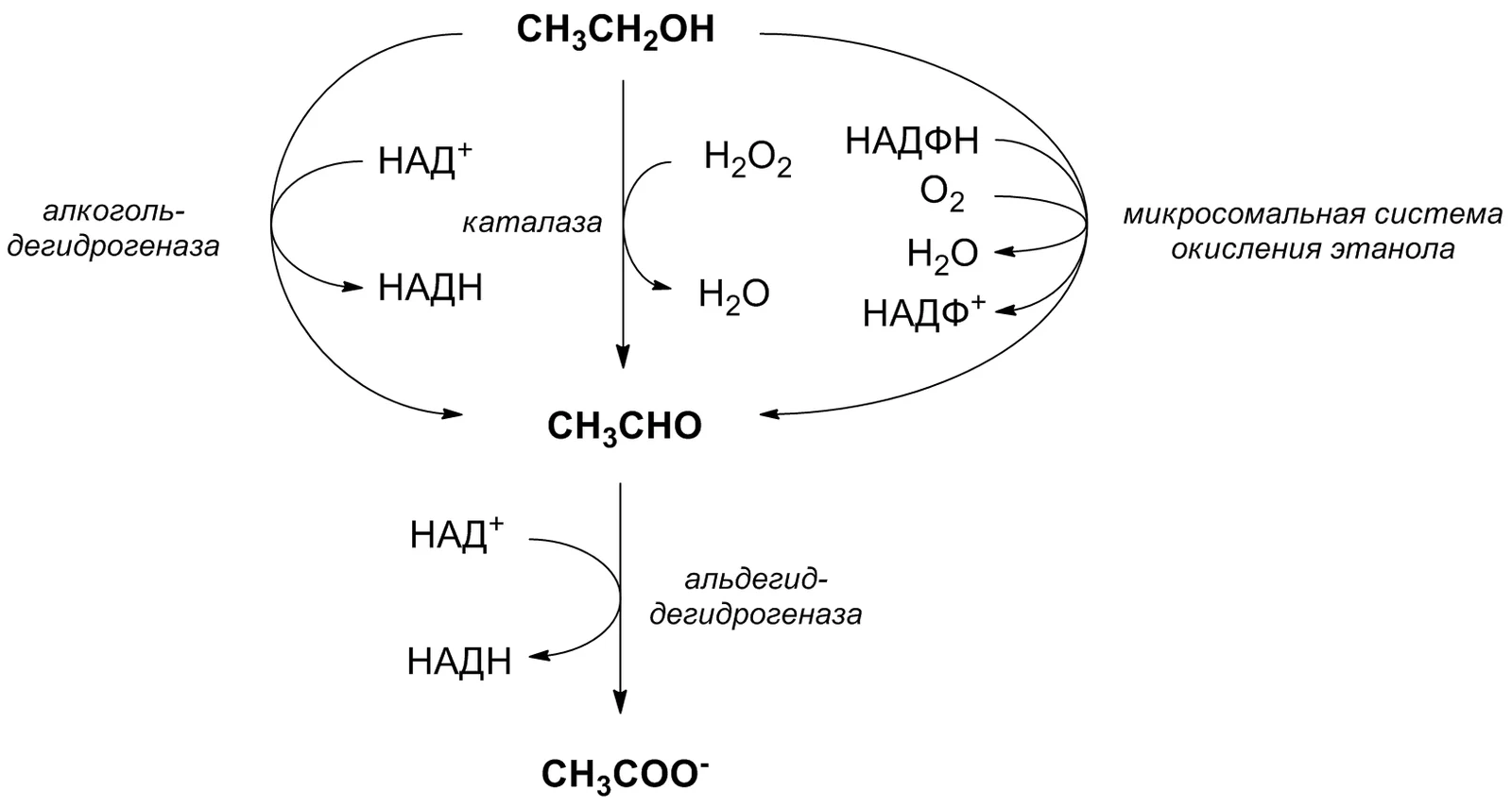
Long description
Ethanol metabolism according to doi:10.1016/0307-4412(90)90174-M

Ethanol metabolism is a central hepatic process that converts the alcohol ingested into non‑toxic metabolites. The liver is the main site, where the reaction proceeds through a series of oxidoreductases that use nicotinamide adenine dinucleotide (NAD⁺) as a co‑factor. The primary product of the first oxidation step is acetaldehyde, a highly reactive compound that is subsequently oxidised to acetate by aldehyde dehydrogenase (ALDH). Acetate is then released into the bloodstream and eventually converted to carbon dioxide and water.
DefinitionKey enzymes
Alcohol dehydrogenase (ADH) catalyses the conversion of ethanol to acetaldehyde; aldehyde dehydrogenase (ALDH) converts acetaldehyde to acetate. Both enzymes use NAD⁺, producing NADH in the process.
The metabolic pathway can be summarised as:
Ethanol → (ADH) → Acetaldehyde → (ALDH) → Acetate → (acetyl‑CoA) → Krebs cycle.
The liver metabolises ethanol mainly through three enzymatic routes: alcohol dehydrogenase (ADH), the microsomal ethanol‑oxidising system (MEOS) involving cytochrome P450 2E1 (CYP2E1), and catalase. ADH1 is the most important enzyme for ethanol oxidation, whereas MEOS and catalase are considered minor alternative pathways. However, recent evidence suggests that the non‑ADH1 pathway may have a greater relevance than previously thought, particularly at higher ethanol concentrations where CYP2E1 activity increases and generates reactive oxygen species (ROS). [2]
The contribution of each pathway varies with ethanol concentration and genetic background. In ADH‑deficient mice, MEOS accounted for a major fraction of ethanol metabolism at low concentrations and remained substantial at high concentrations. In wild‑type mice, MEOS contributed significantly across concentrations, indicating that ADH remains a significant contributor. [2]
ALDH2 is the key enzyme that converts acetaldehyde to acetate. A common polymorphism in East Asian populations produces an inactive ALDH2 enzyme, leading to acetaldehyde accumulation after alcohol consumption. Individuals homozygous for this variant exhibit markedly higher blood alcohol levels and a flushing response, which reduces alcohol intake and lowers the risk of alcoholic cirrhosis, cardiovascular disease, and cancer. [4]
Exam trapGenetic variability matters
Assuming uniform enzyme activity across all individuals ignores the significant impact of ALDH2 polymorphisms on acetaldehyde clearance.
The oxidation of ethanol to acetaldehyde and then to acetate generates NADH, shifting the hepatic redox state towards a more reduced environment. Excess NADH has several downstream effects: [2]
- Inhibition of fatty acid oxidation – High NADH levels inhibit β‑oxidation, leading to hepatic steatosis.
- Promotion of lipogenesis – The surplus NADH drives the conversion of acetyl‑CoA to fatty acids, contributing to fatty liver disease.
- Altered gluconeogenesis – NADH excess suppresses gluconeogenic enzymes, potentially causing hypoglycaemia during chronic alcohol intake.
- Oxidative stress – CYP2E1 activity generates ROS, and the resulting oxidative damage exacerbates liver injury.
These biochemical disturbances underpin the development of alcohol‑related liver disease, including steatosis, hepatitis, fibrosis, and ultimately hepatocellular carcinoma (HCC). To mitigate HCC risk in patients with non‑alcoholic fatty liver disease (NAFLD), smoking cessation, alcohol abstinence, and weight loss are recommended. [1]
Clinical pearlNADH as a metabolic switch
The NADH/NAD⁺ ratio is a critical determinant of hepatic metabolic flux; its elevation by alcohol metabolism drives steatosis and oxidative injury.
| Ethanol concentration (mM) | ADH contribution | MEOS contribution | Catalase contribution |
|---|---|---|---|
| 5–10 | ~0 % (ADH‑deficient) | ~100 % | negligible |
| 40–70 | ~30 % (ADH‑deficient) | ~70 % | negligible |
| 40–70 | ~30 % (ADH‑positive) | ~70 % | negligible |
Ethanol oxidation cascade
- Ethanol intakeIngested alcohol enters bloodstream
- Question: Is ADH active?
Yes
- ADH oxidises ethanol to acetaldehydeUses NAD⁺ → NADH
- ALDH2 oxidises acetaldehyde to acetateUses NAD⁺ → NADH
No
- CYP2E1 (MEOS) oxidises ethanol to acetaldehydeProduces ROS
- ALDH2 oxidises acetaldehyde to acetateUses NAD⁺ → NADH
- Acetate enters systemic circulationConverted to acetyl‑CoA
- Acetyl‑CoA enters Krebs cycleEnergy production
A 35‑year‑old male drinks 40 g of ethanol (≈ 4 mL of 10 % solution). His blood ethanol concentration rises to ~20 mM. ADH oxidises most of this to acetaldehyde, generating ~20 mM NADH. ALDH2 then converts acetaldehyde to acetate, producing an additional ~20 mM NADH. The resulting NADH/NAD⁺ ratio rises sharply, inhibiting fatty acid oxidation and promoting lipid synthesis, which can lead to hepatic steatosis over time.
DefinitionNon‑alcoholic fatty liver disease (NAFLD)
A spectrum of liver conditions characterised by excess triglyceride accumulation in hepatocytes, ranging from simple steatosis to non‑alcoholic steatohepatitis (NASH).
Fatty liver is a common sequela of chronic ethanol consumption. In large epidemiological studies, the prevalence of fatty liver when diagnosed using the fatty liver index (FLI) ranged from 12.6 % to 16.1 %. When transient elastography was employed, the prevalence rose to 42.9 %. In a cohort of 589 living liver donors in Korea, liver biopsy revealed fatty liver in 51 % of cases. These figures underscore the high burden of hepatic steatosis in populations with significant alcohol exposure. [1]
The pathogenesis of ethanol‑induced steatosis involves several interlinked mechanisms:
- Altered lipid metabolism – Ethanol metabolism generates NADH, shifting the redox balance toward fatty acid synthesis and inhibiting β‑oxidation.
- Oxidative stress – Reactive oxygen species (ROS) produced during ethanol oxidation damage mitochondrial function, further impairing fatty acid oxidation.
- Inflammatory signalling – Cytokines such as TNF‑α and IL‑6, released from Kupffer cells, promote lipogenesis and inhibit fatty acid export.
A key enzymatic contributor to ethanol oxidation is catalase (CAT). In the fasted state, when circulating free fatty acids (FFAs) are elevated, CAT activity is up‑regulated. CAT can oxidise ethanol to acetaldehyde via a peroxidatic reaction that depends on hydrogen peroxide (H₂O₂) availability. When H₂O₂ production is low relative to CAT heme content, the peroxidatic pathway dominates, making the rate of ethanol oxidation largely dependent on H₂O₂ generation. In fasted animals, diminished alcohol dehydrogenase (ADH) activity allows CAT to assume a la [2]
The microsomal ethanol‑oxidising system (MEOS) also contributes, especially at higher ethanol concentrations. MEOS has a higher Km for ethanol (~10 mM for CYP2E1 in rats), so it becomes more important when blood ethanol exceeds this threshold. Other CYP isoforms (e.g., CYP1A2) have higher Vmax values but also higher Km, influencing the overall oxidative capacity. [2]
Fructose ingestion potentiates ethanol oxidation, a phenomenon termed the fructose effect. Fructose metabolism increases NADH oxidation, facilitating dissociation of the ADH‑NADH complex, the rate‑limiting step of hepatic ethanol oxidation. However, fructose also reduces H₂O₂ generation, potentially attenuating CAT‑mediated oxidation. [2]
Non‑oxidative pathways, although minor in terms of overall ethanol clearance, generate metabolites such as ethyl glucuronide, ethyl sulfate, phosphatidylethanol (PEth) and fatty acid ethyl esters (FAEE). These compounds persist longer than ethanol itself and serve as biomarkers of alcohol intake. The enzymatic reaction of ethanol with cellular lipids produces FAEE and PEth, which can disrupt cellular signalling and organelle function, contributing to ethanol toxicity. [3]
Clinical pearlKey clinical feature
Ethanol metabolism can precipitate hypoglycaemia and lactic acidosis, particularly in fasting individuals or those with impaired gluconeogenesis.
Ethanol oxidation by ADH and MEOS consumes NAD⁺, producing NADH. The resulting NADH/NAD⁺ imbalance inhibits gluconeogenic enzymes (e.g., pyruvate carboxylase, phosphoenolpyruvate carboxykinase) that require NAD⁺. Consequently, hepatic glucose output falls, leading to hypoglycaemia. In fasting states, when glycogen stores are depleted, the risk is amplified.
The same NADH surplus drives pyruvate conversion to lactate via lactate dehydrogenase, increasing lactate production. Elevated lactate, combined with impaired hepatic clearance, culminates in lactic acidosis. Clinical presentation includes tachypnoea, hypotension, and altered mental status.
- Glucose administration – Intravenous dextrose to correct hypoglycaemia.
- Correction of acidosis – Sodium bicarbonate may be considered in severe cases.
- Monitoring – Serial blood glucose and lactate measurements.
A 35‑year‑old man presents with confusion after binge drinking. Blood glucose is 2.8 mmol/L, lactate 5.0 mmol/L. Immediate IV dextrose (50 % solution) and bicarbonate (1 mmol/kg) are administered. Over 4 h, glucose rises to 5.5 mmol/L and lactate falls to 2.5 mmol/L, with clinical improvement.
| Enzyme | Primary reaction | Key co‑factor | Clinical relevance |
|---|---|---|---|
| ADH | Ethanol → acetaldehyde | NAD⁺ | Rate‑limiting in fed state |
| MEOS (CYP2E1) | Ethanol → acetaldehyde | NADPH | Important at high ethanol |
| CAT | Ethanol → acetaldehyde (peroxidatic) | H₂O₂ | Dominant in fasted state |
| UGT | Ethanol → ethyl glucuronide | UDP‑glucuronic acid | Biomarker formation |
| SULT | Ethanol → ethyl sulfate | PAPS | Biomarker formation |
Ethanol metabolism overview
- Ethanol ingestionEnter bloodstream
- Question: Fed or fasted?
Fed
- ADH oxidationEthanol → acetaldehyde
- MEOS activationCYP2E1, CYP1A2
Fasted
- CAT oxidationEthanol → acetaldehyde via H₂O₂
- Acetaldehyde → acetateALDH
- Acetate → acetyl‑CoAAcetyl‑CoA synthetase
- Metabolic consequencesHypoglycaemia, lactic acidosis, fatty liver
DefinitionEthanol oxidation pathway
Ethanol is oxidised to acetaldehyde by alcohol dehydrogenase (ADH) in the cytosol, then to acetate by aldehyde dehydrogenase (ALDH) in the mitochondria. Minor routes involve the microsomal ethanol‑oxidising system (MEOS) and catalase (CAT).
Disulfiram (Antabuse) is a classic inhibitor of ALDH2. By forming a covalent adduct with the enzyme, it blocks the conversion of acetaldehyde to acetate, causing acetaldehyde to accumulate. The resulting flushing, tachycardia, nausea and headache constitute the “disulfiram reaction” and act as a deterrent to alcohol consumption. Disulfiram is therefore used as a first‑line pharmacological deterrent in alcohol dependence programmes, especially when combined with psychosocial support.
Fomepizole (4‑methylpyrazole) is a potent, selective inhibitor of ADH1. In experimental models it reduces ethanol oxidation by approximately 80 %, leaving the remaining 20 % to be metabolised by non‑ADH1 pathways. Because it does not inhibit ALDH, fomepizole is mainly used in the acute management of methanol or ethylene glycol poisoning, where blocking the initial oxidation step prevents formation of toxic aldehydes and acids. In vitro, fomepizole also inhibits MEOS‑dependent ethanol oxidation by more than 40 % when combined with 1 mM 4‑M‑P. [2] [2]
The most common human polymorphism in ALDH2 is the ALDH2 2 allele, which encodes a catalytically inactive enzyme. Individuals homozygous for 2 (ALDH2*2/2) have virtually no ALDH2 activity, leading to markedly elevated acetaldehyde after alcohol intake. Heterozygotes (ALDH21/*2) have reduced activity (~50 % of normal). The resulting flushing reaction is a protective factor against alcoholism but increases the risk of oesophageal cancer. Genetic testing for ALDH2 variants is increasingly used to personalise alcohol‑related counselling and to predict response to disulfiram therapy. [2]
Exam trapAssuming ADH1 is the sole pathway
Relying solely on ADH1 inhibition to predict ethanol clearance can be misleading. Experimental data show that even when ADH1 is blocked, 20 % of ethanol oxidation persists via non‑ADH1 routes, and at high concentrations (>20 mM) additional enzymes contribute, rendering the process non‑linear.
- Over‑reliance on disulfiram: Disulfiram’s effectiveness depends on patient adherence and the presence of functional ALDH2. In ALDH2*2 carriers, disulfiram offers little additional benefit because the enzyme is already inactive.
- Misinterpreting fomepizole data: Fomepizole’s inhibition of MEOS is significant only at high concentrations and in vitro; in vivo its effect on ethanol metabolism is modest, especially when ADH1 is saturated.
- Ignoring catalase contribution: Although considered minor, CAT can oxidise ethanol in the presence of hydrogen peroxide, particularly when lactate is abundant. Studies show CAT activity can increase ethanol oxidation under certain conditions. [2]
- Assuming linear kinetics: At low ethanol concentrations (<20 mM) metabolism is linear, but above this threshold the rate accelerates disproportionately, reflecting saturation of ADH1 and engagement of other enzymes. [2]
- Neglecting fructose interaction: Fructose can inhibit ethanol metabolism in ADH‑deficient models, suggesting dietary sugars may modulate alcohol clearance. [2]
Clinical pearlClinical indications for fomepizole
Fomepizole is the antidote of choice for methanol or ethylene glycol poisoning, where it blocks the initial oxidation step and prevents formation of toxic metabolites.
- Alcohol dependence treatment: Disulfiram is prescribed in structured programmes for patients with alcohol use disorder, often combined with behavioural therapy. Its deterrent effect is most pronounced in patients who experience the characteristic flushing reaction.
- Acute toxicology: Fomepizole is administered intravenously in cases of methanol or ethylene glycol ingestion. It is preferred over ethanol because it does not require monitoring of blood alcohol levels and has a more predictable pharmacokinetic profile.
- Research settings: Both disulfiram and fomepizole are used experimentally to dissect the relative contributions of ADH1, MEOS and CAT to ethanol metabolism. In animal studies, selective inhibition helps delineate enzyme kinetics and the impact of genetic variants.
- Personalised medicine: Identification of ALDH2 polymorphisms informs risk counselling for alcohol‑related cancers and guides the choice of pharmacological deterrents. Patients with ALDH2*2 may benefit less from disulfiram, whereas heterozygotes may experience a pronounced flushing reaction that can be leveraged therapeutically.
Decision tree for selecting an antidote in alcohol‑related poisoning
- Suspected methanol/ethylene glycol ingestion
- Question: Is the patient stable?
Yes
- Administer fomepizole 15 mg/kg IV
No
- Initiate haemodialysis
- Ethanol is mainly oxidised by ADH1; minor routes involve MEOS and CAT.
- Disulfiram blocks ALDH2, causing acetaldehyde accumulation and a deterrent flushing reaction. [2]
- Fomepizole selectively inhibits ADH1 and is the antidote for methanol/ethylene glycol poisoning.
- ALDH2*2 polymorphism leads to impaired acetaldehyde clearance and increased cancer risk.
- Ethanol metabolism is non‑linear above 20 mM; multiple enzymes contribute. [2]
- Catalase can oxidise ethanol when hydrogen peroxide is available, especially with lactate.
- Fructose can inhibit ethanol oxidation in ADH‑deficient models.
- Genetic testing for ALDH2 variants aids personalised alcohol counselling.
Answer each one in your head first, then open the answer.
What is the primary enzyme responsible for converting ethanol to acetaldehyde?
Show answerHide answer
ADH1Which drug is used to treat methanol poisoning by inhibiting ADH1?
Show answerHide answer
FomepizoleWhat is the consequence of the ALDH2*2 allele after alcohol consumption?
Show answerHide answer
Accumulation of acetaldehyde and flushing reactionAt what ethanol concentration does metabolism become non‑linear?
Show answerHide answer
Above 20 mMWhich minor pathway can oxidise ethanol in the presence of lactate?
Show answerHide answer
Catalase
- Kang SH, Lee HW, Yoo JJ, et al. KASL clinical practice guidelines: Management of nonalcoholic fatty liver disease. Clinical and molecular hepatology 2021;27(3):363-401. doi:10.3350/cmh.2021.0178. Accessed .
- Contreras-Zentella ML, Villalobos-García D, Hernández-Muñoz R. Ethanol Metabolism in the Liver, the Induction of Oxidant Stress, and the Antioxidant Defense System. Antioxidants (Basel, Switzerland) 2022;11(7):1258. doi:10.3390/antiox11071258. Accessed .
- Heier C, Xie H, Zimmermann R. Nonoxidative ethanol metabolism in humans-from biomarkers to bioactive lipids. IUBMB life 2016;68(12):916-923. doi:10.1002/iub.1569. Accessed .
- Shin MJ, Cho Y, Davey Smith G. Alcohol Consumption, Aldehyde Dehydrogenase 2 Gene Polymorphisms, and Cardiovascular Health in Korea. Yonsei medical journal 2017;58(4):689-696. doi:10.3349/ymj.2017.58.4.689. Accessed .
Figure credits
Educational content for medical students and doctors. It is not a substitute for clinical judgement or local guidelines.